Download as PDF, PPTX


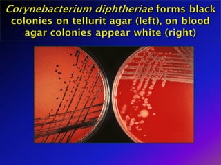
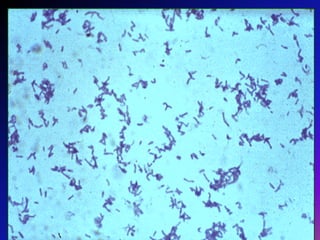
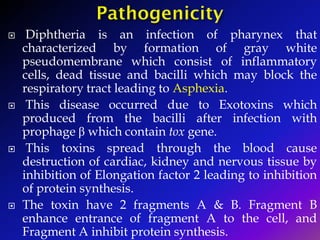
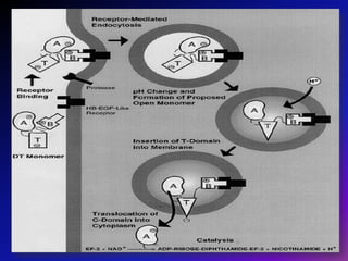

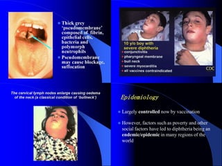
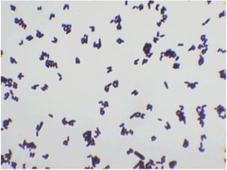


1) In 1884, Loeffler identified Corynebacterium diphtheriae as the bacterial agent that causes diphtheria and described its toxin-producing ability. 2) Diphtheria is an infection of the throat caused by C. diphtheriae that forms a pseudomembrane and releases exotoxins which can damage organs. 3) Diphtheria was largely controlled through vaccination programs using formaldehyde-inactivated toxoid, though remains endemic in some areas due to poverty and social factors.


![CTEV [ clubfoot] DR ARUN LAL ,DR MOHAMED ASHRAF travancore medical college k...](https://cdn.slidesharecdn.com/ss_thumbnails/ctevclubfootdrarunlaldrmohamedashraftravancoremedicalcollegekollamkeralaindia-260208063247-18fc466c-thumbnail.jpg?width=640&height=640&fit=bounds)


