Prescribing Prism for
Diplopia in Neuro-
Ophthalmic Disorders
KELSEY MOODY MILESKI, OD, FAAO
EMORY EYE CENTER
ASSISTANT PROFESSOR OF OPHTHALMOLOGY
SECTION OF OPTOMETRY
 No financial disclosures
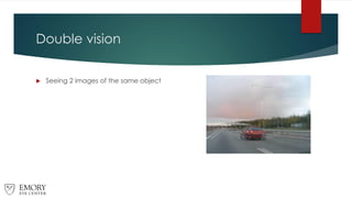
Double vision
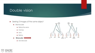
 Seeing 2 images of the same object
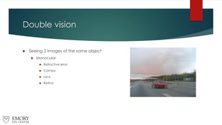
Double vision
 Seeing 2 images of the same object
 Monocular
 Refractive error
 Cornea
 Lens
 Retina
Double vision
 Seeing 2 images of the same object
 Monocular
 Refractive error
 Cornea
 Lens
 Retina
 Binocular
 Not bifoveal
Double vision evaluation
 History is KEY
 BINOCULAR symptom
 Direction
 Duration
 Location
 Associated symptoms
 History of eye turn or abnormal head position
Examination Tools/Techniques
 Sensory
 Motor
Examination Tools/Techniques
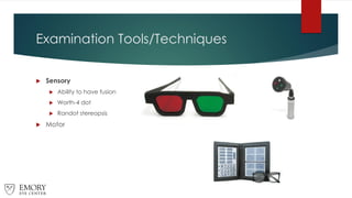
 Sensory
 Ability to have fusion
 Worth-4 dot
 Randot stereopsis
 Motor
Examination Tools/Techniques
 Sensory
 Motor
 Ductions/Versions
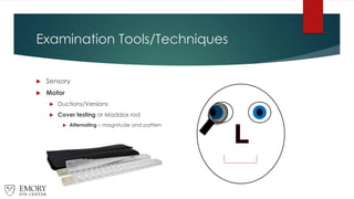
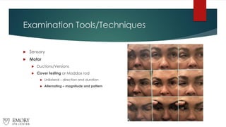
 Cover testing or Maddox rod
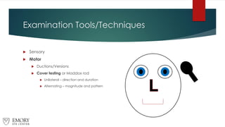
Examination Tools/Techniques
 Sensory
 Motor
 Ductions/Versions
 Cover testing or Maddox rod
 Unilateral – direction and duration
 Alternating – magnitude and pattern
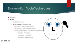
Examination Tools/Techniques
 Sensory
 Motor
 Ductions/Versions
 Cover testing or Maddox rod
 Unilateral – direction and duration
 Alternating – magnitude and pattern
 When eye is covered, it will move to resting position
 ESOTROPIA - EYE RESTS IN
 EXOTROPIA – EYE RESTS OUT
 HYPERTROPIA – EYE RESTS UP
 HYPOTROPIA – EYE RESTS DOWN
Examination Tools/Techniques
 Sensory
 Motor
 Ductions/Versions
 Cover testing or Maddox rod
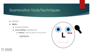
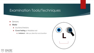
 Unilateral – tells you direction and duration
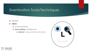
Examination Tools/Techniques
 Sensory
 Motor
 Ductions/Versions
 Cover testing or Maddox rod
 Unilateral – tells you direction and duration
ESOTROPIA
Examination Tools/Techniques
 Sensory
 Motor
 Ductions/Versions
 Cover testing or Maddox rod
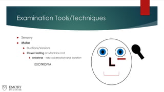
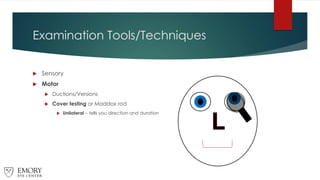
 Unilateral – tells you direction and duration
Examination Tools/Techniques
 Sensory
 Motor
 Ductions/Versions
 Cover testing or Maddox rod
 Unilateral – tells you direction and duration
EXOTROPIA
Examination Tools/Techniques
 Sensory
 Motor
 Ductions/Versions
 Cover testing or Maddox rod
 Unilateral – tells you direction and duration
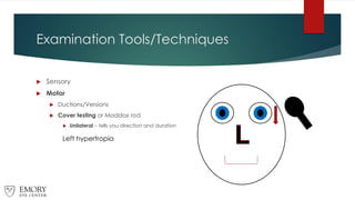
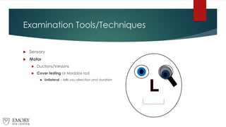
Examination Tools/Techniques
 Sensory
 Motor
 Ductions/Versions
 Cover testing or Maddox rod
 Unilateral – tells you direction and duration
Left hypertropia
Examination Tools/Techniques
 Sensory
 Motor
 Ductions/Versions
 Cover testing or Maddox rod
 Unilateral – tells you direction and duration
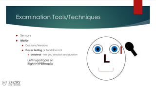
Examination Tools/Techniques
 Sensory
 Motor
 Ductions/Versions
 Cover testing or Maddox rod
 Unilateral – tells you direction and duration
Left hypotropia or
Right HYPERtropia
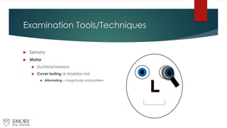
Examination Tools/Techniques
 Sensory
 Motor
 Ductions/Versions
 Cover testing or Maddox rod
 Alternating – magnitude and pattern
Examination Tools/Techniques
 Sensory
 Motor
 Ductions/Versions
 Cover testing or Maddox rod
 Alternating – magnitude and pattern
Examination Tools/Techniques
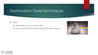
 Prism
 Refracting surface that bends light
 Used in patients with diplopia to move image to the eye
Examination Tools/Techniques
 Prism
 Refracting surface that bends light
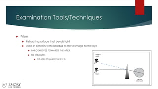
 Used in patients with diplopia to move image to the eye
 IMAGE MOVES TOWARDS THE APEX
 TO MEASURE:
 PUT APEX TO WHERE THE EYE IS
Examination Tools/Techniques
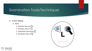
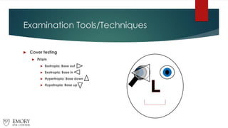
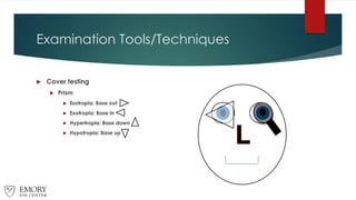
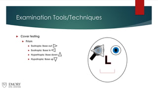
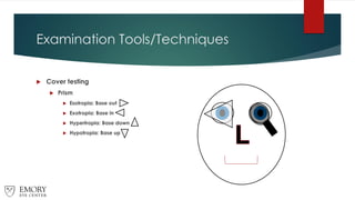
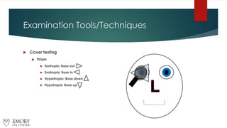
 Cover testing
 Prism
 Esotropia: Base out
 Exotropia: Base in
 Hypertropia: Base down
 Hypotropia: Base up
Examination Tools/Techniques
 Cover testing
 Prism
 Esotropia: Base out
 Exotropia: Base in
 Hypertropia: Base down
 Hypotropia: Base up
Examination Tools/Techniques
 Cover testing
 Prism
 Esotropia: Base out
 Exotropia: Base in
 Hypertropia: Base down
 Hypotropia: Base up
Examination Tools/Techniques
 Cover testing
 Prism
 Esotropia: Base out
 Exotropia: Base in
 Hypertropia: Base down
 Hypotropia: Base up
Examination Tools/Techniques
 Cover testing
 Prism
 Esotropia: Base out
 Exotropia: Base in
 Hypertropia: Base down
 Hypotropia: Base up
Examination Tools/Techniques
 Cover testing
 Prism
 Esotropia: Base out
 Exotropia: Base in
 Hypertropia: Base down
 Hypotropia: Base up
Examination Tools/Techniques
 Sensory
 Motor
 Ductions/Versions
 Cover testing or Maddox rod
 Unilateral – direction and duration
 Alternating – magnitude and pattern
Examination Tools/Techniques
 Sensory
 Motor
 Ductions/Versions
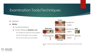
 Cover testing or Maddox rod
 Subjective response from patient
 Exo and hyper’s are crossed
 Eso and hypo are uncrossed
Biousse V, Newman NJ Neuro-ophthalmology Illustrated 2nd ed New York : Thieme; 2016:349-
350
Examination Tools/Techniques
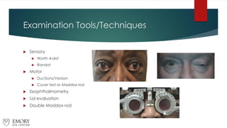
 Sensory
 Worth 4-dot
 Randot
 Motor
 Ductions/Version
 Cover test or Maddox rod
 Exophthalmometry
 Lid evaluation
 Double Maddox rod
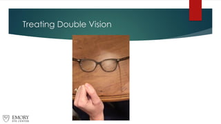
Treating Double Vision
 Rule out pathologic cause
 Neuro-imaging
 Bloodwork
 Nonsurgical
 Surgical
Treating Double Vision
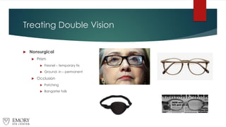
 Nonsurgical
 Prism
 Fresnel – temporary fix
 Ground- in – permanent
 Occlusion
 Patching
 Bangarter foils
Treating Double Vision
 Prism
 Goal is binocular vision in primary gaze
 Apex of prism is where the eye is
 Exotropia: base in prism
 Esotropia: base out prism
 Right hypertropia: base down prism
 Right hypotropia: base up prism
 May need 2 pairs of glasses if deviation is different at distance and near
Treating Double Vision
 Fresnel prism
 Press-on lens for alleviation
of diplopia
 Can use initially to
determine if patient can
adapt to prism
 Good to use if etiology is still
under investigation or
alignment may change
 Advantages
 Can be put on glasses
immediately in clinic for
relief
 Easy to change
 Lightweight
 Can be prescribed up to 40
PD! – Ideal for large
deviations
 Inexpensive
 Disadvantages
 Blur
 Only use in front of one
eye
 Visible
 Hard (but possible) to
correct both horizontal and
vertical deviations
simultaneously
Treating Double Vision
 Ground-in prism
 Prism built into glasses
prescription
 Good to use when
deviation is stable
 Advantages
 Two clear lenses
 Can be split between
right and left eye to
make lenses symmetric
 Not easily visible
 Easily corrects vertical
and horizontal
 Disadvantages
 Can only make up to 20
PD total
 Time to be made
 Expensive
 May need multiple
changes
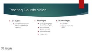
Treating Double Vision
 Occlusion
 Goal is monocular
vision to alleviate
diplopia
 Advantages
 Diplopia resolves in
all positions of gaze
 No additional
glasses needed
 Immediate relief
 Inexpensive
 Disadvantages
 Cosmesis
 Lack of binocular
vision
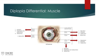
Diplopia Differential: Muscle
1. Elevation
2. Intorsion
3. Adduction
1. Adduction
1. Depression
2. Extorsion
3. Adduction
1. Extorsion
2. Depression (on abduction)
3. Abduction
1. Abduction
1. Intorsion
2. Depression (on
adduction)
3. Abduction
Diplopia Differential: Nerve
1. Elevation – SR and IO
2. Depression – IR
3. Adduction – MR
4. Eyelid elevation: levator
5. Pupil: constriction
1. Intorsion
2. Depression (on adduction)
3. Abduction
1. Abduction
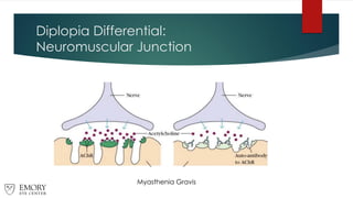
Diplopia Differential:
Neuromuscular Junction
Myasthenia Gravis
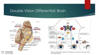
Double Vision Differential: Brain
Dinkin M. Diagnostic Approach to Diplopia. Continuum (Minneap Minn)
2014;20(4):942-965
Double Vision Differential
 Brain
 Internuclear ophthalmoplegia
 Gaze palsy
 Skew deviation
 Dorsal midbrain syndrome
 Neurodegenerative disease
 Nerve
 CN III
 CN IV
 CN VI
 Neuromuscular junction
 Myasthenia gravis
 Muscle
 Thyroid eye disease
 Orbital fracture
 Orbital mass
 Inflammation
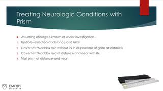
Treating Neurologic Conditions with
Prism
 Assuming etiology is known or under investigation…
1. Update refraction at distance and near
2. Cover test/Maddox rod without Rx in all positions of gaze at distance
3. Cover test/Maddox rod at distance and near with Rx
4. Trial prism at distance and near
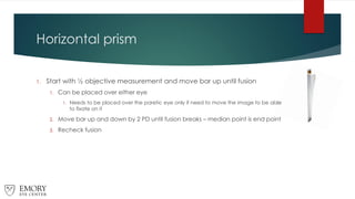
Horizontal prism
1. Start with ½ objective measurement and move bar up until fusion
1. Can be placed over either eye
1. Needs to be placed over the paretic eye only if need to move the image to be able
to fixate on it
2. Move bar up and down by 2 PD until fusion breaks – median point is end point
3. Recheck fusion
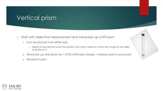
Vertical prism
1. Start with objective measurement and move bar up until fusion
1. Can be placed over either eye
1. Needs to be placed over the paretic eye only if need to move the image to be able
to fixate on it
2. Move bar up and down by 1-2 PD until fusion breaks – median point is end point
3. Recheck fusion
Horizontal + Vertical Prism
1. Neutralize larger deviation first (will still be diplopic)
1. Goal: Move image until they are lined up vertically or horizontally
2. Add in second prism over fellow eye
3. If end point is difficult, trial frame Rx and prism and make small changes (1
PD)
4. Can be challenging and time consuming but obtainable
Treating Neurologic Conditions with
Prism
 Start with Fresnel first
 Etiology unknown
 Deviation may change
 Consider ground-in prism
 If any deficit remains
 Once deficit is stable
 Consider referral for surgical intervention
Treating Neurologic Conditions with
Prism
 When to refer for surgery
 Constant diplopia
 Cover testing has been stable for ~1 year
 Unlikely to progress
 Not undergoing treatment, etc.
 Able to achieve fusion with prism
 Instead of moving the image to the eye, surgery moves the eye to the image
 If unable to fuse with prism, likely won’t be able to fuse after surgery
 Goal is single vision in primary gaze – may still be diplopic in other gazes
Yoked prism?
 Goal: move the image out of defect
 Hemianopsia
 Nystagmus
 Abnormal head posture
 Generally a 2 D prism shifts the image by 1 degree
 Max 20PD in ground-in glasses
 Shift the field by only 10 degrees
 Higher amounts with Fresnel
 Will require 2 in front of each eye which creates blur and distortion
 Better to place sectorially
Yoked prism?
Yoked prism?
 Visual neglect
 Common in right brain stroke
 Patients ignore left side
 Turn head to right
 Extinction phenomenon when presented with simultaneous stimulus
 Prism adaption
 Yoked prism to right and perform exercises
 Overcompensate to the right
 With practice, adapt
 Remove prism and naturally move into neglected field
Prism challenges
 Other ocular comorbidities
 Maculopathies
 Asymmetric visual acuity
 Glaucoma and/or other visual field defects
 Not wanting to come out of progressive lenses or have separate glasses
 >40D of prism needed
 Nystagmus
 Suppression
CASES
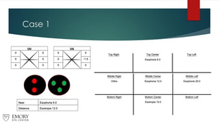
Case 1
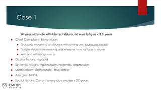
54 year old male with blurred vision and eye fatigue x 2.5 years
 Chief Complaint: Blurry vision:
 Gradually worsening at distance with driving and looking to the left
 Double vision in the evening and when he turns his face to shave
 With and without glasses on
 Ocular history: myopia
 Systemic history: Hypercholesterolemia, depression
 Medications: Atorvastatin, duloxetine
 Allergies: NKDA
 Social history: Current every day smoker x 27 years
Case 1
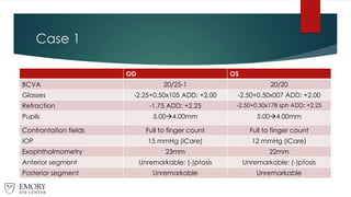
OD OS
BCVA 20/25-1 20/20
Glasses -2.25+0.50x10⁵ ADD: +2.00 -2.50+0.50x007 ADD: +2.00
Refraction -1.75 ADD: +2.25 -2.50+0.50x178 sph ADD: +2.25
Pupils 5.00→4.00mm 5.00→4.00mm
Confrontation fields Full to finger count Full to finger count
IOP 15 mmHg (iCare) 12 mmHg (iCare)
Exophthalmometry 23mm 22mm
Anterior segment Unremarkable; (-)ptosis Unremarkable; (-)ptosis
Posterior segment Unremarkable Unremarkable
Case 1
Case 1
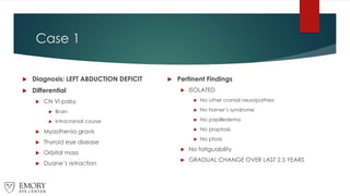
 Diagnosis: LEFT ABDUCTION DEFICIT
 Differential
 CN VI palsy
 Brain
 Intracranial course
 Myasthenia gravis
 Thyroid eye disease
 Orbital mass
 Duane’s retraction
 Pertinent Findings
 ISOLATED
 No other cranial neuropathies
 No horner’s syndrome
 No papilledema
 No proptosis
 No ptosis
 No fatiguability
 GRADUAL CHANGE OVER LAST 2.5 YEARS
Case 1: POLL
54 year old male with blurred vision and eye fatigue x 2.5 years
What type of prism would you like to prescribe?
1. Base in Fresnel prism
2. Base out Fresnel prism
3. Base in ground-in prism
4. Base out ground-in prism
Case 1
 Started with 6 BO prism
 Still diplopic
 Increased prism to 10 BO to obtain fusion
 Improved clarity when increased to 12 BO
 Blurrier when increased to 14 BO
Case 1
 Fit with 12 BO Fresnel prism
 Needs temporary fix
 Able to tolerate at distance and near
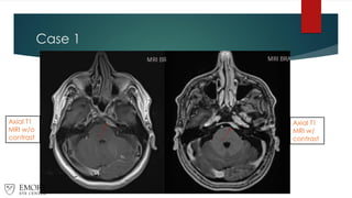
 Order MRI of the brain and orbits W/W/O contrast
Case 1
Axial T1
MRI w/o
contrast
Axial T1
MRI w/
contrast
Case 1
 4-month F/U visit
 Referred to neurosurgery
 MRI monitoring vs radiotherapy
 Plans to monitor with MRI only
 Motility and cover testing stable
 Bothered by cosmesis of Fresnel and requests ground in prism
Case 1
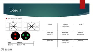
 8-month F/U visit
Case 1
 Fit with 20 BO Fresnel prism OVER GROUND IN PRISM (12 BO)
 Needs temporary fix
 Able to tolerate at distance. Will remove glasses for reading
 Refer to radiation oncology
 F/U after treatment
Case 1
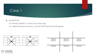
 4 month F/U
 Finished radiation 1 month and 10 days ago
 Hopeful for ground-in prism.- took off 20 BO Fresnel and still diplopic
Case 1
 Fit with 12 BO Fresnel prism OVER GROUND IN PRISM (12 BO)
 24 BO too much for ground-in prism
 Refer for strabismus surgery consult
 Educated that surgery will not be recommended for several months
Case 2
Case 2
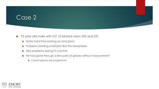
 72 year old male with CC of blurred vision OD and OS
 Notes hard time looking up and down
 Problems reading small print like the newspaper
 Also problems seeing TV controls
 He had gone through a few pairs of glasses without improvement
 Current glasses are progressives
Case 2
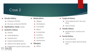
 Ocular history:
 Cataract OD/OS
 Dry eye syndrome OD/OS
 Ophthalmic meds: none
 Systemic history:
 Arthritis
 Atrial fibrillation
 Hypertension
 Hypercholesterolemia
 Obstructive sleep apnea
 Progressive supranuclear palsy
 Medications
 Allegra
 Amlodipine
 Carvedilol
 Eliquis
 Furosemide
 Losartan
 Lovastatin
 Memantine
 Allergies:
 NKDA
 Surgical history:
 Joint replacement: hip and
knee
 Hernia repair
 Family history:
 Hypertension: mother and
father
 Social history
 Social alcohol
 Former smoker: Quit 33 years
ago
Case 2
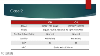
OD OS
BCVA 20/30-2 PH: 20/20 20/25 PH: 20/20
Pupils Equal, round, reactive to light; no RAPD
Confrontation Fields Normal Normal
Motility Restricted Restricted
IOP 14 15
NPC Reduced at 20 cm
Case 2
Near
Case 2
OD OS
Eyelids
2+ blepharitis and
vascularization
2+ blepharitis and
vascularization
Cornea
Tear debris
1+ SPK
Tear debris
1+ SPK
Lens 1-2+ NS 1-2+ NS
Optic disc 0.30/0.30 0.35/0.35
Retina Normal Normal
Case 2 POLL
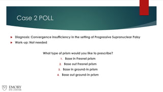
 Diagnosis: Convergence insufficiency in the setting of Progressive Supranuclear Palsy
 Work-up: Not needed
What type of prism would you like to prescribe?
1. Base in Fresnel prism
2. Base out Fresnel prism
3. Base in ground-in prism
4. Base out ground-in prism
Case 2
 Updated glasses Rx and trial framed near Rx
 Started with 10 BI prism at near
 Still diplopic
 Increased prism to 14 BI to obtain fusion
 Blurrier when increased to 12 BI or decreased to 16 BI
 Yoked prism attempted but no benefit
 Trialed 2 BI prism at distance and uncomfortable
Case 2
72 year old male with blurry vision
 Take out of progressive lenses
 Fit with 14 BI ground-in prism in NVO
 Hold reading material higher
 Aids to bring reading material higher
 Prescribed DVO without prism
 F/U in 4-6 weeks
Case 3
Case 3
55 year old female with double vision and headache
 Chief complaint: double vision
 Started 3 days ago while driving
 Constant
 Associated symptoms
 Mild blurry vision OD
 Headache
 Ocular history: Dry eye OU
 Systemic history: diabetes (HbA1c 5.8%), hypertension (200/131), asthma
 Medications: albuterol, amlodipine, aspirin, atorvastatin, duloxetine,
hydrochlorothiazide-losartan, metformin, nortriptyline, zantac
 Surgical history: spinal stimulator, hemorrhoidectomy, C-sectionx2
 Allergies: latex, codeine, penicillins, sulfadiazine
 Social history: unremarkable
Case 3
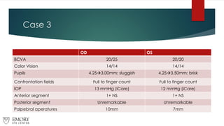
OD OS
BCVA 20/25 20/20
Color Vision 14/14 14/14
Pupils 4.25→3.00mm; sluggish 4.25→3.50mm; brisk
Confrontation fields Full to finger count Full to finger count
IOP 13 mmHg (iCare) 12 mmHg (iCare)
Anterior segment 1+ NS 1+ NS
Posterior segment Unremarkable Unremarkable
Palpebral aperatures 10mm 7mm
Case 3
Case 3
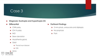
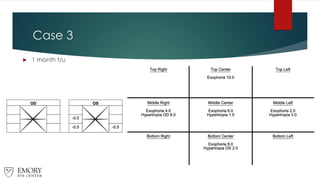
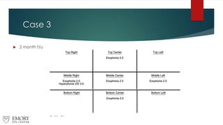
 Diagnosis: Exotropia and hypertropia OS
 Differential
 CNIII palsy
 CN VI palsy
 INO
 Skew deviation
 Myasthenia gravis
 Orbit
 Thyroid eye disease
 IOIS
 GCA
 Pertinent Findings
 3 mm ptosis, anisocoria and diplopia
 No proptosis
 Pain
Case 3: POLL
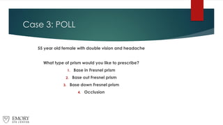
55 year old female with double vision and headache
What type of prism would you like to prescribe?
1. Base in Fresnel prism
2. Base out Fresnel prism
3. Base down Fresnel prism
4. Occlusion
Case 3
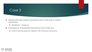
 Started with 8 BI rotated clockwise in front of left eye to correct
hypotropia
 Still diplopic – diagonal
 Increased to 10 BI rotated clockwise in front of left eye
 Fusion in primary gaze but diplopic with small eye movements
Case 3
55 year old female with double vision and headache
 Recommend occlusion
 Non-comitant
 Unable to fuse with prism
 Concern for CN III with pupil involvement
 Order CTA head to rule out PCOM aneurysm and MRI brain and orbits W/W/O
contrast
 Pending normal imaging consider work-up for MG
Neuro-imaging NORMAL!
Case 3
 1 month f/u
Case 3
 2 month f/u
Clinical Pearls
• Prism can be challenging and time consuming
• Schedule appropriate time slots
• Look for neuro-ophthalmic etiology is patient has not undergone work-
up
• Set patient expectations for prism
• Always bring the patient back for follow-up
• Check to make sure glasses were made correctly
• Don’t be afraid to make changes
• It’s okay to sometimes recommend occlusion
• Eye patch, bangarter or occlusion CL
References
 Cornblath WT. Diplopia due to ocular motor cranial neuropathies. Continuum (Minneap
Minn) 2014;20(4):966-980
 Dinkin M. Diagnostic Approach to Diplopia. Continuum (Minneap Minn) 2014;20(4):942-
965
 Eggenberger ER. Supranuclear eye movement abnormalities. Continuum (Minneap Minn)
2014;20(4):981-992
 Nerrant E, Tilikete C. Ocular Motor Manifestations of Multiple Sclerosis. J Neuroophthalmol.
2017 Sep;37(3):332-340
 Peragallo JH, Newman NJ. Diplopia-An Update. Semin Neurol. 2016 Aug;36(4):357-361
 Tamhankar MA, Biouuse V, Gui-Shuang Y, et al. Isolated third, fourth and sixth cranial
nerve palsies from presumed microvascular versus other causes: a prospective study.
Ophthalmology. 2013 Nov; 120(11):1-13