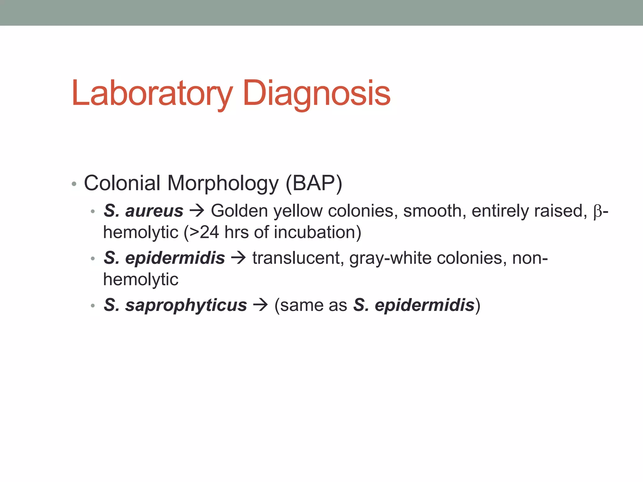
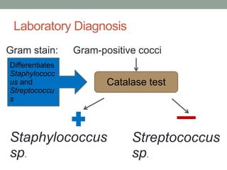
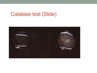
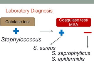
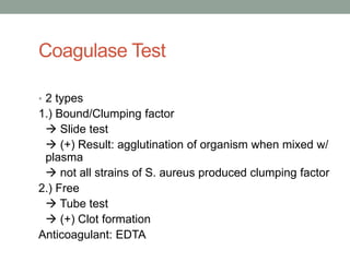
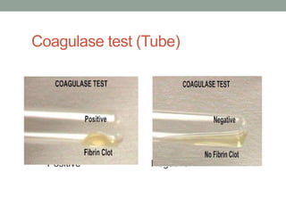
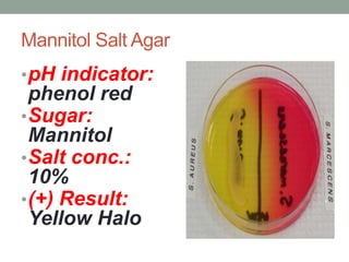
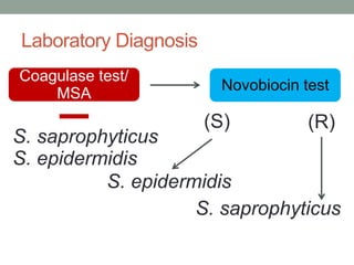
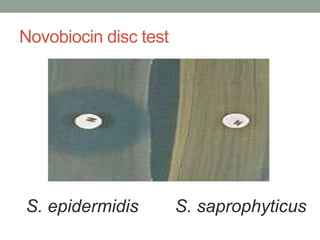
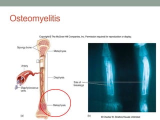
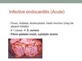
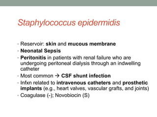
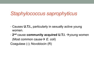
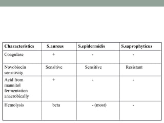
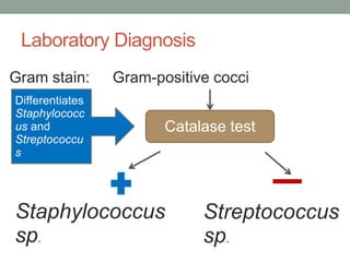
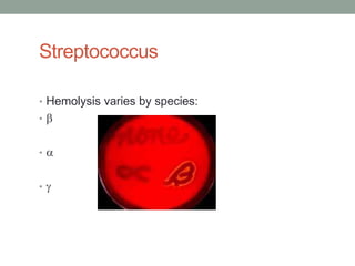
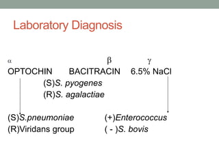
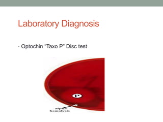
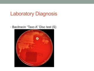
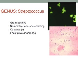
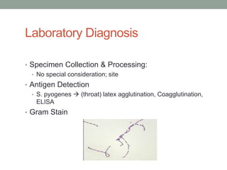
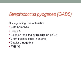
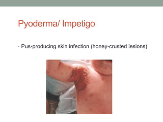
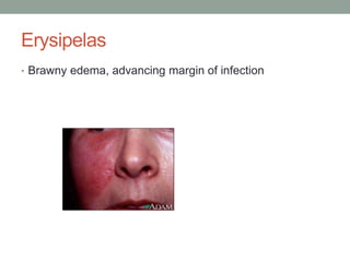
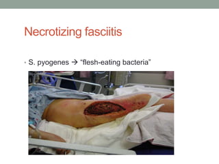
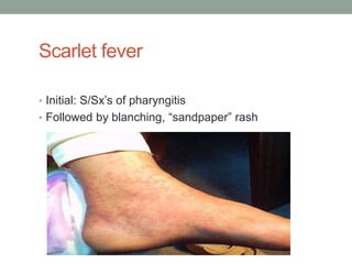
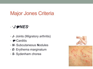
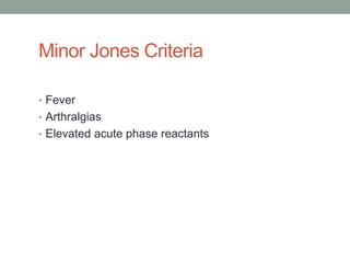
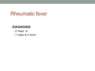
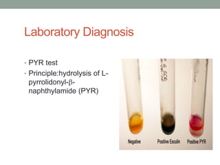
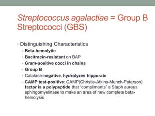
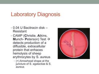
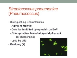
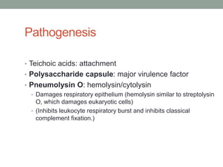
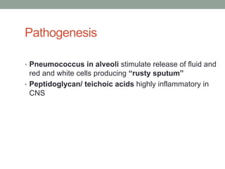
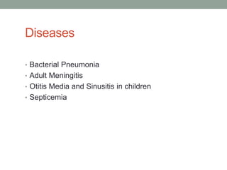
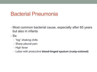
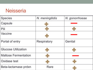
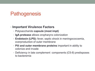
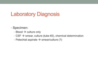
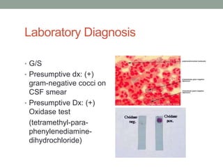
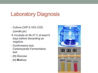
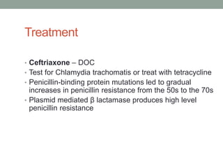
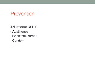
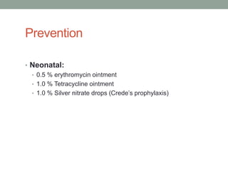
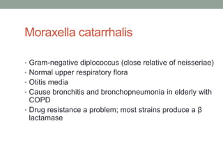
Bangalore Call Girls Marathahalli 📞 9907093804 High Profile Service 100% Safe
Cocci 2011
1. Laboratory Diagnosis
• Colonial Morphology (BAP)
• S. aureus Golden yellow colonies, smooth, entirely raised, -
hemolytic (>24 hrs of incubation)
• S. epidermidis translucent, gray-white colonies, non-
hemolytic
• S. saprophyticus (same as S. epidermidis)
2. Laboratory Diagnosis
Gram stain: Gram-positive cocci
Differentiates
Staphylococc
us and Catalase test
Streptococcu
s
Staphylococcus Streptococcus
sp. sp.
6. Coagulase Test
• 2 types
1.) Bound/Clumping factor
Slide test
(+) Result: agglutination of organism when mixed w/
plasma
not all strains of S. aureus produced clumping factor
2.) Free
Tube test
(+) Clot formation
Anticoagulant: EDTA
15. Transmission
• Spread via the hands and sneezing
• Fomites
• Surgical wounds
• Lungs of cystic fibrosis patients
• Foods associated with food poisoning
(Ham/Canned meats, Custard pastries and potato
salad)
16. Predisposing Factors for Infections
• Any break in skin (sx)
• Any foreign body (sx packing, sutures, tampons)
• Ventilators
• WBC <500/ L
• Dse: CF, CGD
• IV drug abuse
27. Infective endocarditis (Acute)
• Fever, malaise, leukocytosis, heart murmur (may be
absent initially)
• # 1 cause S. aureus
• Fibrin platelet mesh, cytolytic toxins
28. B) INTOXICATIOINS:
The disease is caused by the bacterial exotoxins,
which are produced either in the infected host
or preformed in vitro.
There are 3 types-
1. Food poisoning
2. Toxic shock syndrome
3. Staphylococcal scalded skin syndrome
29. Gastroenteritis (food poisoning)
• 1-8 hours after ingesting toxin
• Nausea
• abdominal pain
• Vomiting
• followed by diarrhea
• No fever
• Enterotoxins A-F preformed in food (heat-stable)
32. Laboratory Diagnosis:
Specimens collected: Depends on the type of infection.
• Suppurative lesion- Pus,
• Respiratory infection- Sputum,
• Bacteremia & septicemia- Blood,
• Food poisoning- Feces, vomit & the remains of
suspected food,
• For the detection of carriers- Nasal swab.
33. Treatment
• Methicillin/ Nafcillin/ Oxacillin/ Cloxacillin
• Methicillin-resistant S. aureus (MRSA) (due to changes
in major penicillin-binding proteins) is commonly
resistant to all antibiotics EXCEPT Vancomycin and
Fusidic acid.
• Topical mupirocin reduces nasal colonization.
35. Staphylococcus epidermidis
• Reservoir: skin and mucous membrane
• Neonatal Sepsis
• Peritonitis in patients with renal failure who are
undergoing peritoneal dialysis through an indwelling
catheter
• Most common CSF shunt infection
• Infxn related to intravenous catheters and prosthetic
implants (e.g., heart valves, vascular grafts, and joints)
• Coagulase (-); Novobiocin (S)
36. Staphylococcus saprophyticus
• Causes U.T.I., particularly in sexually active young
women.
• 2nd cause community acquired U.T.I. young women
(Most common cause E. coli)
Coagulase (-); Novobiocin (R)
38. Laboratory Diagnosis
Gram stain: Gram-positive cocci
Differentiates
Staphylococc
us and Catalase test
Streptococcu
s
Staphylococcus Streptococcus
sp. sp.
45. Streptococci
• Are serogrouped using known antibodies to the cell wall
carbohydrates
(Lancefield’s Group A-H, K-U)
• Group A- Rhamnose-N-acetylglucosamine
• Group B-Rhamnose-glucosamine polysaccharide
• Group C-Rhamnose-N-acetylgalactosamine
• Group D- Glycerol teichoic acid
• Group F- Glucopyranosyl-N-acetylgalactosamine
46. Laboratory Diagnosis
• Specimen Collection & Processing:
• No special consideration; site
• Antigen Detection
• S. pyogenes (throat) latex agglutination, Coagglutination,
ELISA
• Gram Stain
49. Streptococcus pyogenes (GABS)
Distinguishing Characteristics
Beta-hemolytic
Group A
Colonies inhibited by Bacitracin on BA
Gram-positive cocci in chains
Catalase-negative
PYR (+)
53. Cell wall components
• Hyaluronic acid capsule (a polysaccharide) is non-
immunogenic; inhibits phagocytic uptake
• M-protein: major virulence factor, hair-like projections;
antiphagocytic, used to type group A Strep
55. Exotoxins A-C
(pyrogenic/erythrogenic)
• Phage-coded (e.g., the cells are lysogenized by a
phage)
• Cause fever and the rash of Scarlet fever
• Inhibit liver clearance of endotoxin (from normal
flora), creating shock-like conditions
• Superantigens: activate many helper T cells by bridging
T cell receptors and MHC class II markers without
processed antigen
56. Spreading factors:
• Streptokinase (fibrinolysin): breaks down fibrin clot
• Streptococcal Dnase (Streptodornase): liquefies pus,
extension of lesion
• Hyaluronidase: hydrolyzes the ground substances of
the connective tissues; important to spread in cellulitis
66. Non-suppurative Sequelae to Group
A Streptococcal Infections
• Rheumatic fever
• Acute glomerulonephritis (M12 serotype)
67. Rheumatic fever
• Sequelae to : Pharyngitis with group A Strep
(not group C)
• Mechanism: in genetically susceptible individuals, the
infection results in production of antibodies that cross-
react with cardiac antigens
68. Rheumatic fever
• Symptoms occurs 2-3 weeks after a pharyngeal
infection
• Lab: elevated ASO titers (>200)
• Jones Criteria
76. Streptococcus agalactiae = Group B
Streptococci (GBS)
• Distinguishing Characteristics
• Beta-hemolytic
• Bacitracin-resistant on BAP
• Gram-positive cocci in chains
• Group B
• Catalase-negative, hydrolyzes hippurate
• CAMP test-positive: CAMP(Christie-Atkins-Munch-Peterson)
factor is a polypeptide that “compliments” a Staph aureus
sphingomyelinase to make an area of new complete beta-
hemolysis
80. Diseases
• Neonatal septicemia
• Neonatal meningitis (Neonate – 2 mths)
• Most common causative agent ( GEL)
# 1 – S. agalactiae (GBS)
2 – E. coli
Rare: L. monocytogenes
81. Laboratory Diagnosis
• 0.04 U Bacitracin disk –
Resistant
• CAMP (Christie, Atkins,
Munch- Peterson) Test
detects production of a
diffusible, extracellular
protein that enhaces
hemolysis of sheep
erythrocytes by S. aureus
• (+) Arrowhead shape at the
juncture of S. agalactiae & S.
aureus
83. Prevention
• Treat mother prior to delivery if she had a previous baby
with GBS, has documented GBS colonization, or
prolonged rupture of membranes
88. Pathogenesis
• Teichoic acids: attachment
• Polysaccharide capsule: major virulence factor
• Pneumolysin O: hemolysin/cytolysin
• Damages respiratory epithelium (hemolysin similar to streptolysin
O, which damages eukaryotic cells)
• (Inhibits leukocyte respiratory burst and inhibits classical
complement fixation.)
89. Pathogenesis
• Pneumococcus in alveoli stimulate release of fluid and
red and white cells producing “rusty sputum”
• Peptidoglycan/ teichoic acids highly inflammatory in
CNS
91. Bacterial Pneumonia
• Most common bacterial cause, especially after 65 years
but also in infants
• Sx:
• “big” shaking chills
• Sharp pleural pain
• High fever
• Lobar with productive blood-tinged sputum (rusty-colored)
92. Predisposing Conditions for
Pneumonia
• Antecedent influenza or measles infection:
damage to mucociliary elevator
• Chronic obstructive pulmonary disorders
• Congestive heart failure
• Alcoholism
• Asplenia predisposes to septicemia
96. Treatment
• Penicillin G DOC
• Resistance (both low level and high level) is
chromosomal (altered penicillin-binding proteins); major
concern in meningitis (Vancomycin Rifampin used)
98. Viridans Streptococci (S. sanguis, S.
mutans, etc.)
• Distinguishing Characteristics
• Alpha-hemolytic, resistant to optochin
• Gram-positive cocci in chains
• NOT bile soluble
103. Pathogenesis
• Dextran (biofilm)-mediated adherence onto tooth
enamel or damaged heart valve and to each other
(vegetation). Growth in vegetation protects organism
from immune system.
109. Pathogenesis/ Predisposing
Conditions
• Bile/ Salt tolerance allows survival in bowel and gall
bladder
• During medical procedures on GI or GU tract:
E. faecalis bloodstream previously damaged
valves ENDOCARDITIS (SBE)
111. Treatment
• All strains carry some drug resistance
• Some vancomycin-resistant strains of Enterococcus
faecium or E. faecalis: no reliably effective treatment
112. Prevention
• Prophylactic use of penicillin and gentamicin in patients
with damaged heart valves prior to intestinal or urinary
tract manipulation
116. Neisseria
Species N. meningitidis N. gonorrhoeae
Capsule
Pili
Vaccine
Portal of entry Respiratory Genital
Glucose Utilization
Maltose Fermentation
Oxidase test
Beta-lactamase prdxn Rare
117. Neisseria meningitidis
(meningococcus)
• Distinguishing Characteristics
• Gram-negative kidney bean-shaped diplococci
• Large capsule
• Grows on chocolate (not blood) agar in 5-10% CO2
• Ferments maltose
• Oxidase positive
• 13 Serogroups: A, B, C, D,29E, H, I, K,L,X,Y,Z & W-135
119. Transmission
• Respiratory droplets
• Oropharyngeal colonization
• Spread to the meninges via the bloodstream
• Disease occurs in only small percent of colonized
120. Pathogenesis
• Important Virulence Factors
• Polysaccharide capsule (most impt)
• IgA protease allows oropharynx colonization
• Endotoxin (LPS): fever, septic shock in meningococcemia,
overproduction of outer membrane
• Pili and outer membrane proteins important in ability to
colonize and invade
• Deficiency in late complement components (C5-8) predisposes
to bacteremia
125. Laboratory Diagnosis
• Culture CAP 5-10% CO2
(candle jar)
Incubate at 36-37 C at least 5
days before discarding as
negative
• Confirmatory test:
Carbohydrate Fermentation
test
• (+) Glucose
• (+) Maltose
129. Neisseria gonorrhoeae
• Distinguishing Characteristics
• Gram-negative kidney bean-shaped
diplococci
• Intracellular Gram-negative diplococci
in PMNs from urethral smear is
suggestive of N.g.
• Sensitive to drying and cold
132. Pathogenesis
• Pili
• Attachment to mucosal surfaces
• Inhibit phagocytic uptake
• Antigenic (immunogenic) variation
• Most impt
133. Pathogenesis
• Outer membrane Proteins
• OMP I: Structural, antigen used in serotyping
• OPA proteins (opacity): antigenic variation, adherence
• IgA protease: aids in colonization and cellular uptake
135. Laboratory Diagnosis
• Specimen
• Discharge from the GUT
• Discharge from the rectal mucosa
• Discharge from the throat/ oropharynx
• Skin lesions
• Eye/ Conjuntival Discharge
• Synovial Fluid
136. Laboratory Diagnosis
• Collection:
• Use Non-toxic cotton swabs (treated with charcoal to absorb toxic fatty
acid present in the cotton fiber)
• Swabs should be plated immediately (best method) or within 6 hours
• Specimen from sterile sites requires no special method in transport like
synovial fluids in the syringes, they should be transpotred immediately to
the laboratory
• Blood culture is an exception, N. gonorrheae and N. meningitidis are
sensitive to SPS (Sodium Polyanetholsulfate) which is present in
vacutainer tubes, if present should < 0.025%
• Transport media:
• Amie’s charcoal transport medium
• Transgrow medium
• New York City medium
• JEMBEC
137. Laboratory Diagnosis
• G/S & C/S of d/c
• Presumptive test – (+) gram-negative intracellular
diplococci
• Presumptive test – Oxidase test
139. Media Used:
• Chocolate agar plate (CAP)
• Sterile sites
• Thayer-Martin Chocolate (T M) medium
• Modified medium of CAP
• Non-sterile sites
• Vancomycin, Colistin, Nystatin
• Modified Thayer-Martin (MTM)
• T-M + trimetroprim to (-) swarming Proteus
• M-Lewis Agar
• Same as T-M but instead of Nystatin, Anisomycin is use
140. Treatment
• Ceftriaxone – DOC
• Test for Chlamydia trachomatis or treat with tetracycline
• Penicillin-binding protein mutations led to gradual
increases in penicillin resistance from the 50s to the 70s
• Plasmid mediated β lactamase produces high level
penicillin resistance
143. Moraxella catarrhalis
• Gram-negative diplococcus (close relative of neisseriae)
• Normal upper respiratory flora
• Otitis media
• Cause bronchitis and bronchopneumonia in elderly with
COPD
• Drug resistance a problem; most strains produce a β
lactamase