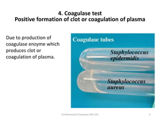
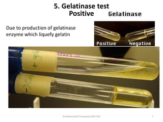
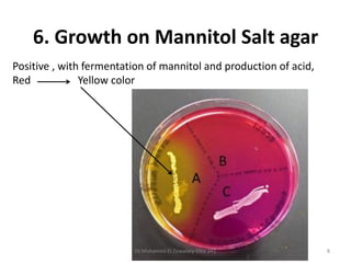
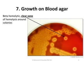
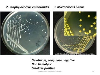
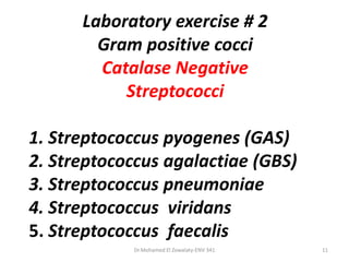
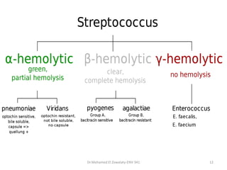
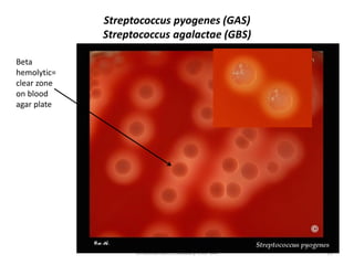
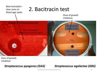
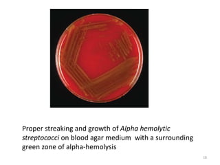
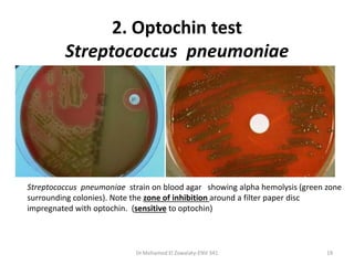
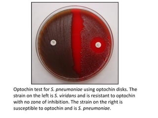
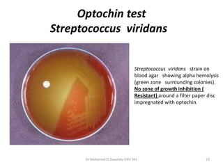
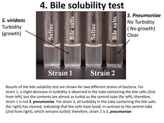
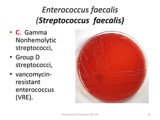
Practical Microbiology
ENV 341
Dr Mohamed El Zowalaty
Dr.Mohamed El Zowalaty-ENV 341 1
Laboratory exercise # 1
Gram positive cocci
Catalase Positive
1 – Staphylococcus aureus
2. Staphylococcus epidermidis
3. Micrococcus luteus
Dr.Mohamed El Zowalaty-ENV 341 2
1. Microscopical examination
Staphylococcus aureus Sputum smear
Gram positive cocci arranged in clusters or bunches
Dr.Mohamed El Zowalaty-ENV 341 3
2. Growth on nutrient agar medium
Golden yellow colonies
Dr.Mohamed El Zowalaty-ENV 341 4
Dr.Mohamed El Zowalaty-ENV 341 5
Catalase test
Negative and positive catalase (O2 is evolved from H2O2) test results. The absence of
bubbling from a transferred colony indicates a negative test.
3. Catalase test
Positive formation of bubbles or effervescence
Dr.Mohamed El Zowalaty-ENV 341 6
Due to production of
coagulase enzyme which
produces clot or
coagulation of plasma.
4. Coagulase test
Positive formation of clot or coagulation of plasma
Due to production of gelatinase
enzyme which liquefy gelatin
5. Gelatinase test
Positive
Dr.Mohamed El Zowalaty-ENV 341 7
Positive , with fermentation of mannitol and production of acid,
Red Yellow color
6. Growth on Mannitol Salt agar
Dr.Mohamed El Zowalaty-ENV 341 8
Beta hemolytic, clear zone
of hemolysis around
colonies
7. Growth on Blood agar
Dr.Mohamed El Zowalaty-ENV 341 9
Dr.Mohamed El Zowalaty-ENV 341 10
2. Staphylococcus epidermidis 3. Micrococcus luteus
Gelatinase, coagulase negative
Non hemolytic
Catalase positive
Laboratory exercise # 2
Gram positive cocci
Catalase Negative
Streptococci
1. Streptococcus pyogenes (GAS)
2. Streptococcus agalactiae (GBS)
3. Streptococcus pneumoniae
4. Streptococcus viridans
5. Streptococcus faecalis
Dr.Mohamed El Zowalaty-ENV 341 11
Dr.Mohamed El Zowalaty-ENV 341 12
1. Gram stain
Dr.Mohamed El Zowalaty-ENV 341 13
Gram positive cocci arranged in chains
A. Beta hemolytic, clear
zone of hemolysis around
colonies
2. Growth on Blood agar
Dr.Mohamed El Zowalaty-ENV 341 14
Streptococcus pyogenes (GAS)
Streptococcus agalactae (GBS)
Dr.Mohamed El Zowalaty-ENV 341 15
Beta
hemolytic=
clear zone
on blood
agar plate
2. Bacitracin test
Dr.Mohamed El Zowalaty-ENV 341 16
Beta hemolytic=
clear zone on
blood agar plate
Zone of growth
inhibition
Zone of growth
inhibition
Streptococcus pyogenes (GAS) Streptococcus agalactae (GBS)
B. Alpha hemolytic,
greenish zone of
hemolysis around colonies
2. Growth on Blood agar
Dr.Mohamed El Zowalaty-ENV 341 17
Dr Mohamed Ezzat-ENV 341 18
Proper streaking and growth of Alpha hemolytic
streptococci on blood agar medium with a surrounding
green zone of alpha-hemolysis
Dr.Mohamed El Zowalaty-ENV 341 19
2. Optochin test
Streptococcus pneumoniae
Streptococcus pneumoniae strain on blood agar showing alpha hemolysis (green zone
surrounding colonies). Note the zone of inhibition around a filter paper disc
impregnated with optochin. (sensitive to optochin)
• The quellung reaction (swelling
reaction) forms the basis of
serotyping and depends on the
swelling of the capsule upon
binding of homologous
antibody.
• The test consists of mixing a
loopful of colony with equal
quantity of specific antiserum
and then examining
microscopically at 1000X for
capsular swelling.
Dr Mohamed Ezzat-ENV 341 20
3. Capsule swelling test
Dr Mohamed Ezzat-ENV 341 21
Optochin test for S. pneumoniae using optochin disks. The
strain on the left is S. viridans and is resistant to optochin
with no zone of inhibition. The strain on the right is
susceptible to optochin and is S. pneumoniae.
Dr.Mohamed El Zowalaty-ENV 341 22
Optochin test
Streptococcus viridans
Dr.Mohamed El Zowalaty-ENV 341 23
Streptococcus viridans strain on
blood agar showing alpha hemolysis
(green zone surrounding colonies).
No zone of growth inhibition (
Resistant) around a filter paper disc
impregnated with optochin.
Dr Mohamed Ezzat-ENV 341 24
Results of the bile solubility test are shown for two different strains of bacteria. For
strain 1, a slight decrease in turbidity is observed in the tube containing the bile salts (2nd
from left), but the contents are almost as turbid as the control tube (far left); therefore,
strain 1 is not S. pneumoniae. For strain 2, all turbidity in the tube containing the bile salts
(far right) has cleared, indicating that the cells have lysed, in contrast to the control tube
(2nd from right), which remains turbid; therefore, strain 2 is S. pneumoniae
4. Bile solubility test
S. Pneumoniae
No Turbidity
( No growth)
Clear
S. viridans
Turbidity
(growth)
Enterococcus faecalis
(Streptococcus faecalis)
• C. Gamma
Nonhemolytic
streptococci,
• Group D
streptococci,
• vancomycin-
resistant
enterococcus
(VRE).
Dr.Mohamed El Zowalaty-ENV 341 25
Acknowledgment
• I, Dr Mohamed El Zowalaty, would like to
express special thankfulness to the US CDC
Public Health Image Library, NIH/NIAID, Dr
Kenneth Toda, and others for providing the
photomicrographs available in the public
domain.
Dr.Mohamed El Zowalaty-ENV 341 26

Practical microbiology Gram positive cocci

  • 1.
    Practical Microbiology ENV 341 DrMohamed El Zowalaty Dr.Mohamed El Zowalaty-ENV 341 1
  • 2.
    Laboratory exercise #1 Gram positive cocci Catalase Positive 1 – Staphylococcus aureus 2. Staphylococcus epidermidis 3. Micrococcus luteus Dr.Mohamed El Zowalaty-ENV 341 2
  • 3.
    1. Microscopical examination Staphylococcusaureus Sputum smear Gram positive cocci arranged in clusters or bunches Dr.Mohamed El Zowalaty-ENV 341 3
  • 4.
    2. Growth onnutrient agar medium Golden yellow colonies Dr.Mohamed El Zowalaty-ENV 341 4
  • 5.
    Dr.Mohamed El Zowalaty-ENV341 5 Catalase test Negative and positive catalase (O2 is evolved from H2O2) test results. The absence of bubbling from a transferred colony indicates a negative test. 3. Catalase test Positive formation of bubbles or effervescence
  • 6.
    Dr.Mohamed El Zowalaty-ENV341 6 Due to production of coagulase enzyme which produces clot or coagulation of plasma. 4. Coagulase test Positive formation of clot or coagulation of plasma
  • 7.
    Due to productionof gelatinase enzyme which liquefy gelatin 5. Gelatinase test Positive Dr.Mohamed El Zowalaty-ENV 341 7
  • 8.
    Positive , withfermentation of mannitol and production of acid, Red Yellow color 6. Growth on Mannitol Salt agar Dr.Mohamed El Zowalaty-ENV 341 8
  • 9.
    Beta hemolytic, clearzone of hemolysis around colonies 7. Growth on Blood agar Dr.Mohamed El Zowalaty-ENV 341 9
  • 10.
    Dr.Mohamed El Zowalaty-ENV341 10 2. Staphylococcus epidermidis 3. Micrococcus luteus Gelatinase, coagulase negative Non hemolytic Catalase positive
  • 11.
    Laboratory exercise #2 Gram positive cocci Catalase Negative Streptococci 1. Streptococcus pyogenes (GAS) 2. Streptococcus agalactiae (GBS) 3. Streptococcus pneumoniae 4. Streptococcus viridans 5. Streptococcus faecalis Dr.Mohamed El Zowalaty-ENV 341 11
  • 12.
  • 13.
    1. Gram stain Dr.MohamedEl Zowalaty-ENV 341 13 Gram positive cocci arranged in chains
  • 14.
    A. Beta hemolytic,clear zone of hemolysis around colonies 2. Growth on Blood agar Dr.Mohamed El Zowalaty-ENV 341 14
  • 15.
    Streptococcus pyogenes (GAS) Streptococcusagalactae (GBS) Dr.Mohamed El Zowalaty-ENV 341 15 Beta hemolytic= clear zone on blood agar plate
  • 16.
    2. Bacitracin test Dr.MohamedEl Zowalaty-ENV 341 16 Beta hemolytic= clear zone on blood agar plate Zone of growth inhibition Zone of growth inhibition Streptococcus pyogenes (GAS) Streptococcus agalactae (GBS)
  • 17.
    B. Alpha hemolytic, greenishzone of hemolysis around colonies 2. Growth on Blood agar Dr.Mohamed El Zowalaty-ENV 341 17
  • 18.
    Dr Mohamed Ezzat-ENV341 18 Proper streaking and growth of Alpha hemolytic streptococci on blood agar medium with a surrounding green zone of alpha-hemolysis
  • 19.
    Dr.Mohamed El Zowalaty-ENV341 19 2. Optochin test Streptococcus pneumoniae Streptococcus pneumoniae strain on blood agar showing alpha hemolysis (green zone surrounding colonies). Note the zone of inhibition around a filter paper disc impregnated with optochin. (sensitive to optochin)
  • 20.
    • The quellungreaction (swelling reaction) forms the basis of serotyping and depends on the swelling of the capsule upon binding of homologous antibody. • The test consists of mixing a loopful of colony with equal quantity of specific antiserum and then examining microscopically at 1000X for capsular swelling. Dr Mohamed Ezzat-ENV 341 20 3. Capsule swelling test
  • 21.
    Dr Mohamed Ezzat-ENV341 21 Optochin test for S. pneumoniae using optochin disks. The strain on the left is S. viridans and is resistant to optochin with no zone of inhibition. The strain on the right is susceptible to optochin and is S. pneumoniae.
  • 22.
  • 23.
    Optochin test Streptococcus viridans Dr.MohamedEl Zowalaty-ENV 341 23 Streptococcus viridans strain on blood agar showing alpha hemolysis (green zone surrounding colonies). No zone of growth inhibition ( Resistant) around a filter paper disc impregnated with optochin.
  • 24.
    Dr Mohamed Ezzat-ENV341 24 Results of the bile solubility test are shown for two different strains of bacteria. For strain 1, a slight decrease in turbidity is observed in the tube containing the bile salts (2nd from left), but the contents are almost as turbid as the control tube (far left); therefore, strain 1 is not S. pneumoniae. For strain 2, all turbidity in the tube containing the bile salts (far right) has cleared, indicating that the cells have lysed, in contrast to the control tube (2nd from right), which remains turbid; therefore, strain 2 is S. pneumoniae 4. Bile solubility test S. Pneumoniae No Turbidity ( No growth) Clear S. viridans Turbidity (growth)
  • 25.
    Enterococcus faecalis (Streptococcus faecalis) •C. Gamma Nonhemolytic streptococci, • Group D streptococci, • vancomycin- resistant enterococcus (VRE). Dr.Mohamed El Zowalaty-ENV 341 25
  • 26.
    Acknowledgment • I, DrMohamed El Zowalaty, would like to express special thankfulness to the US CDC Public Health Image Library, NIH/NIAID, Dr Kenneth Toda, and others for providing the photomicrographs available in the public domain. Dr.Mohamed El Zowalaty-ENV 341 26