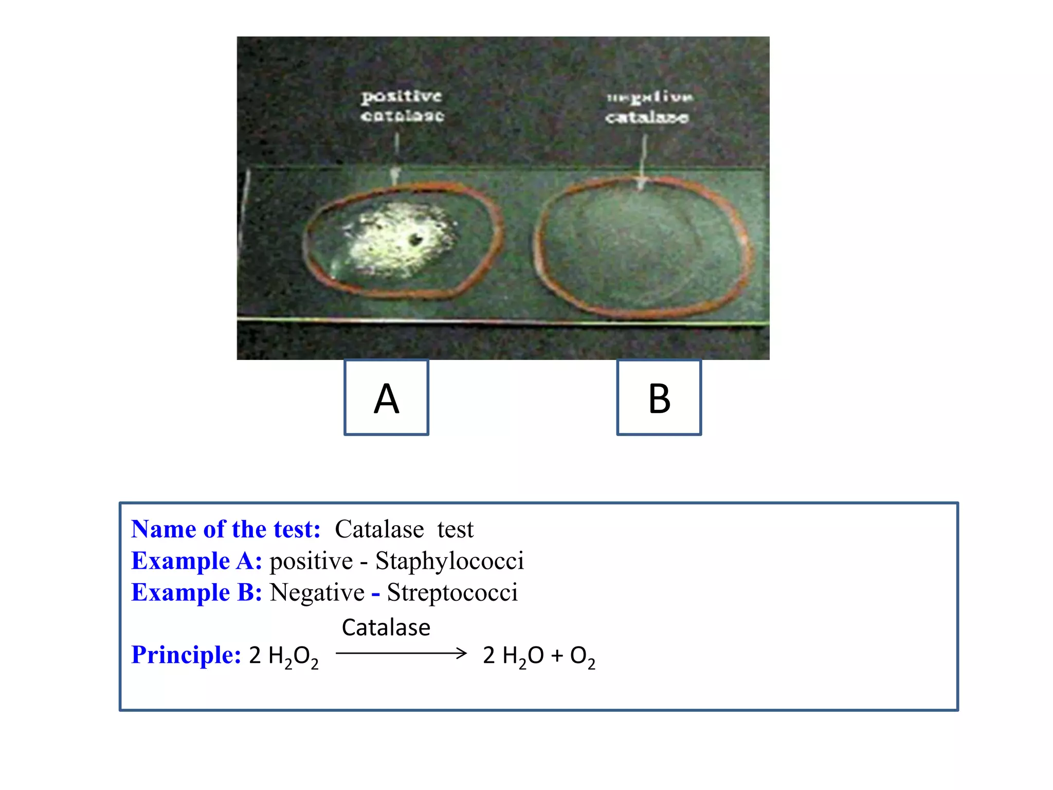
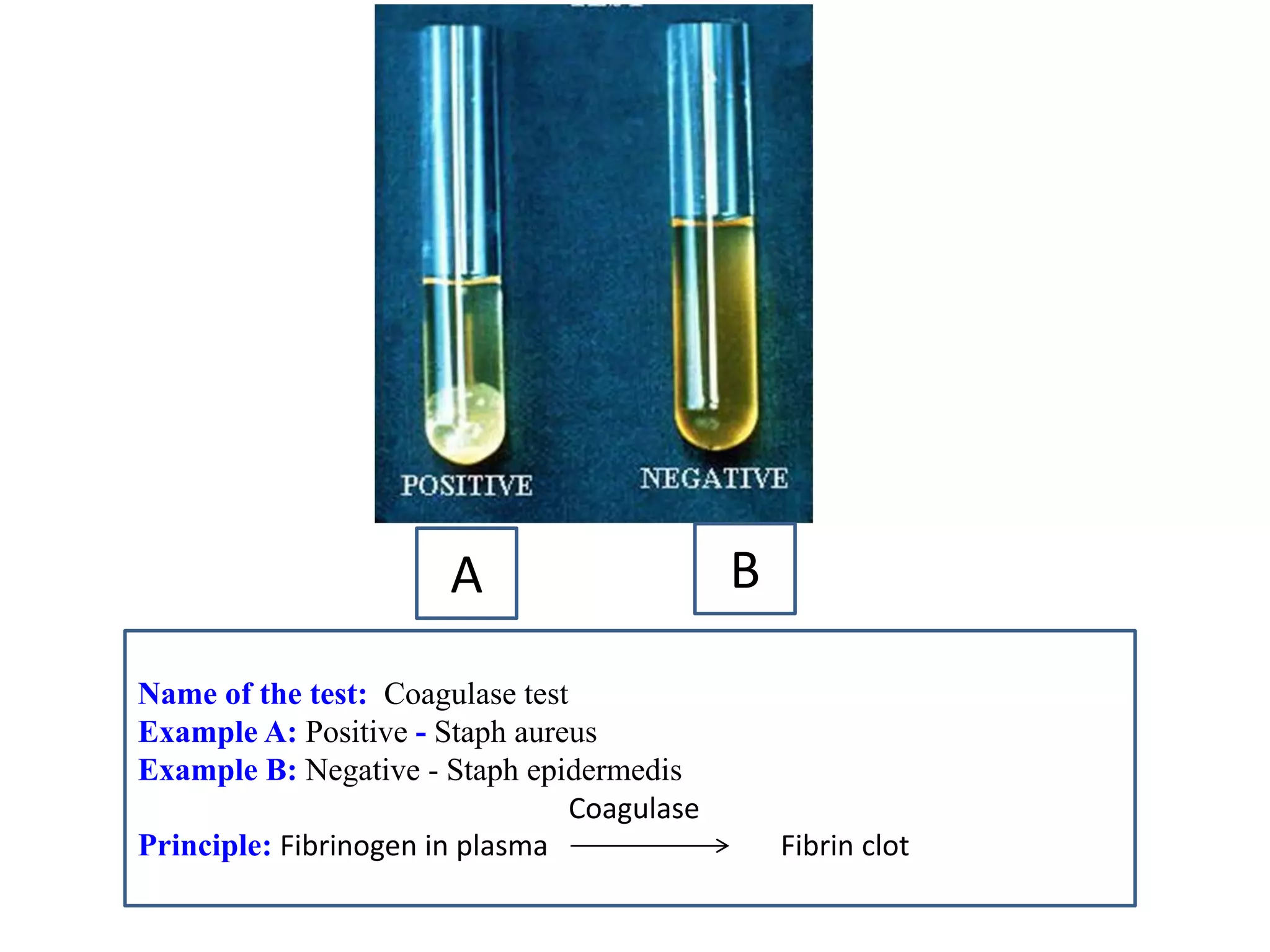
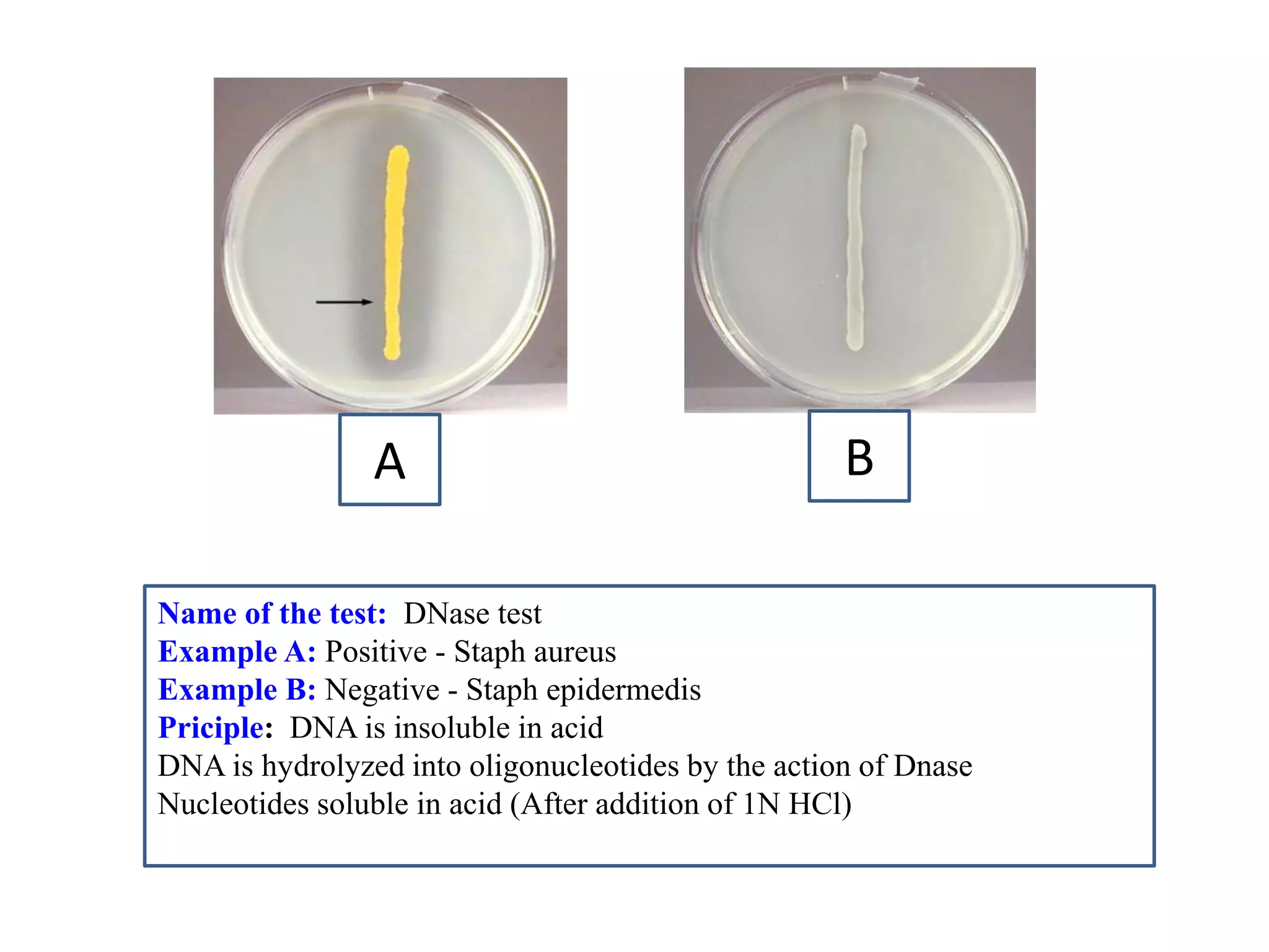
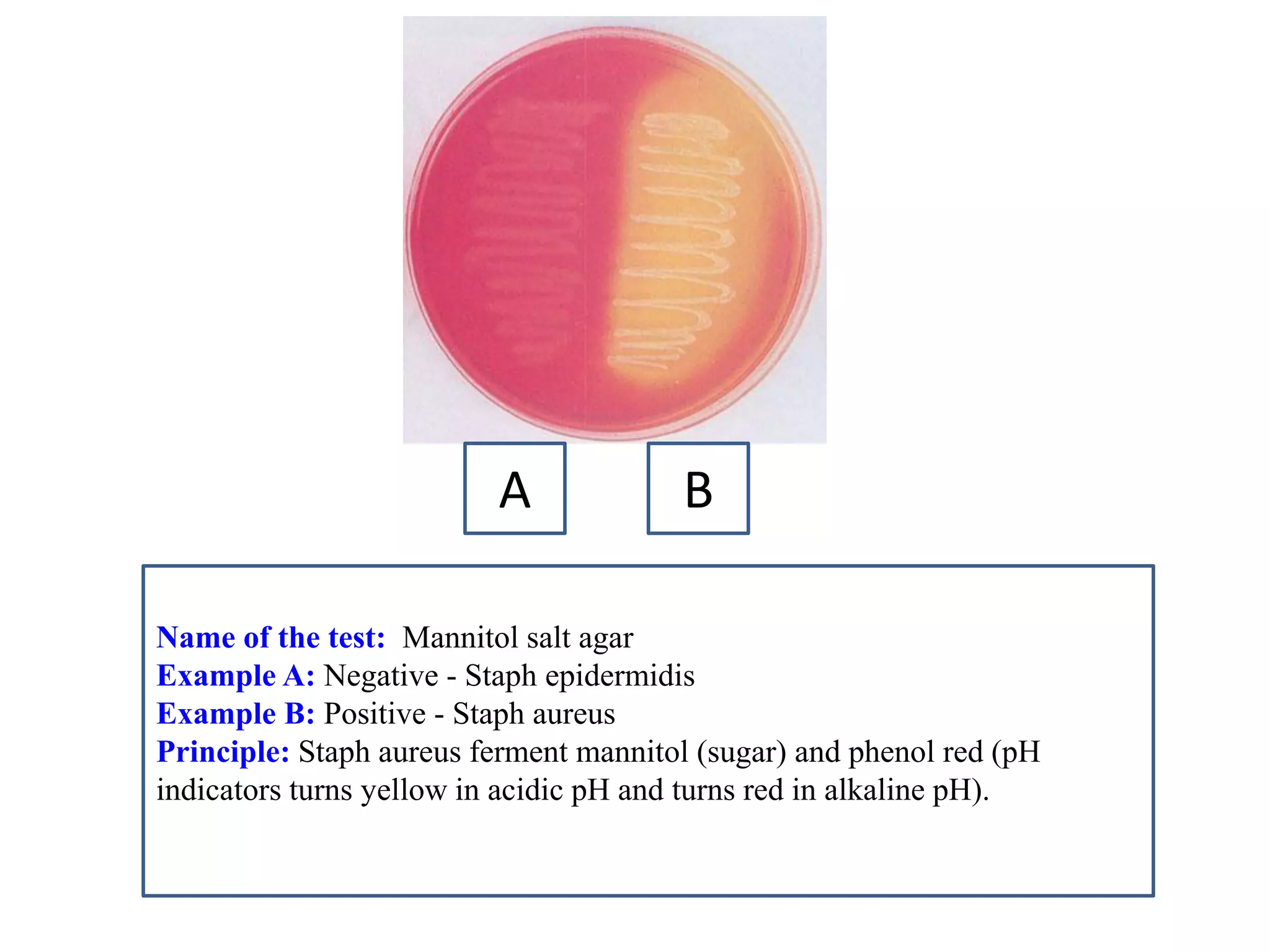
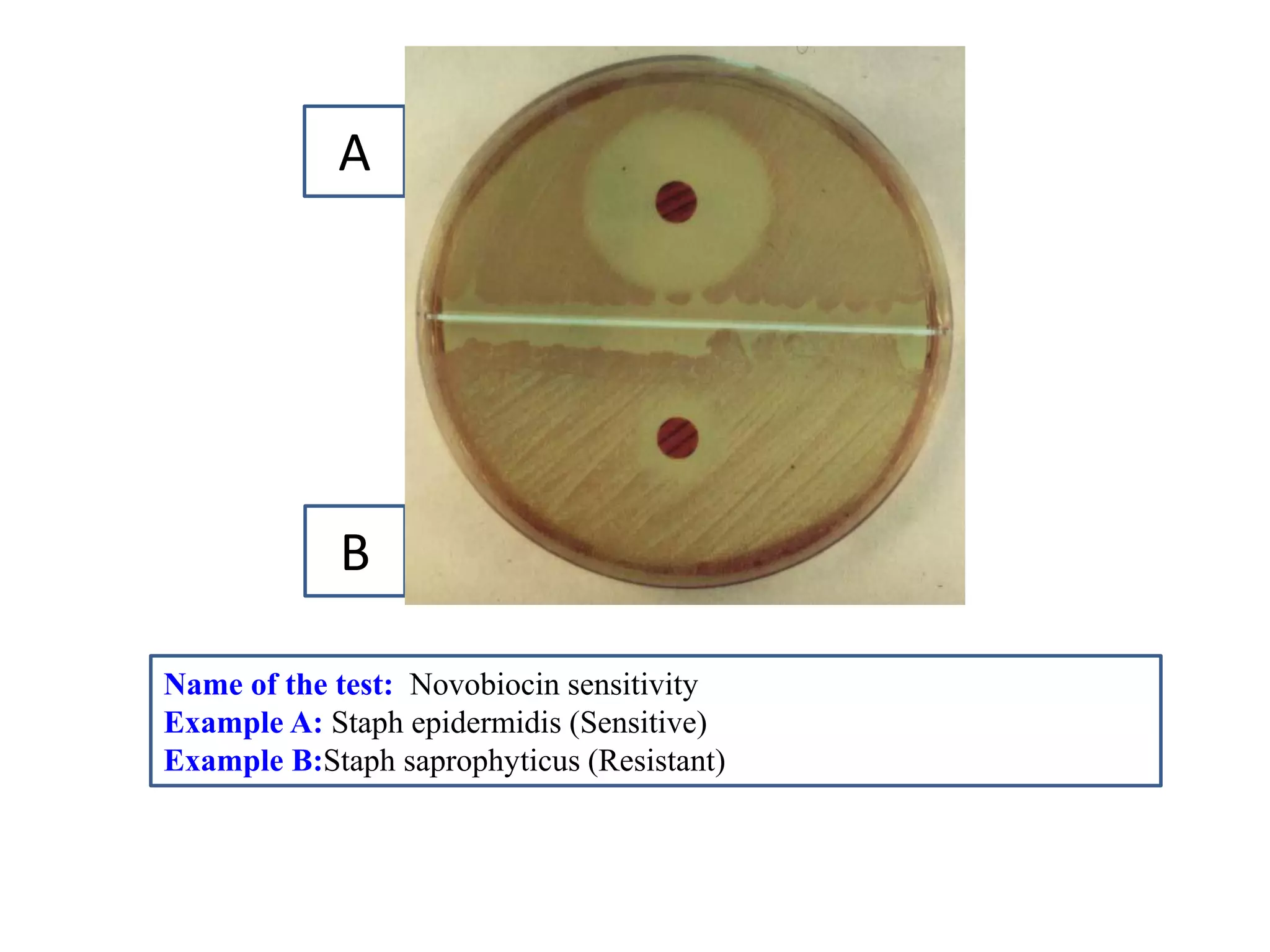
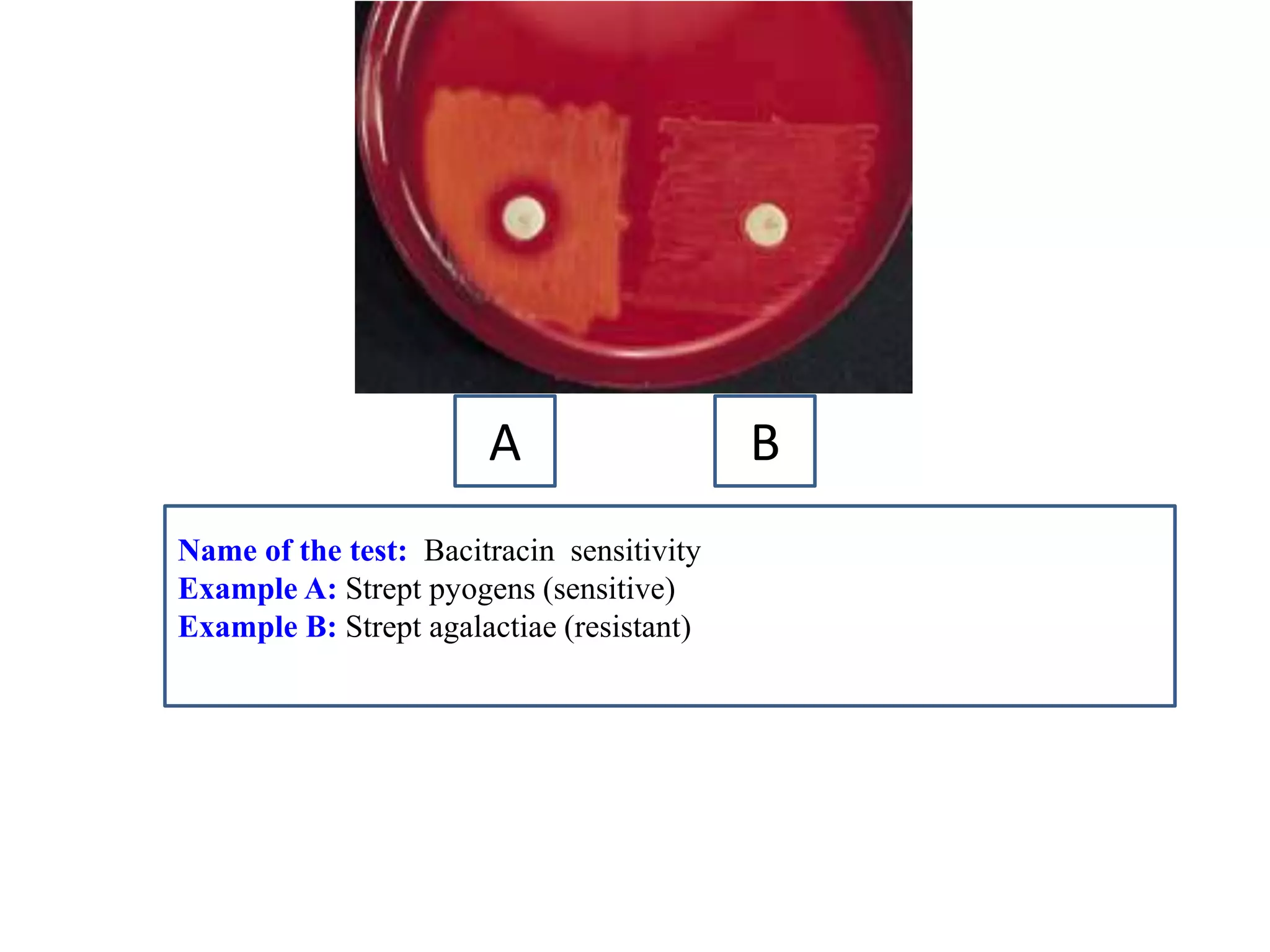
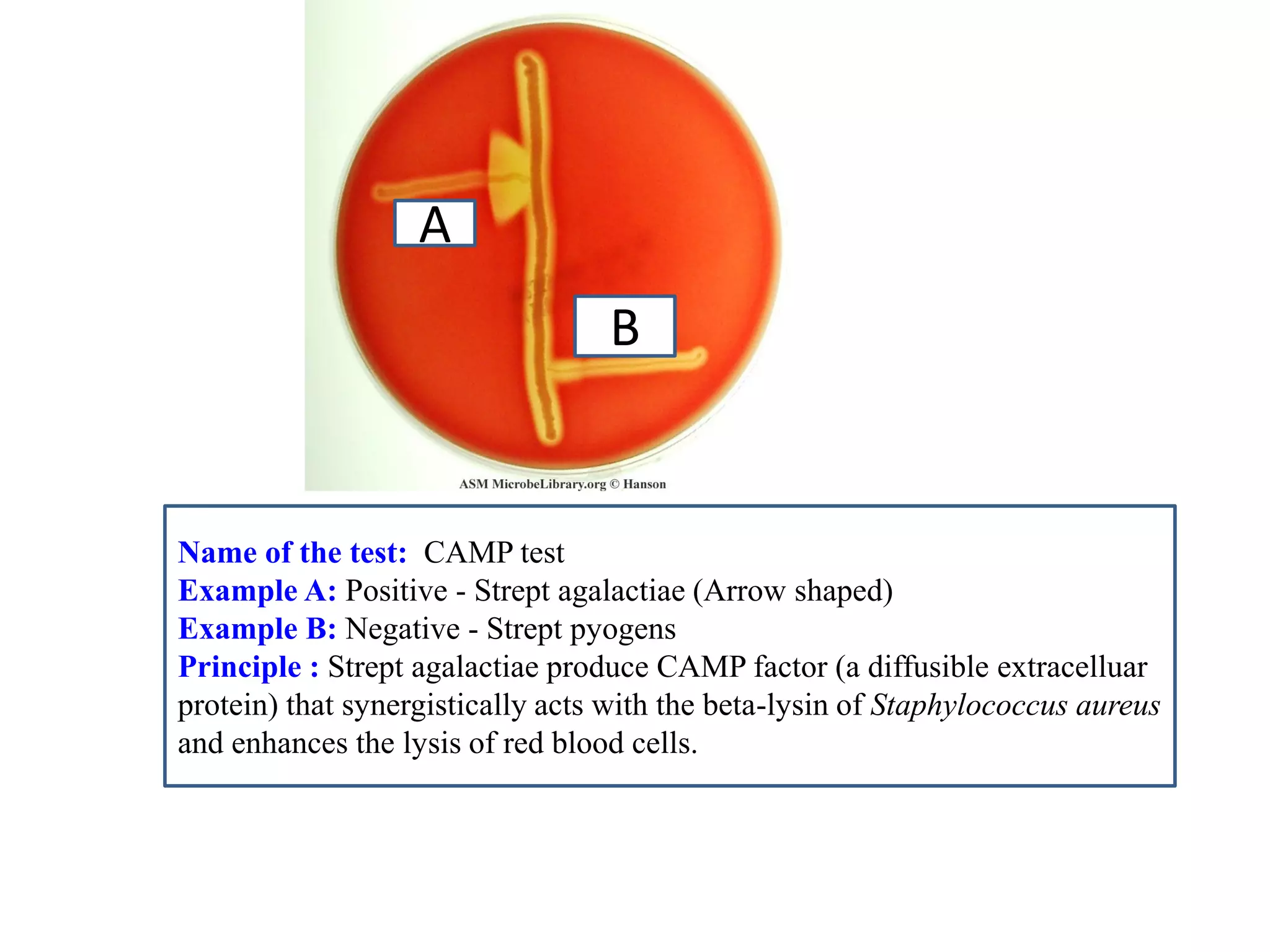
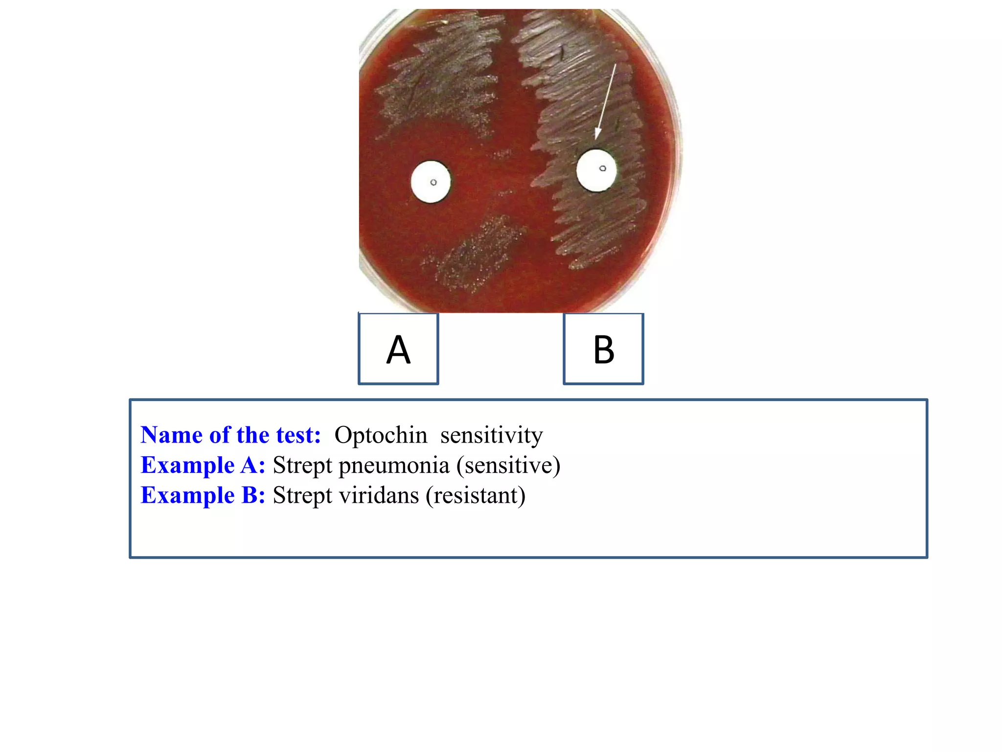
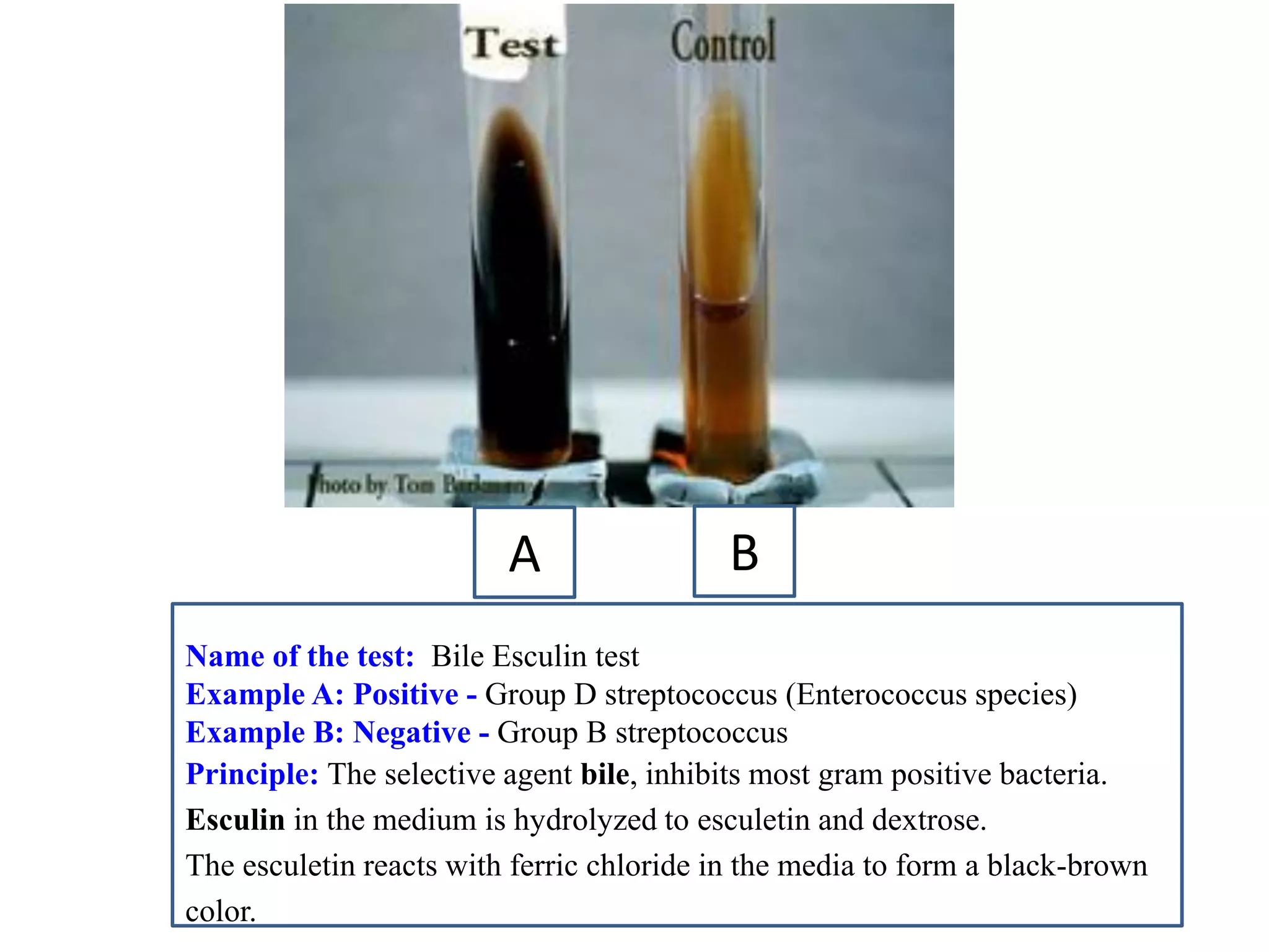
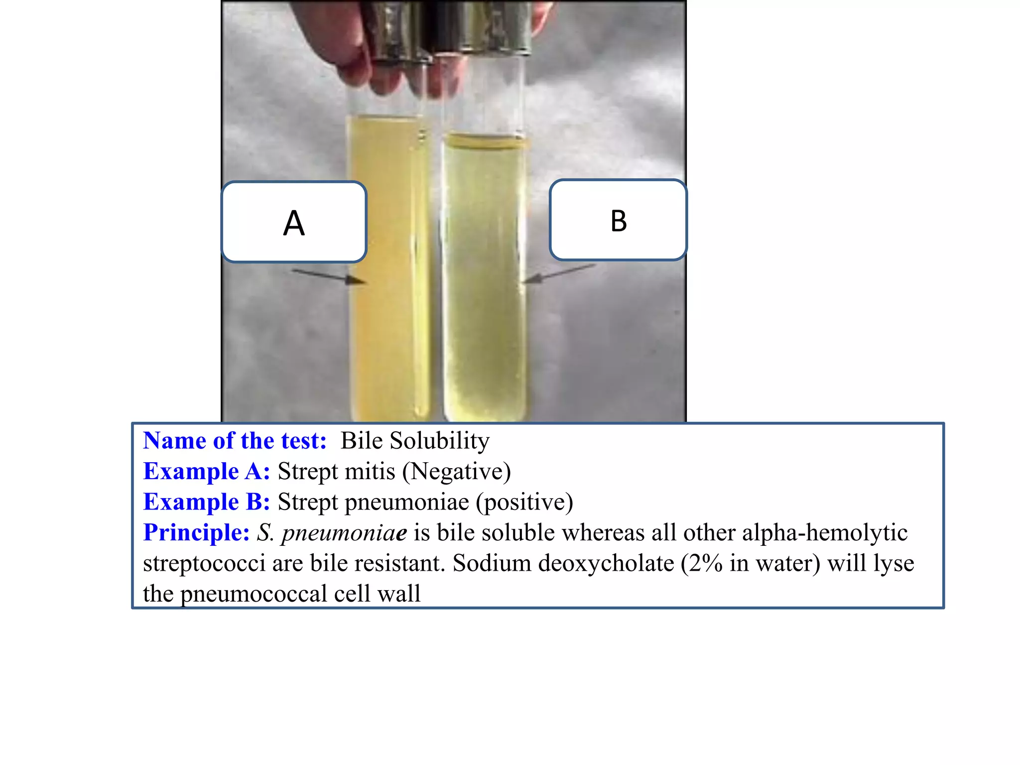
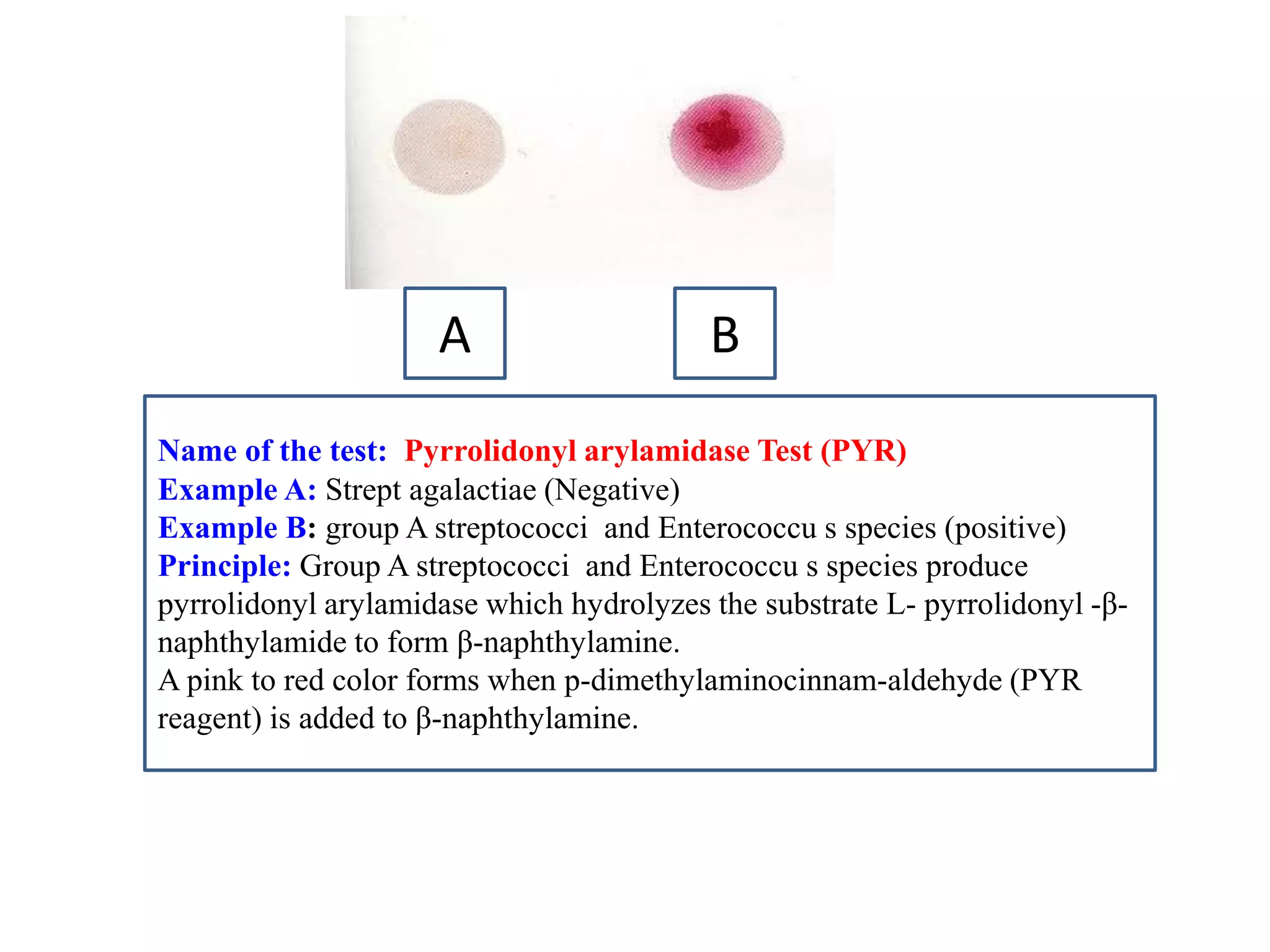
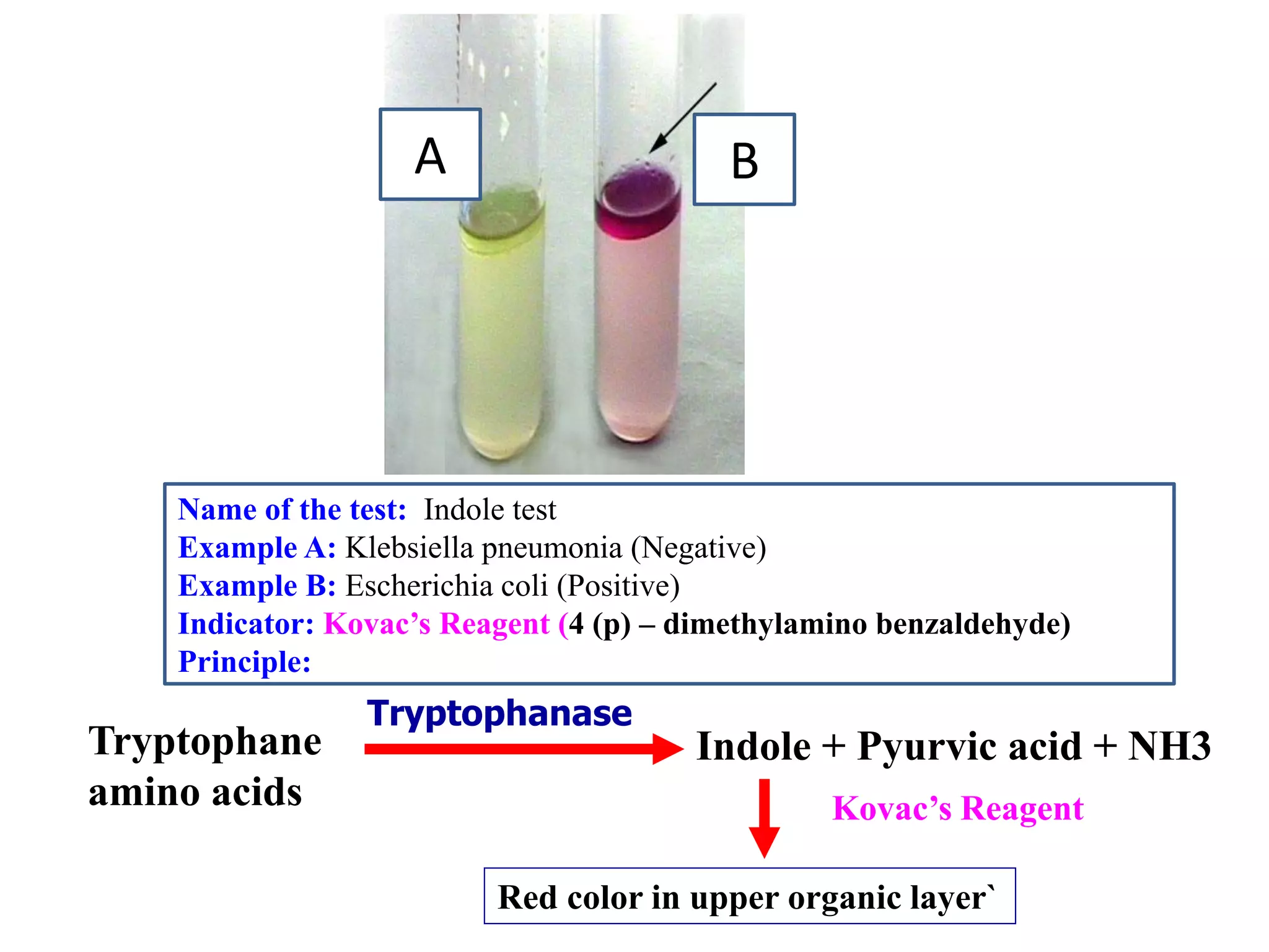
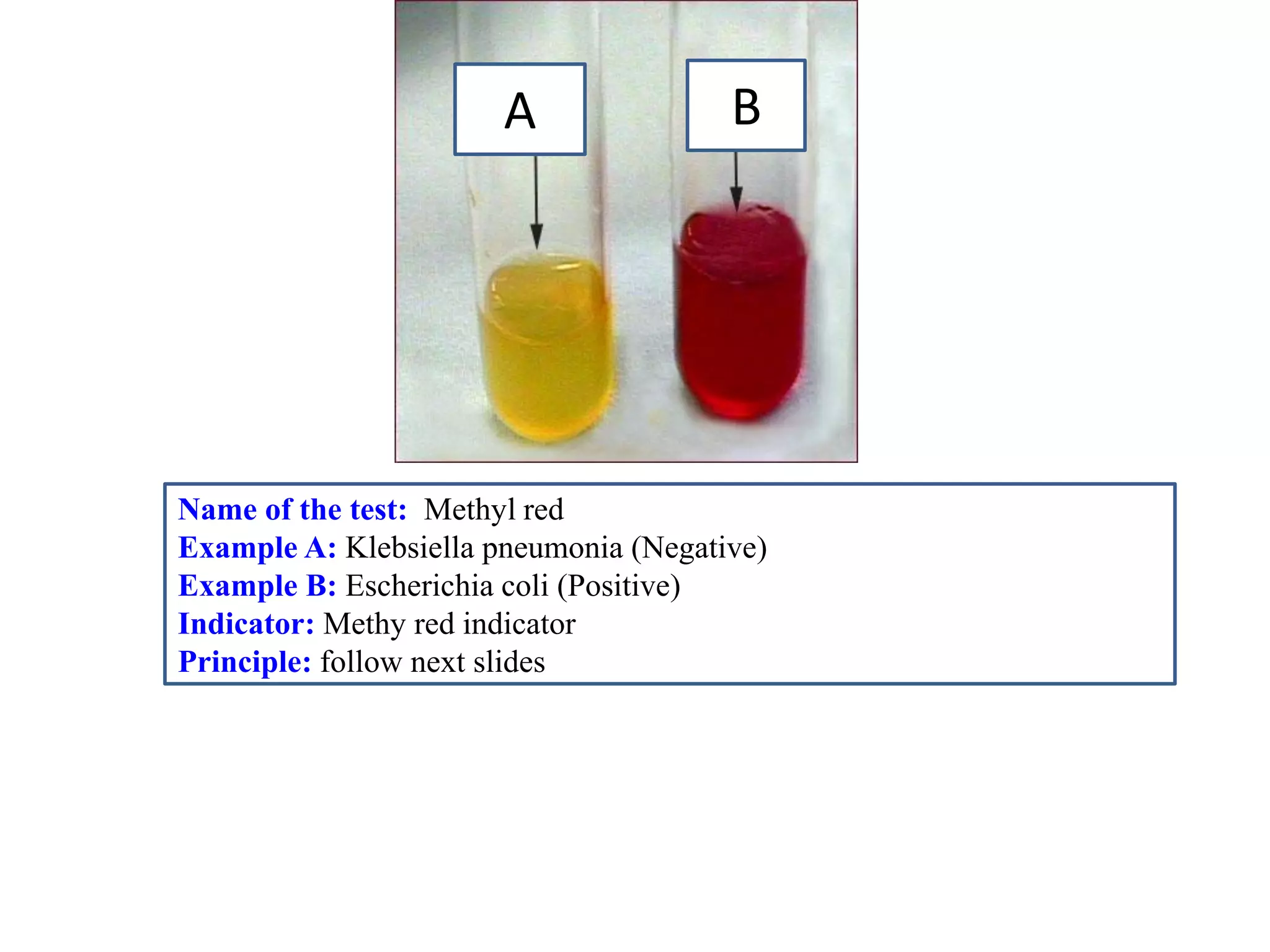
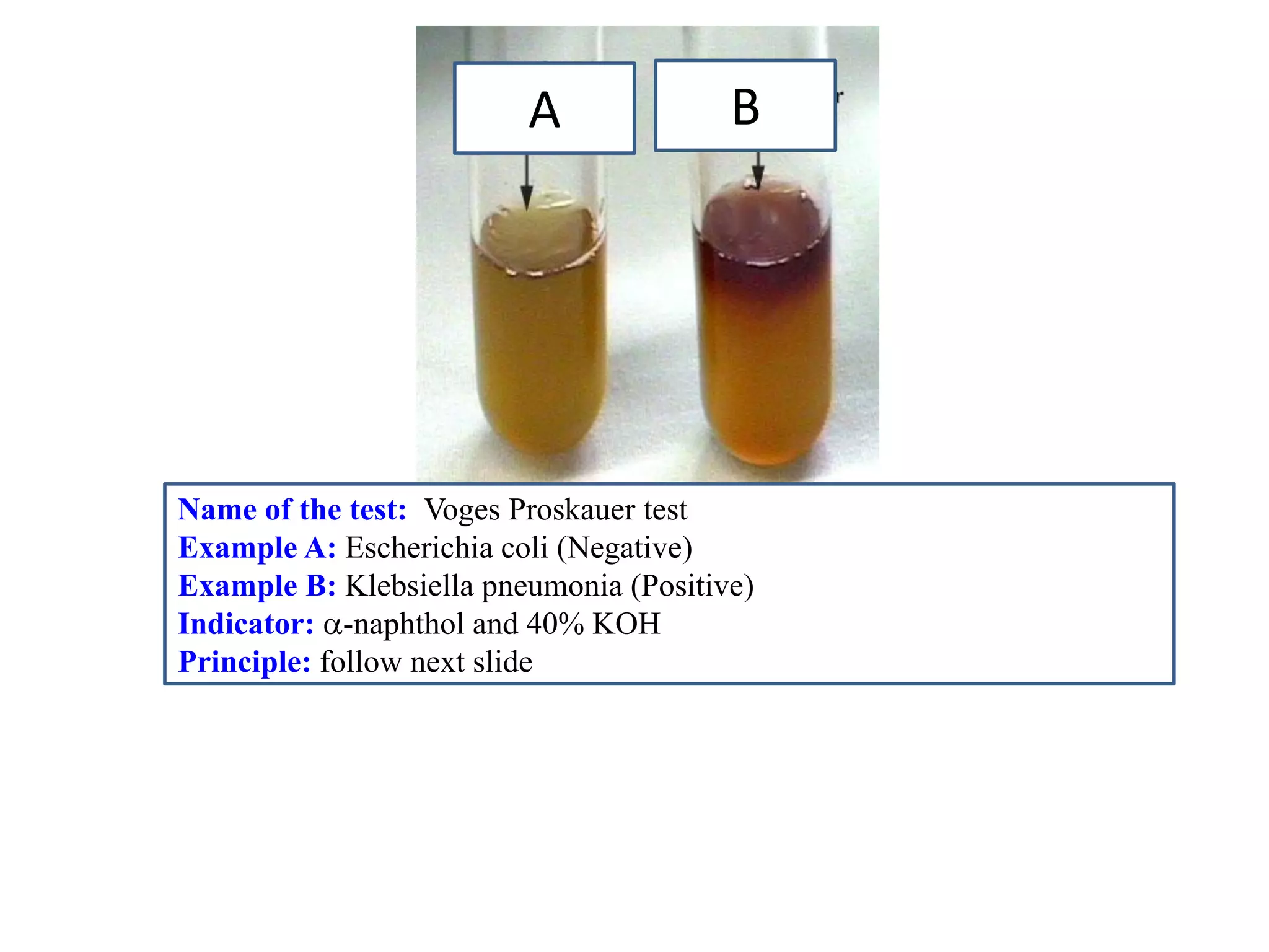
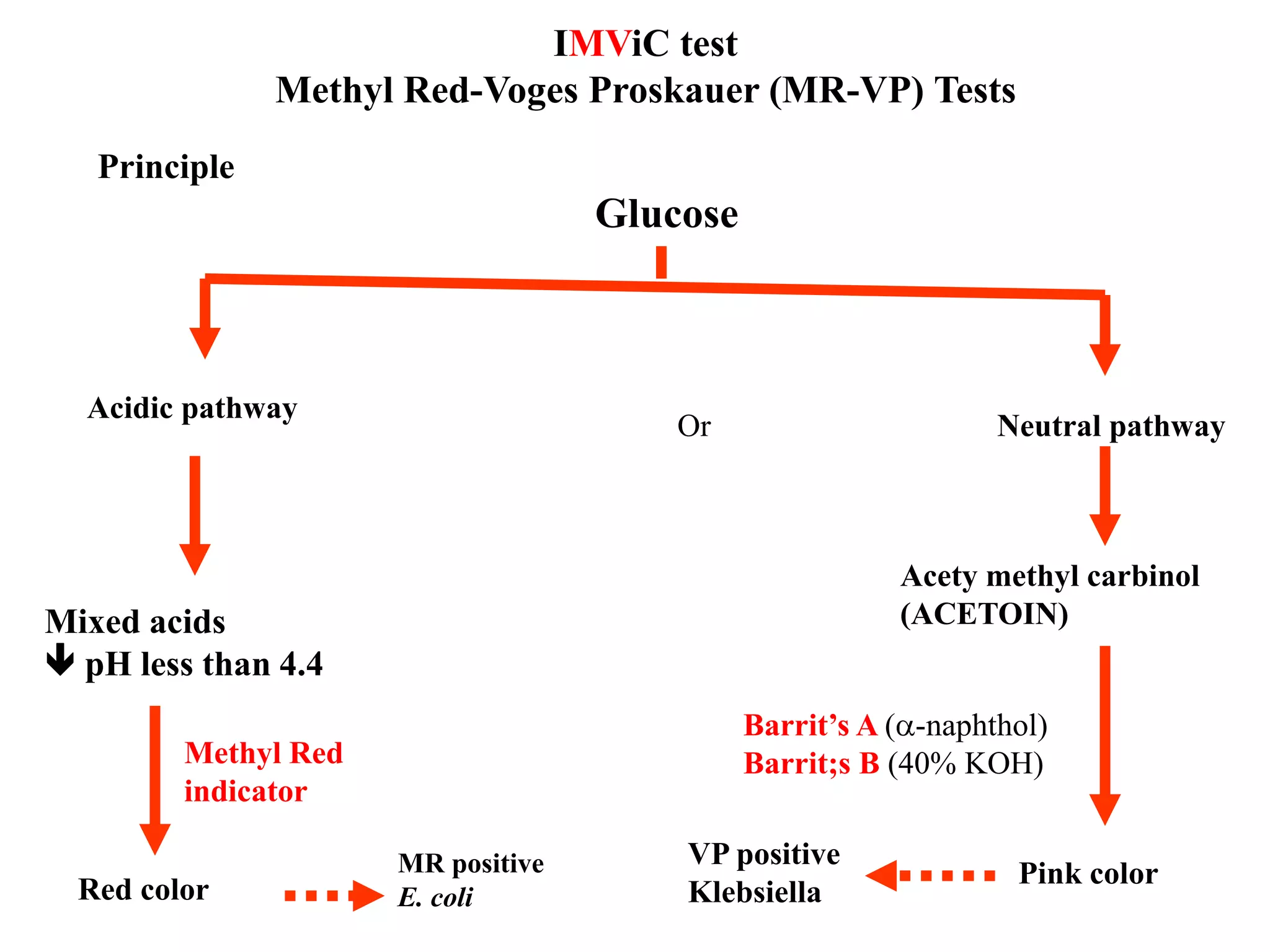
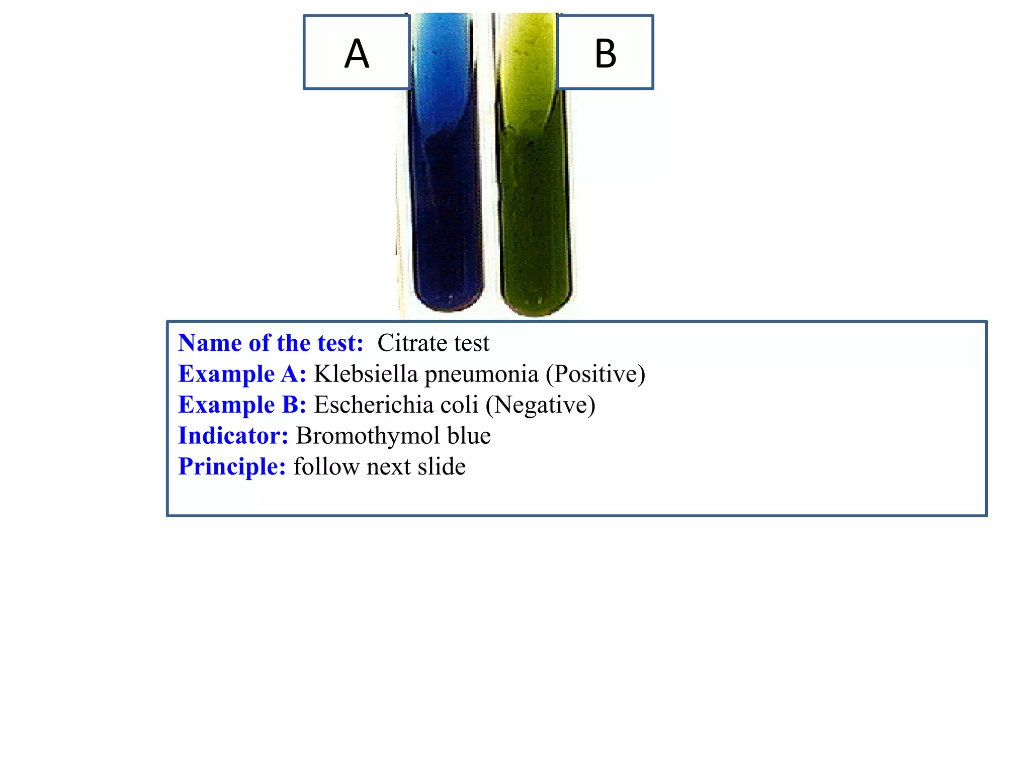
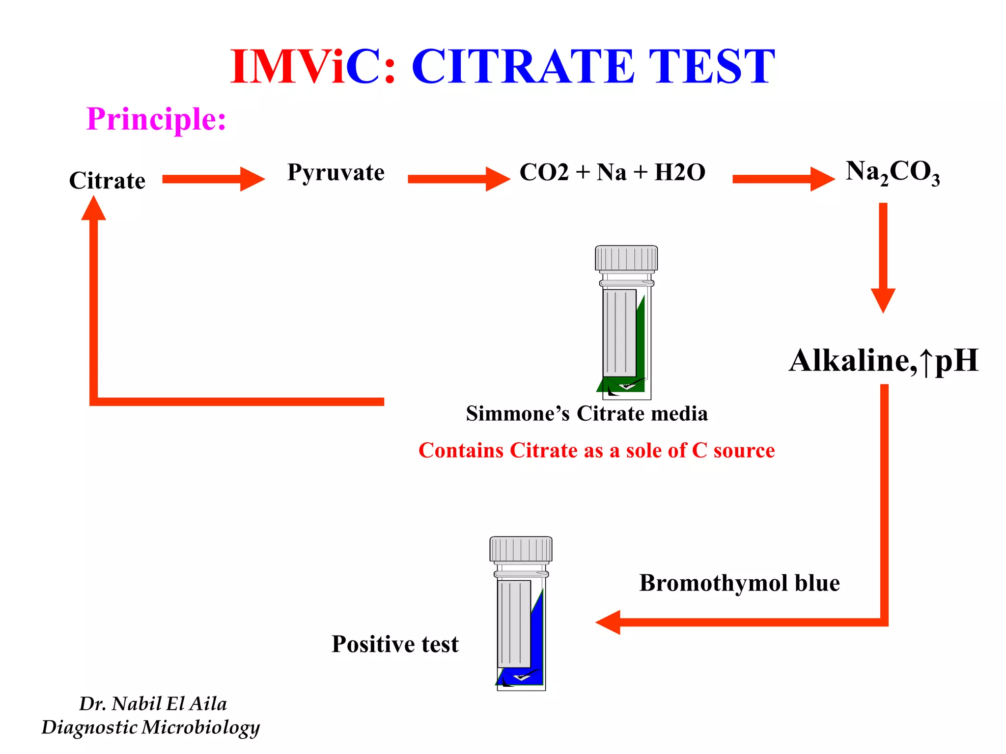
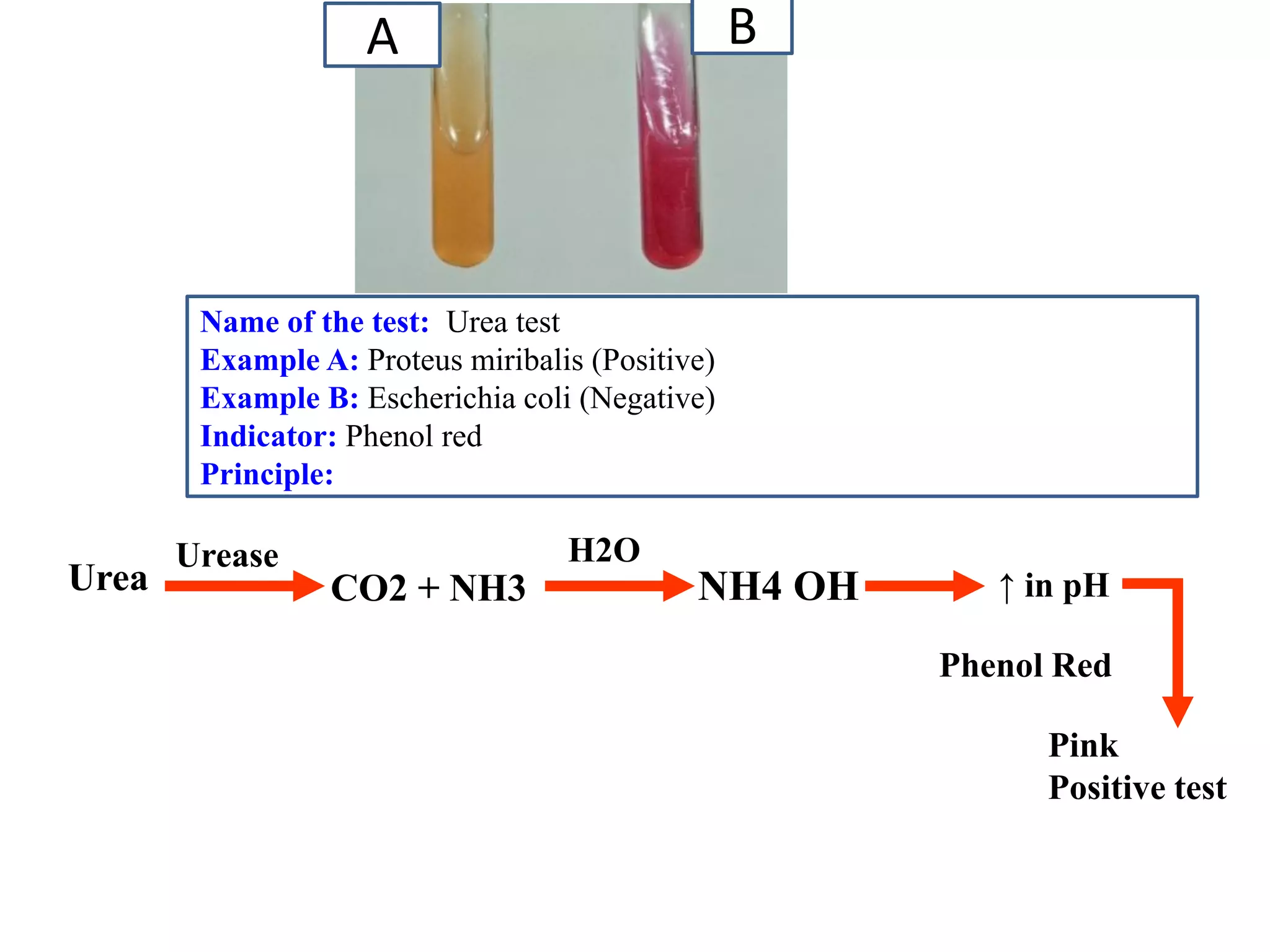
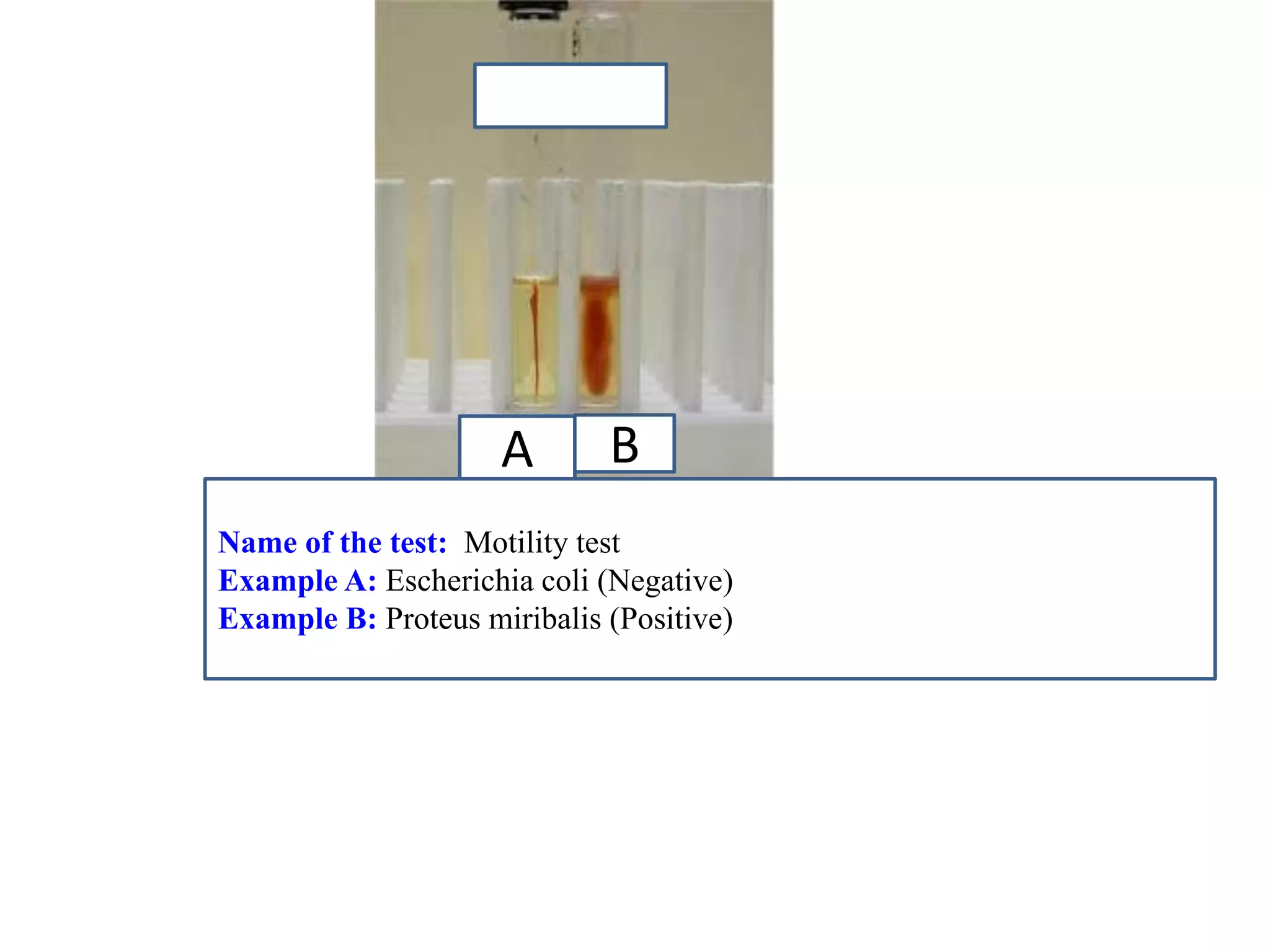
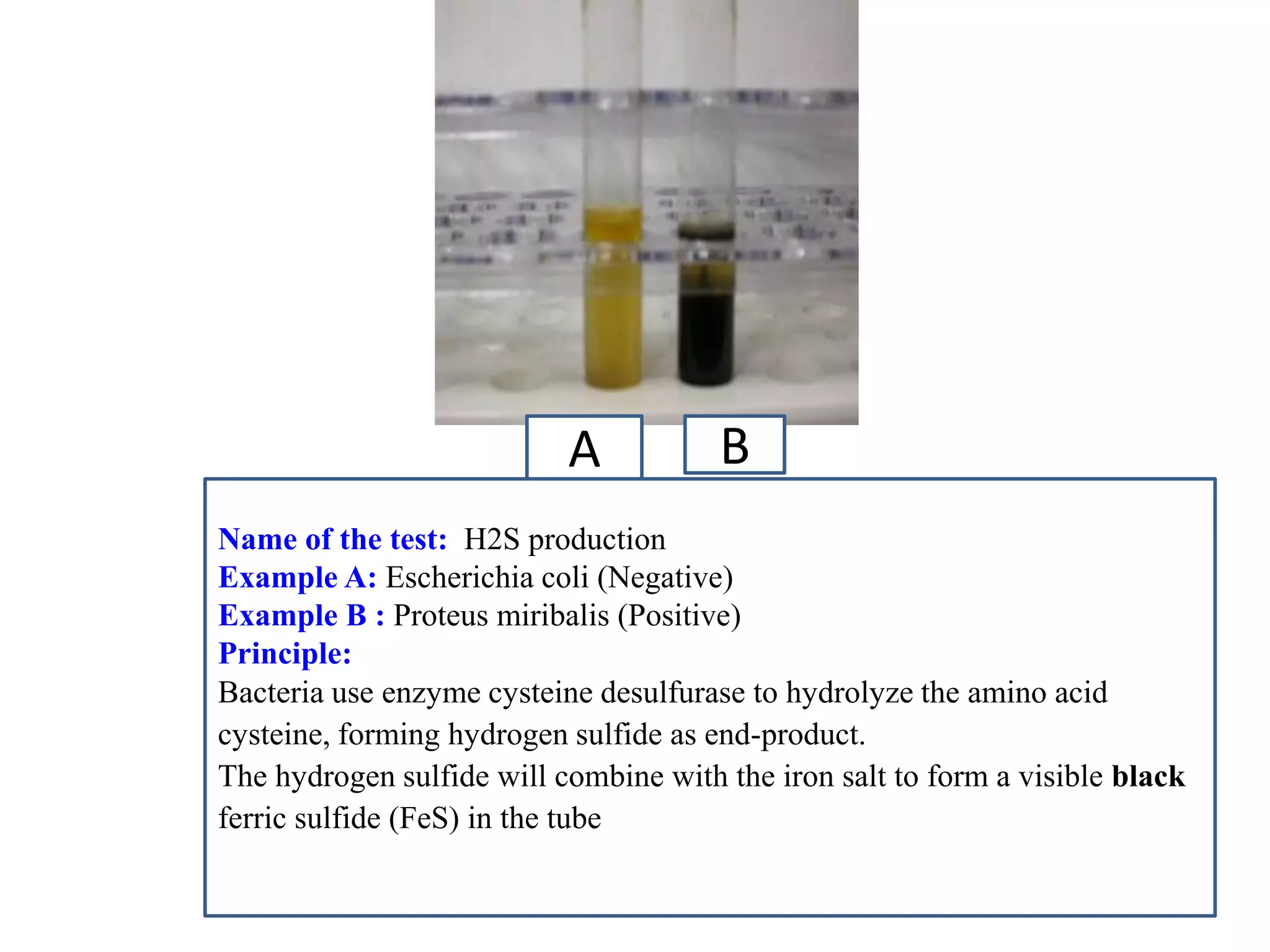
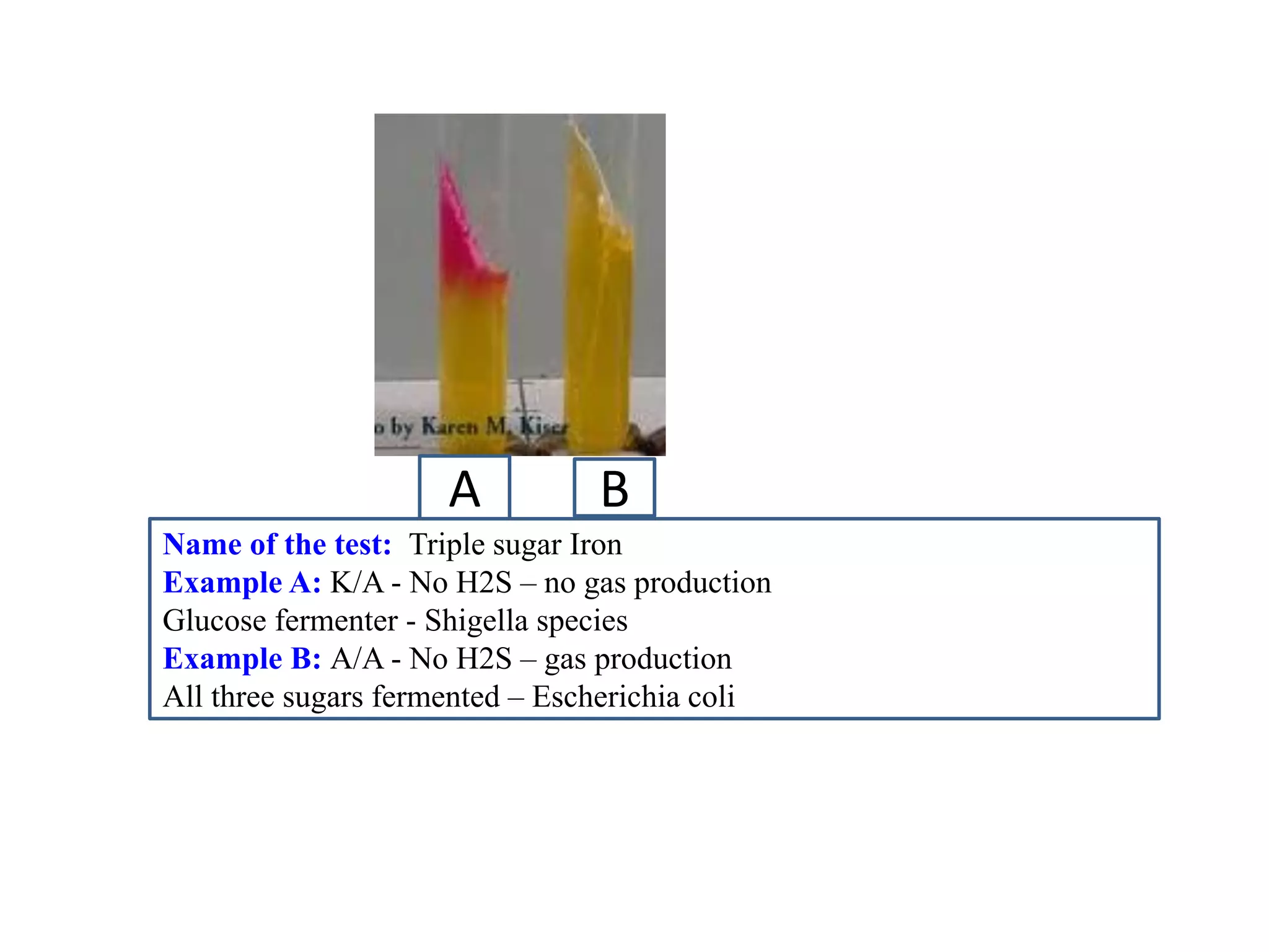
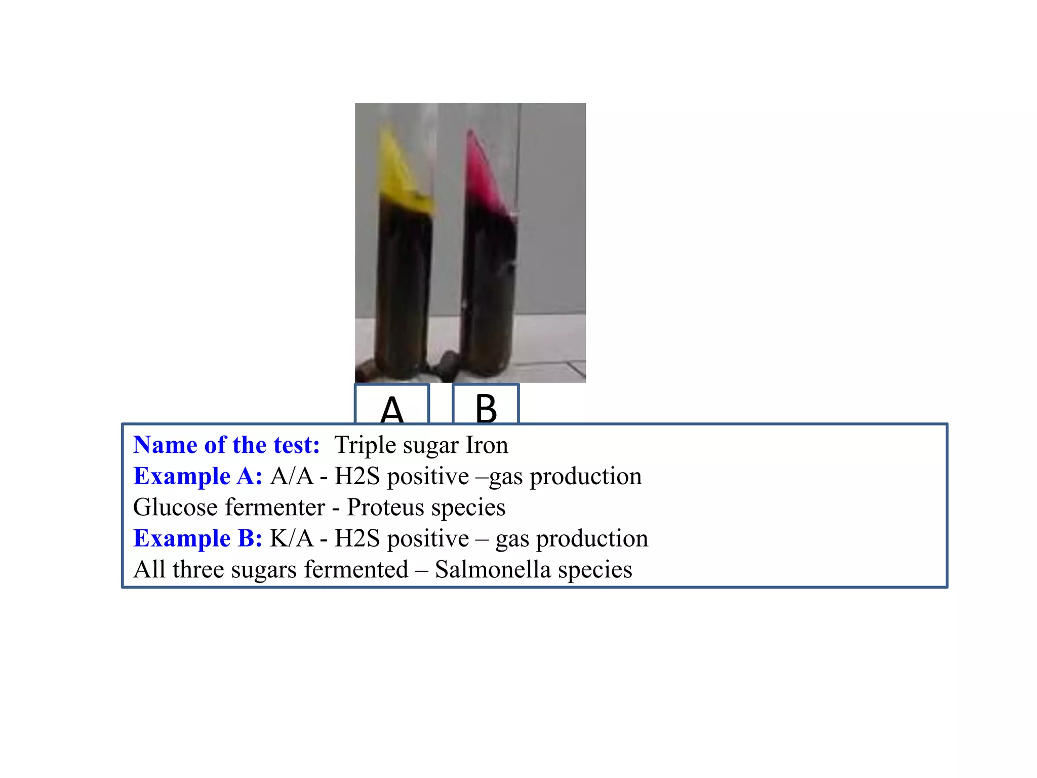
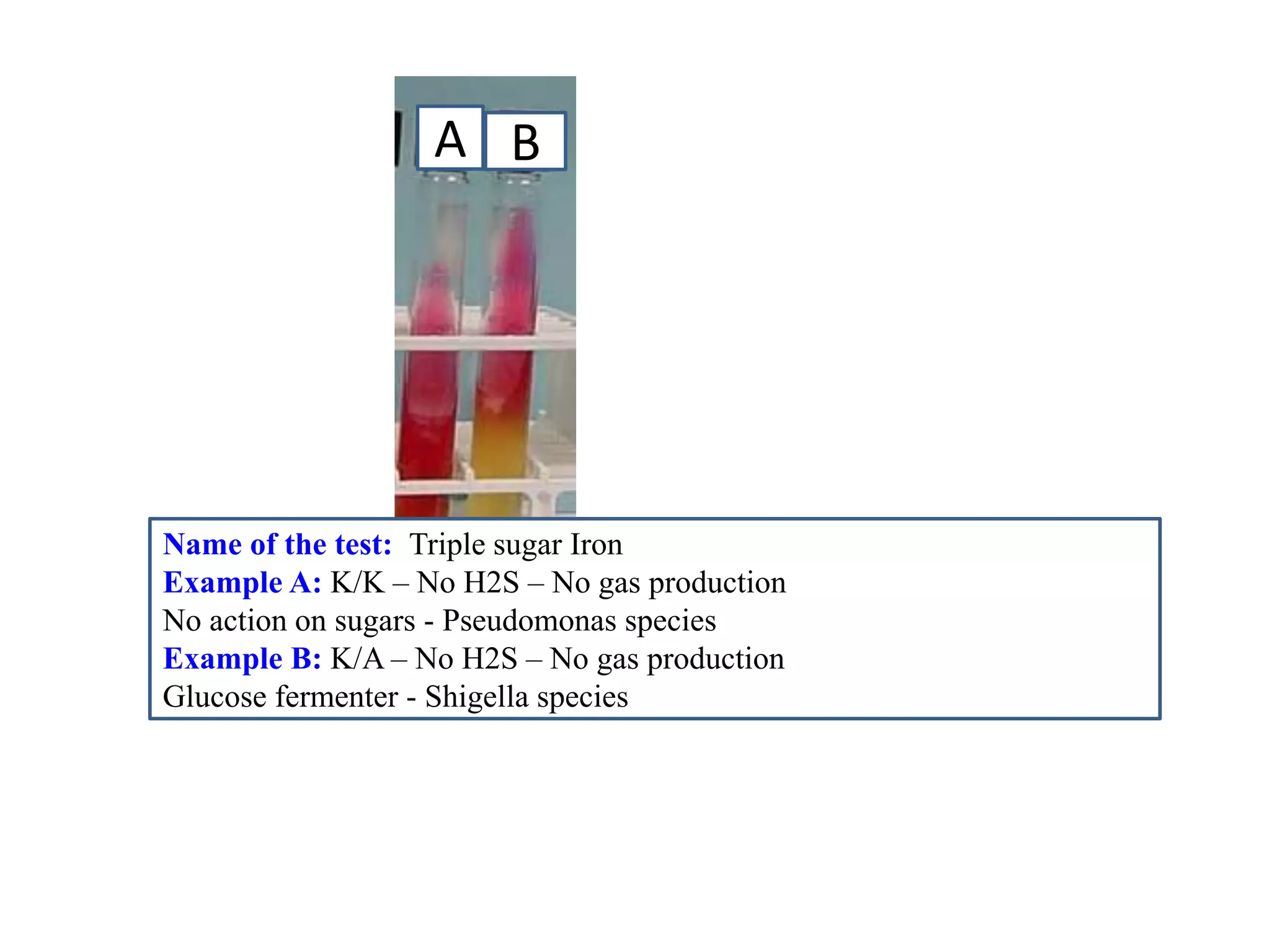
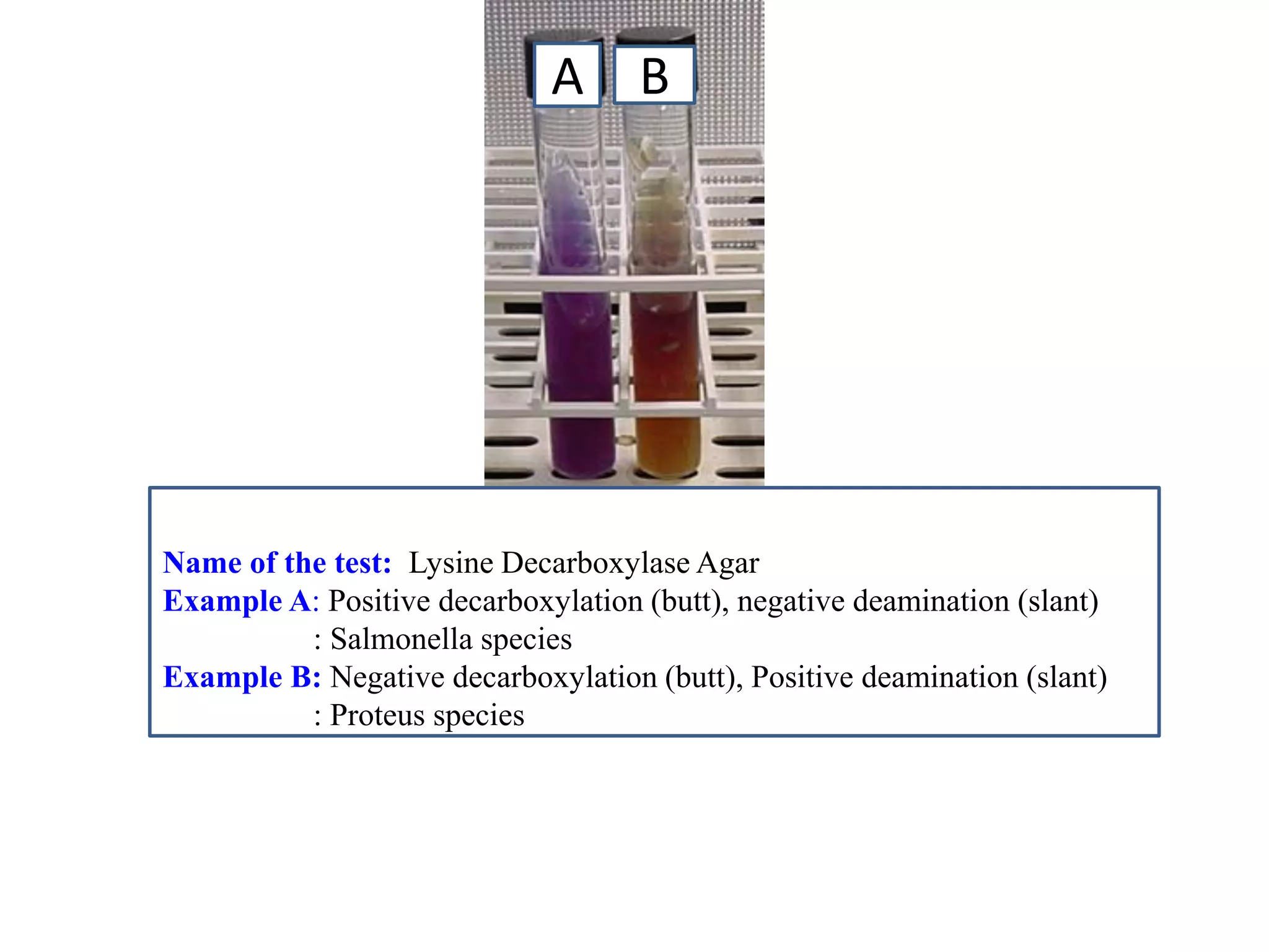
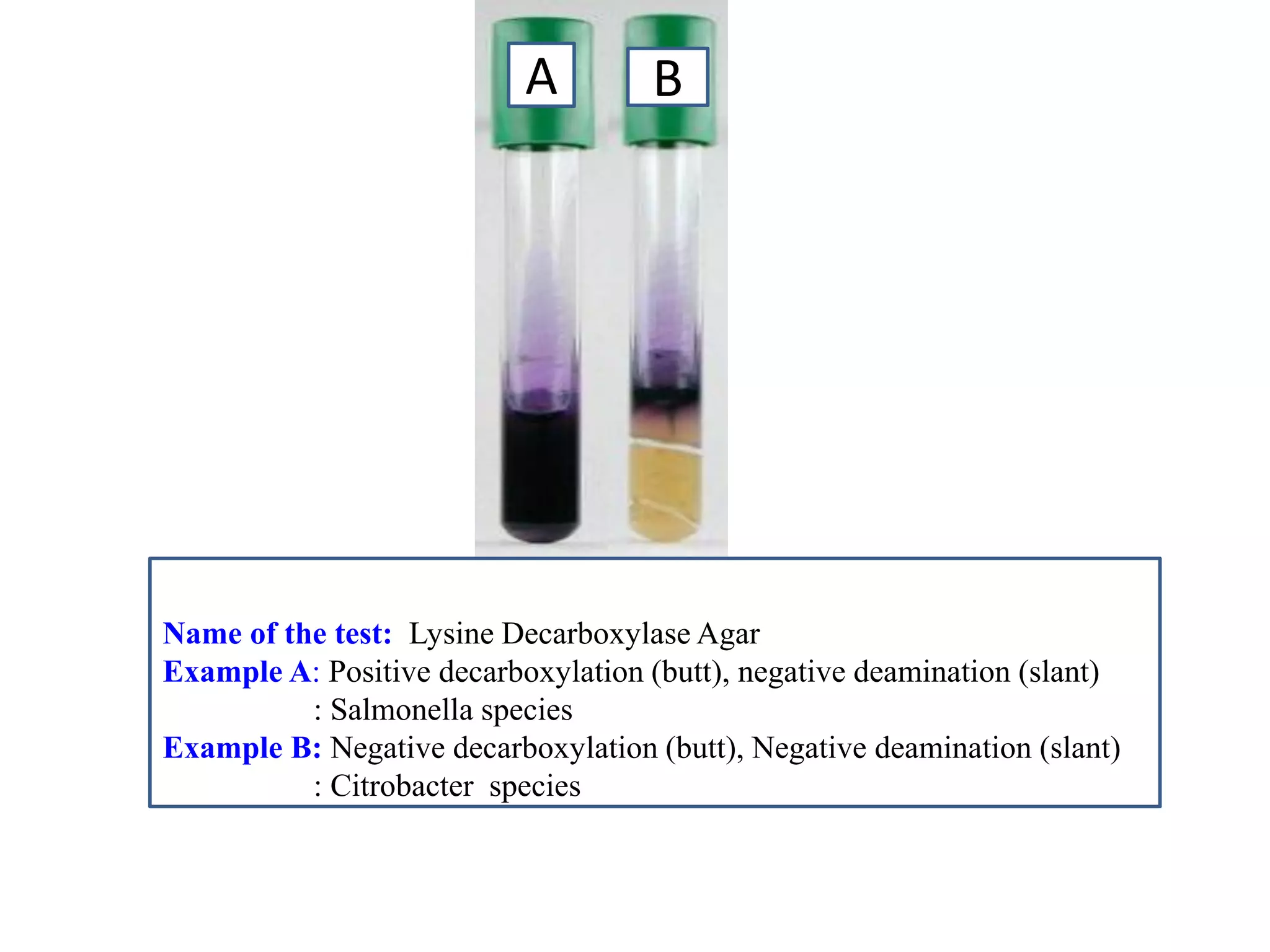
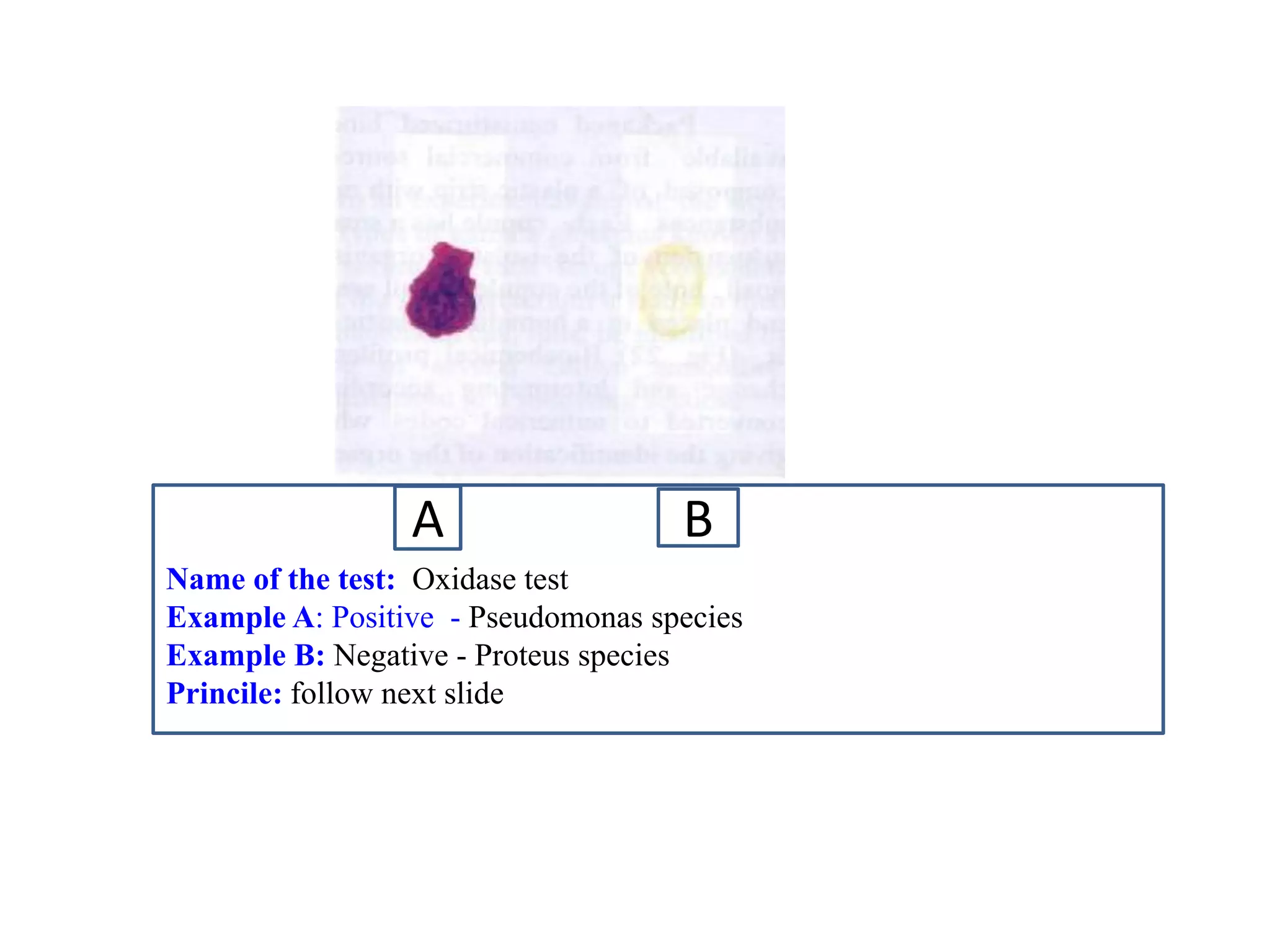
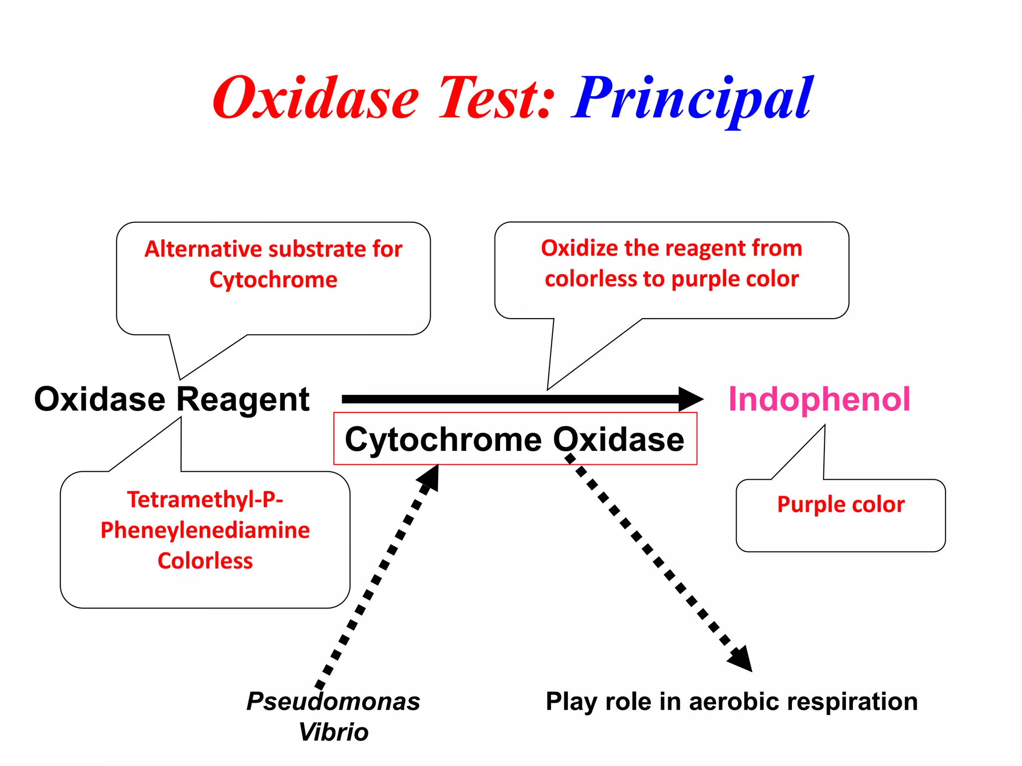
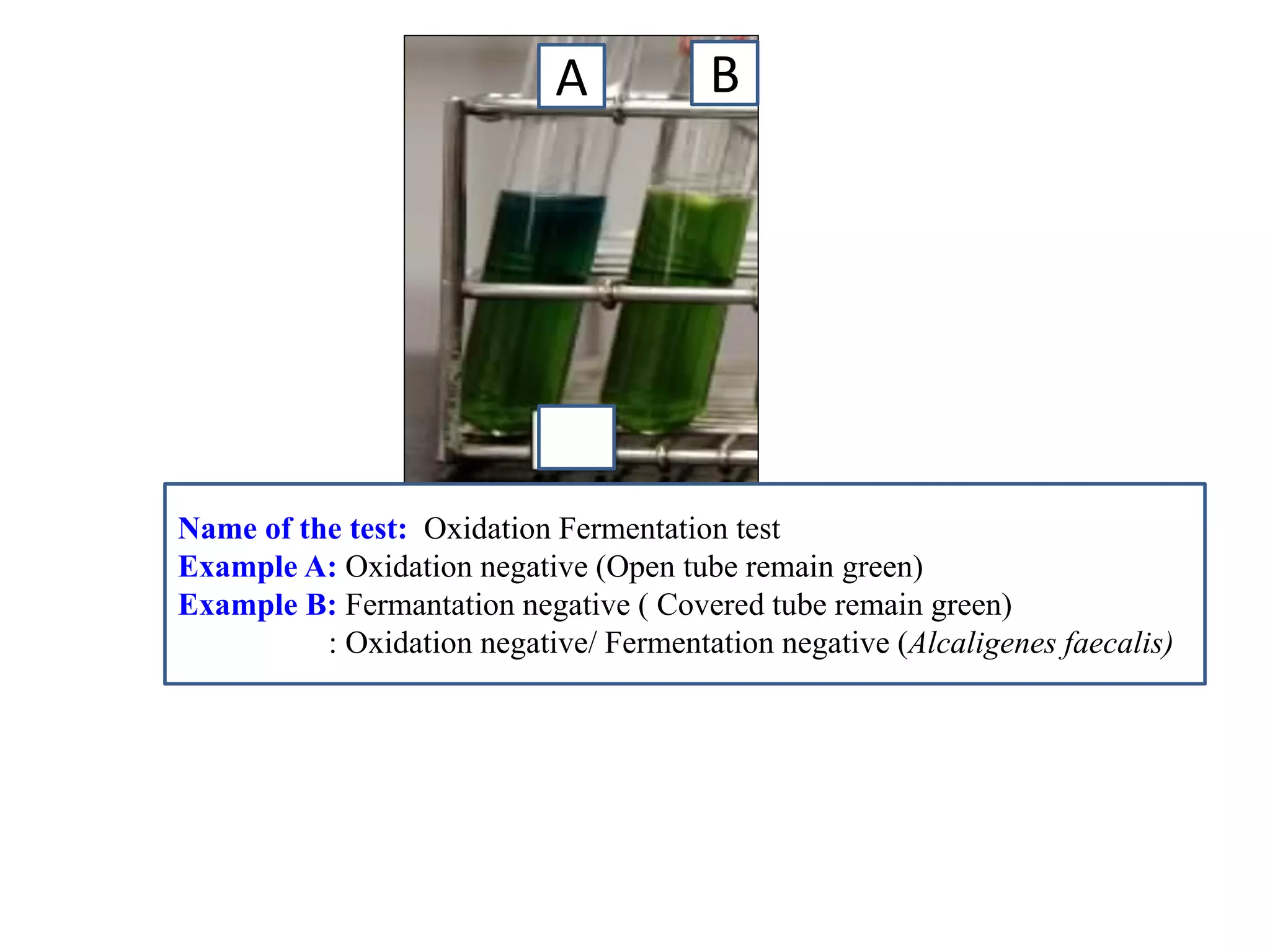
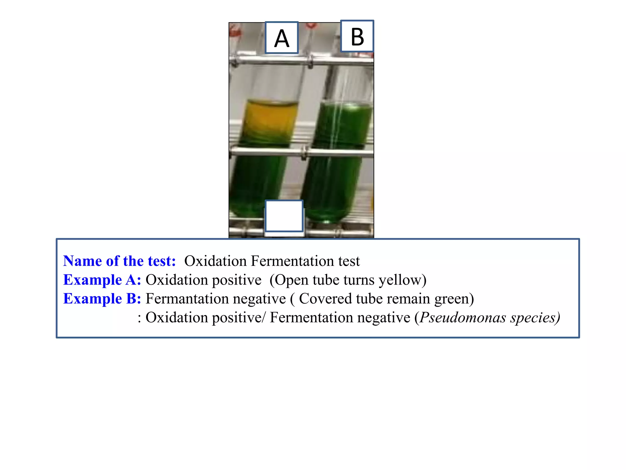
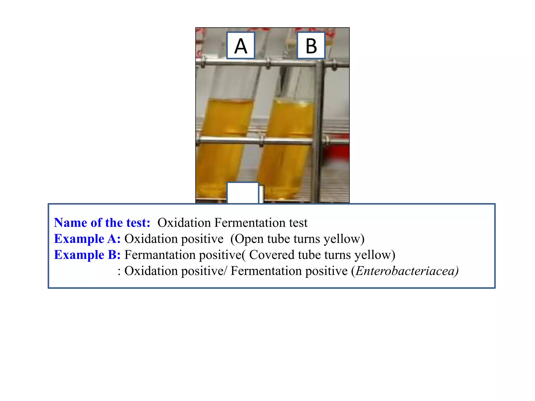
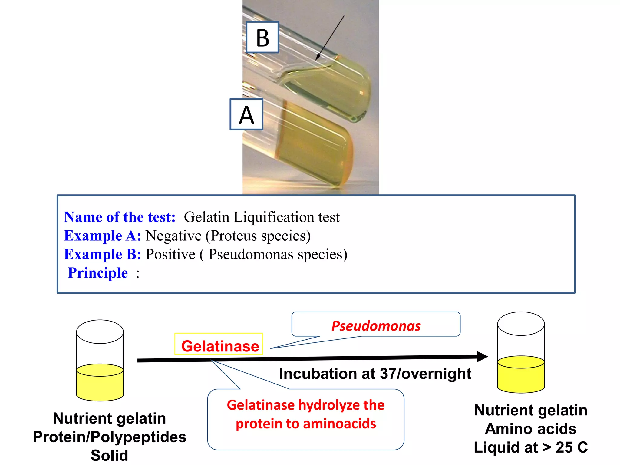
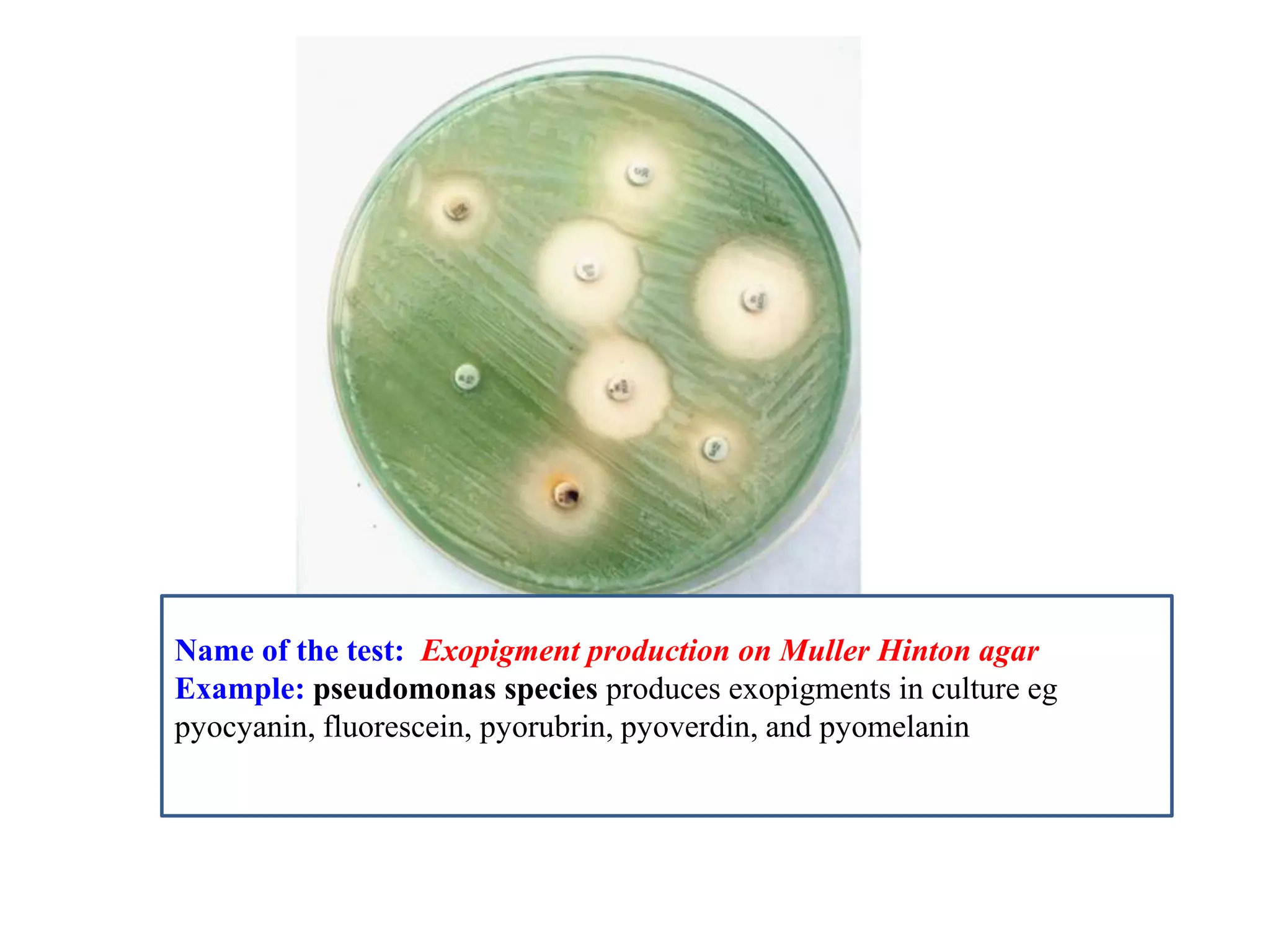
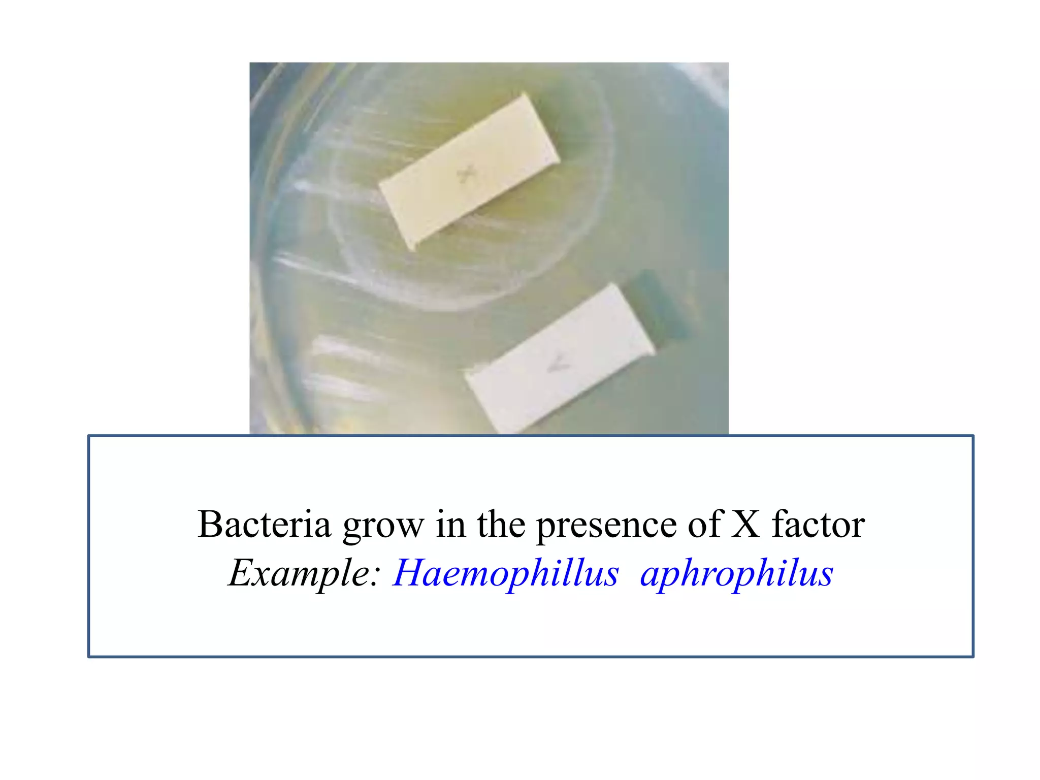
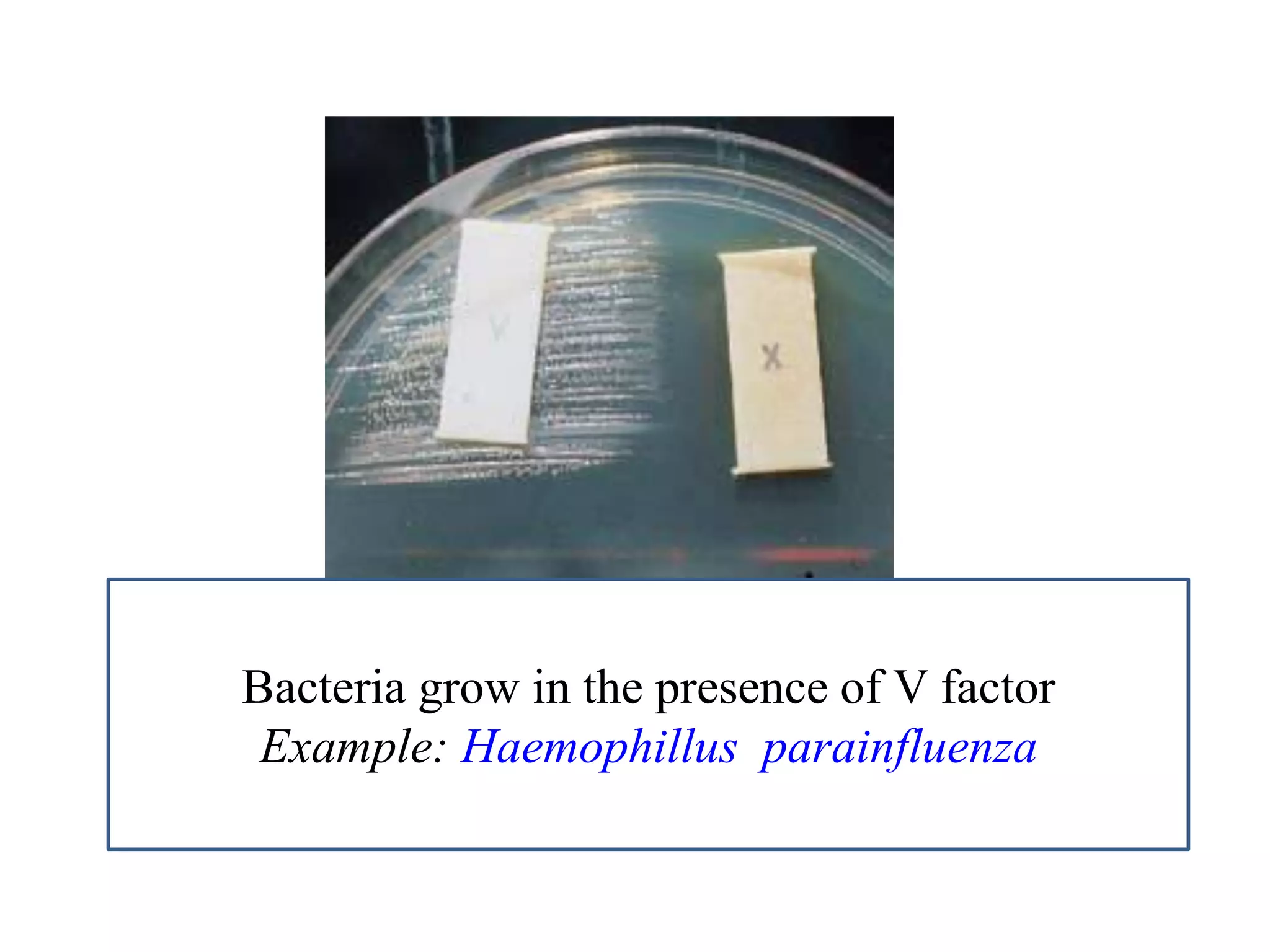
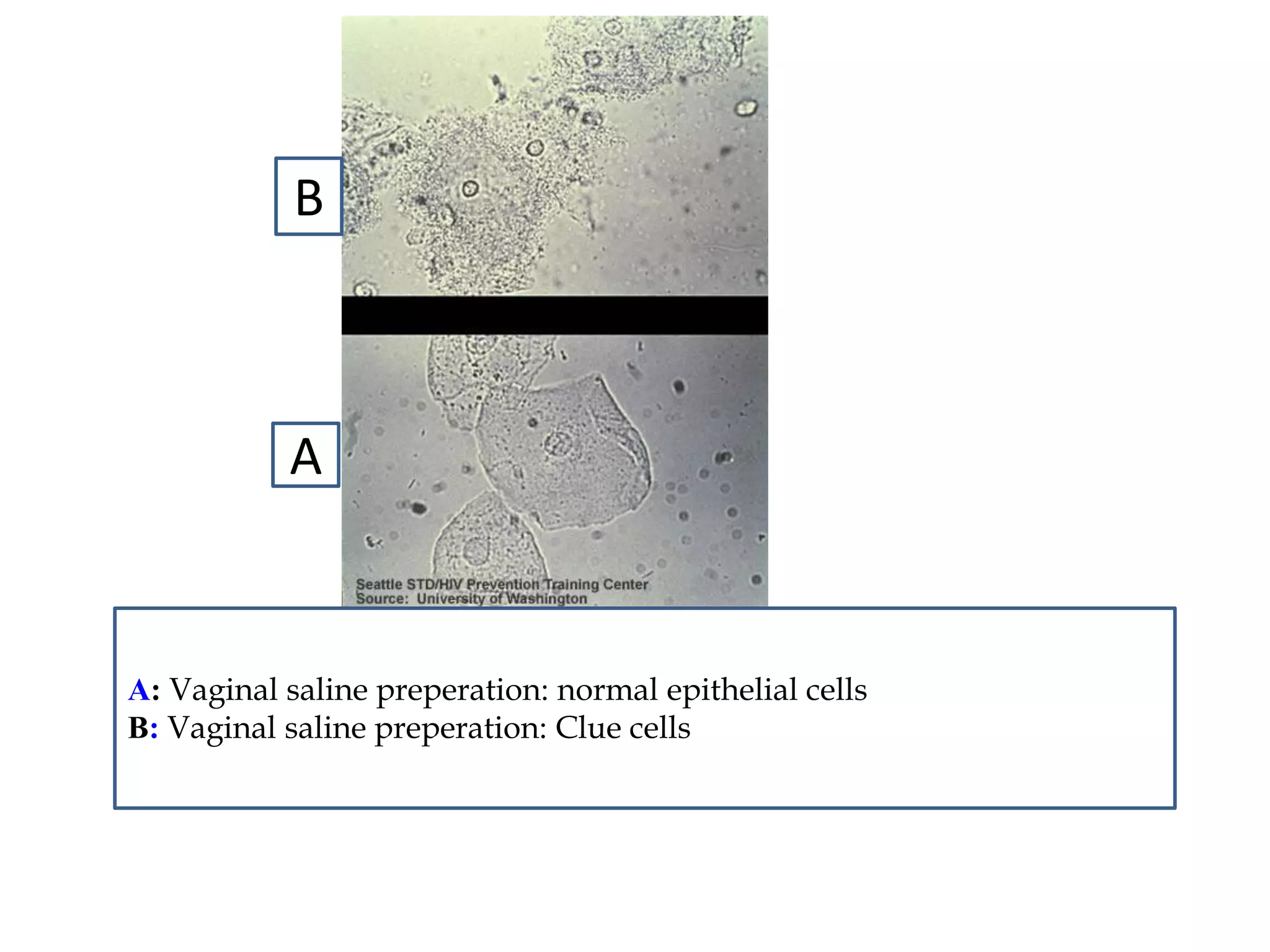
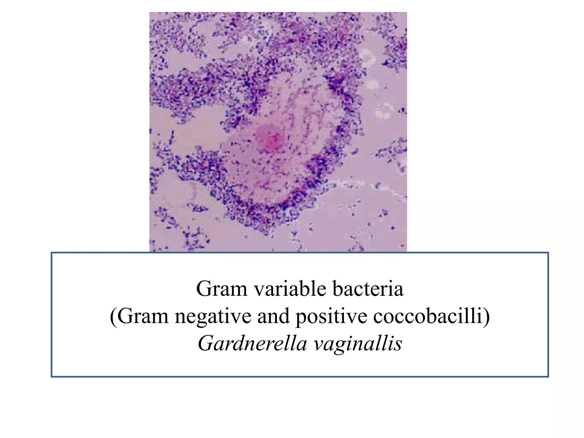
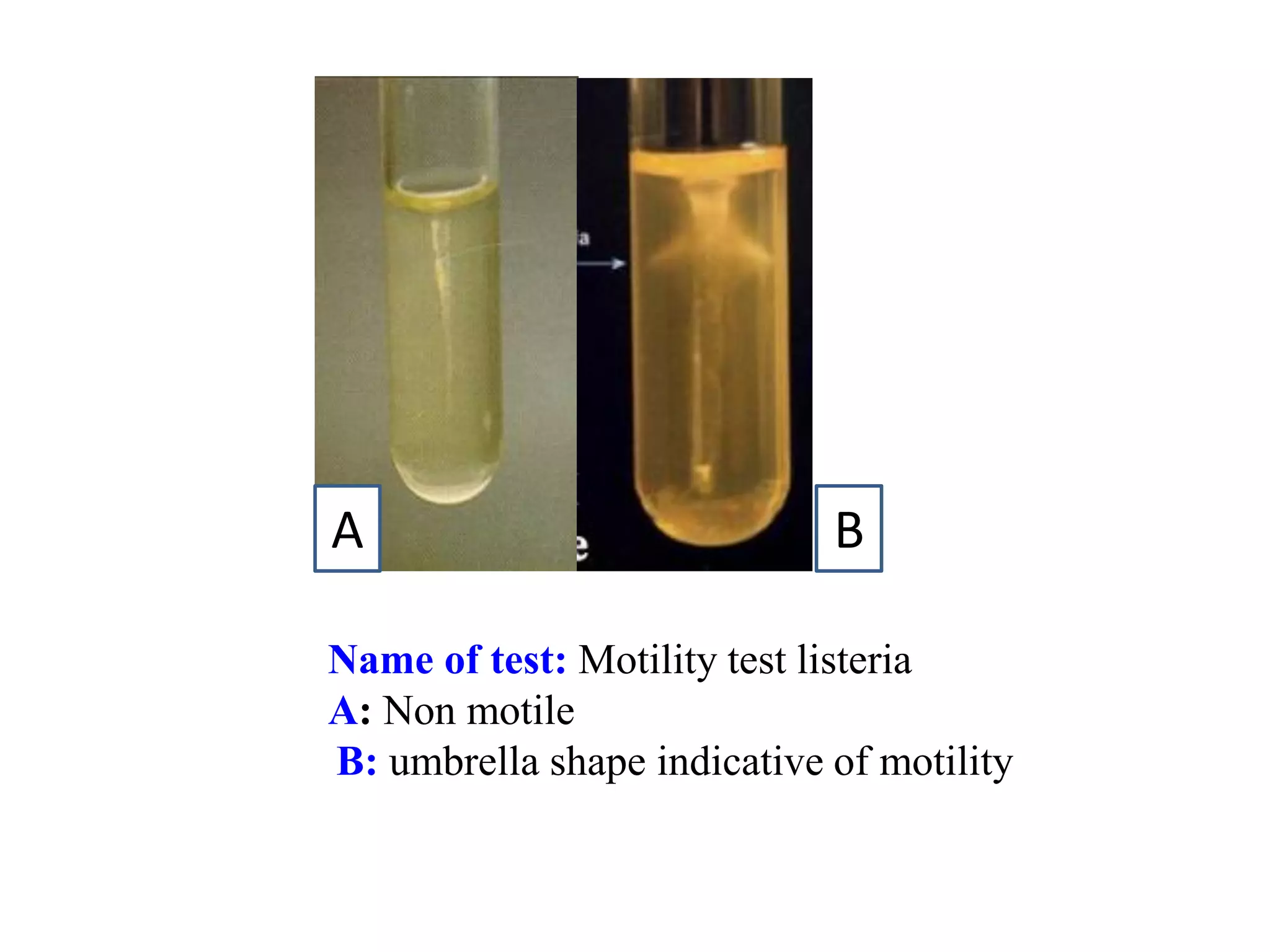
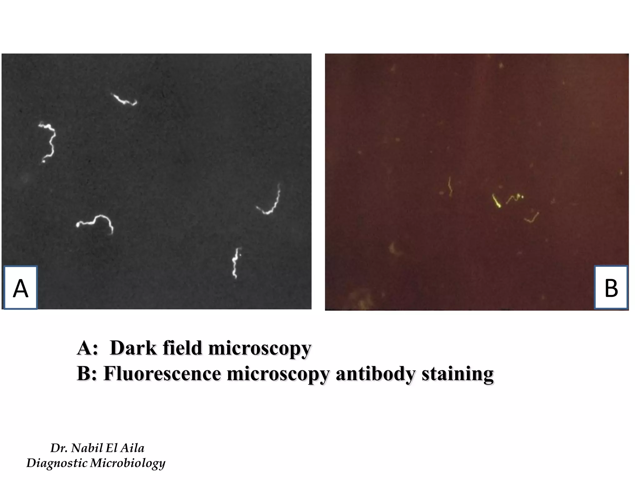
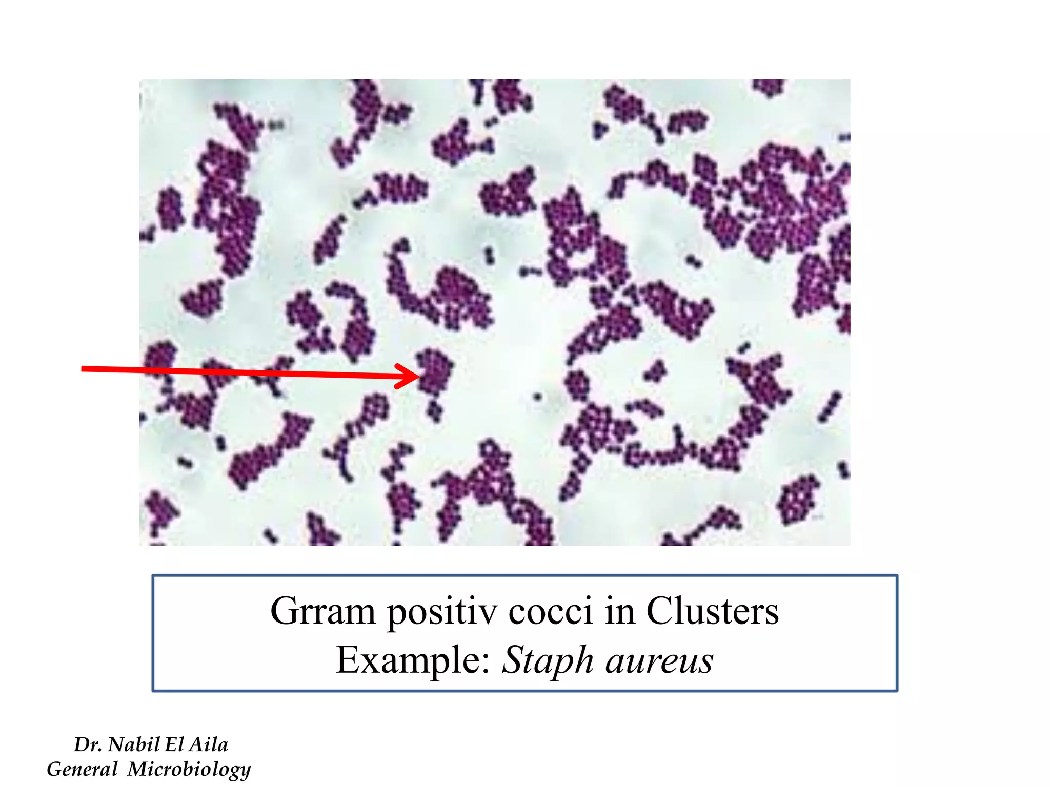
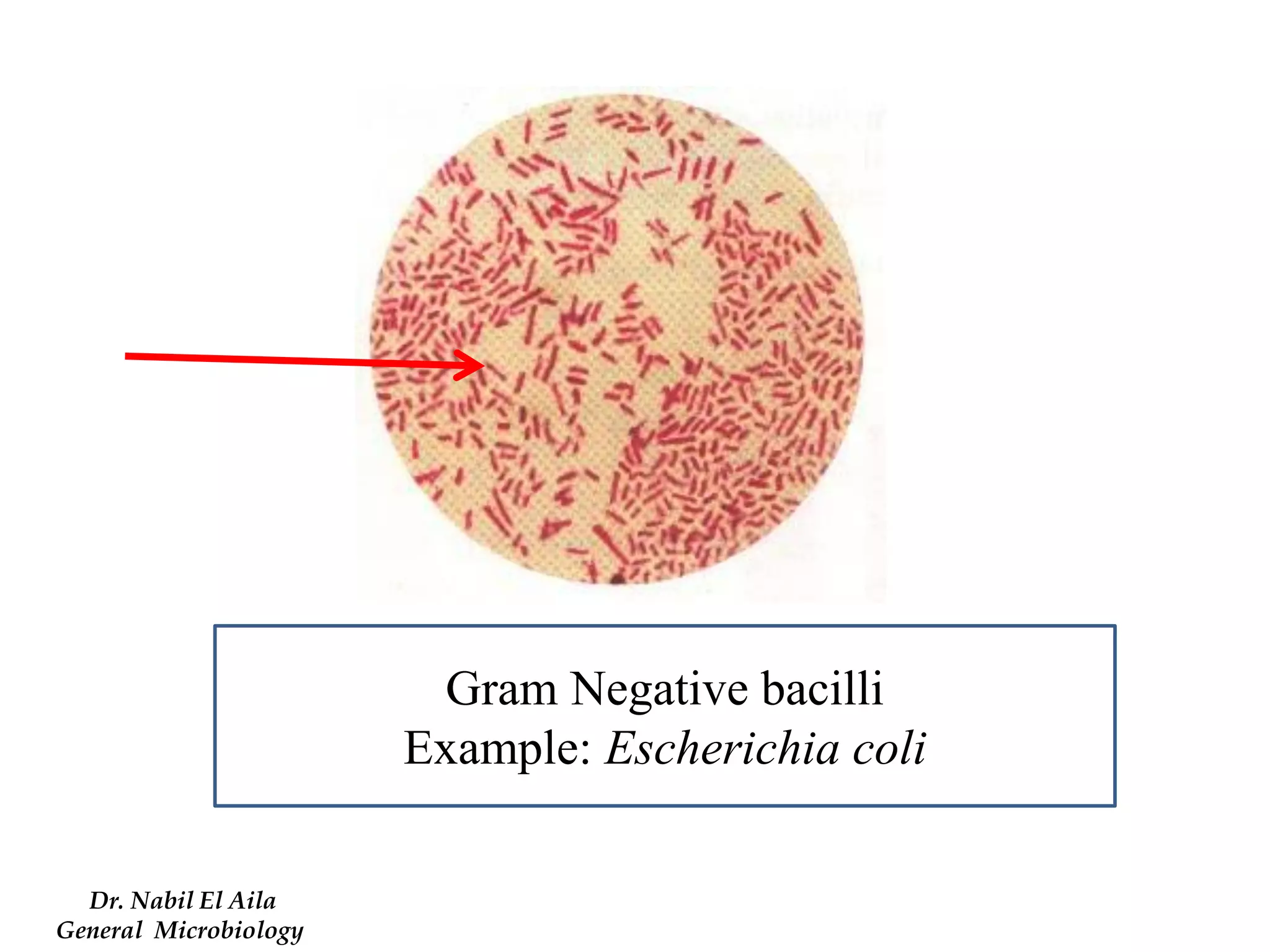
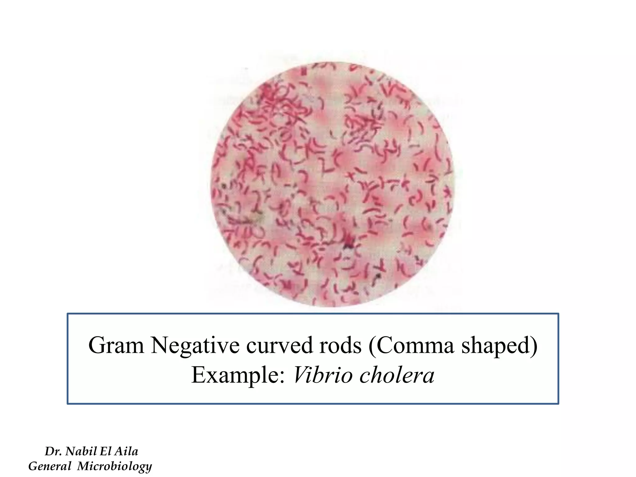
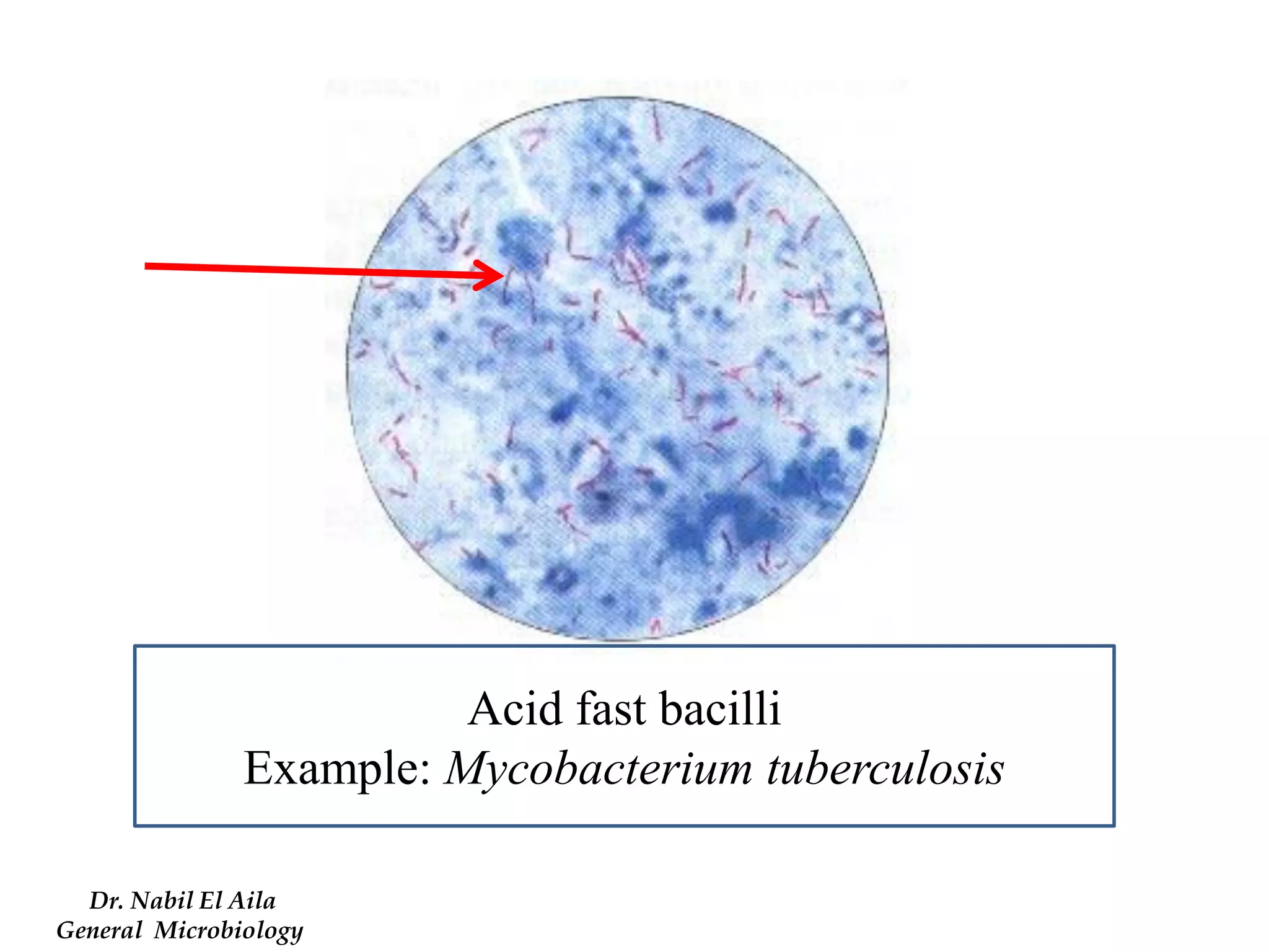
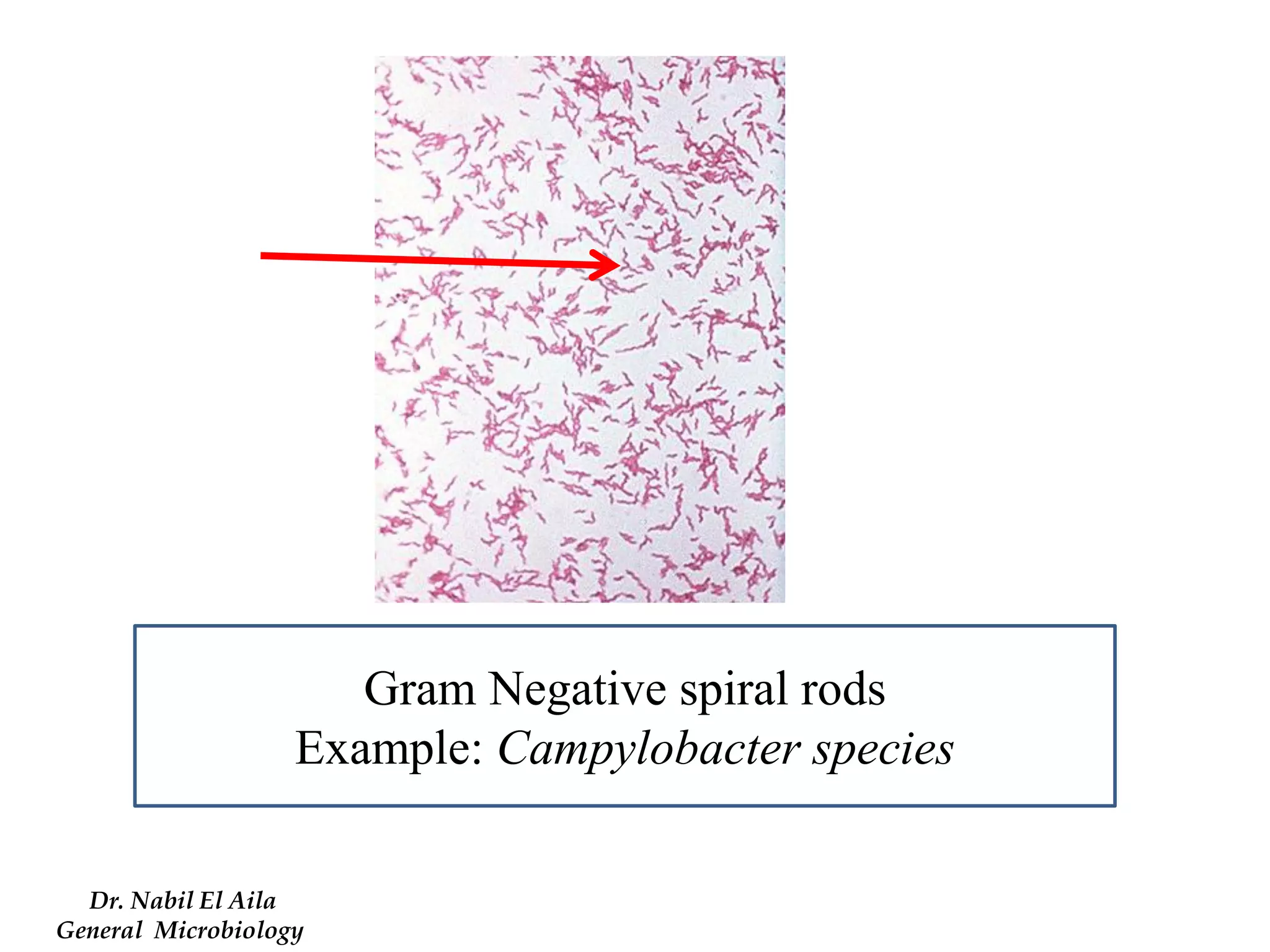
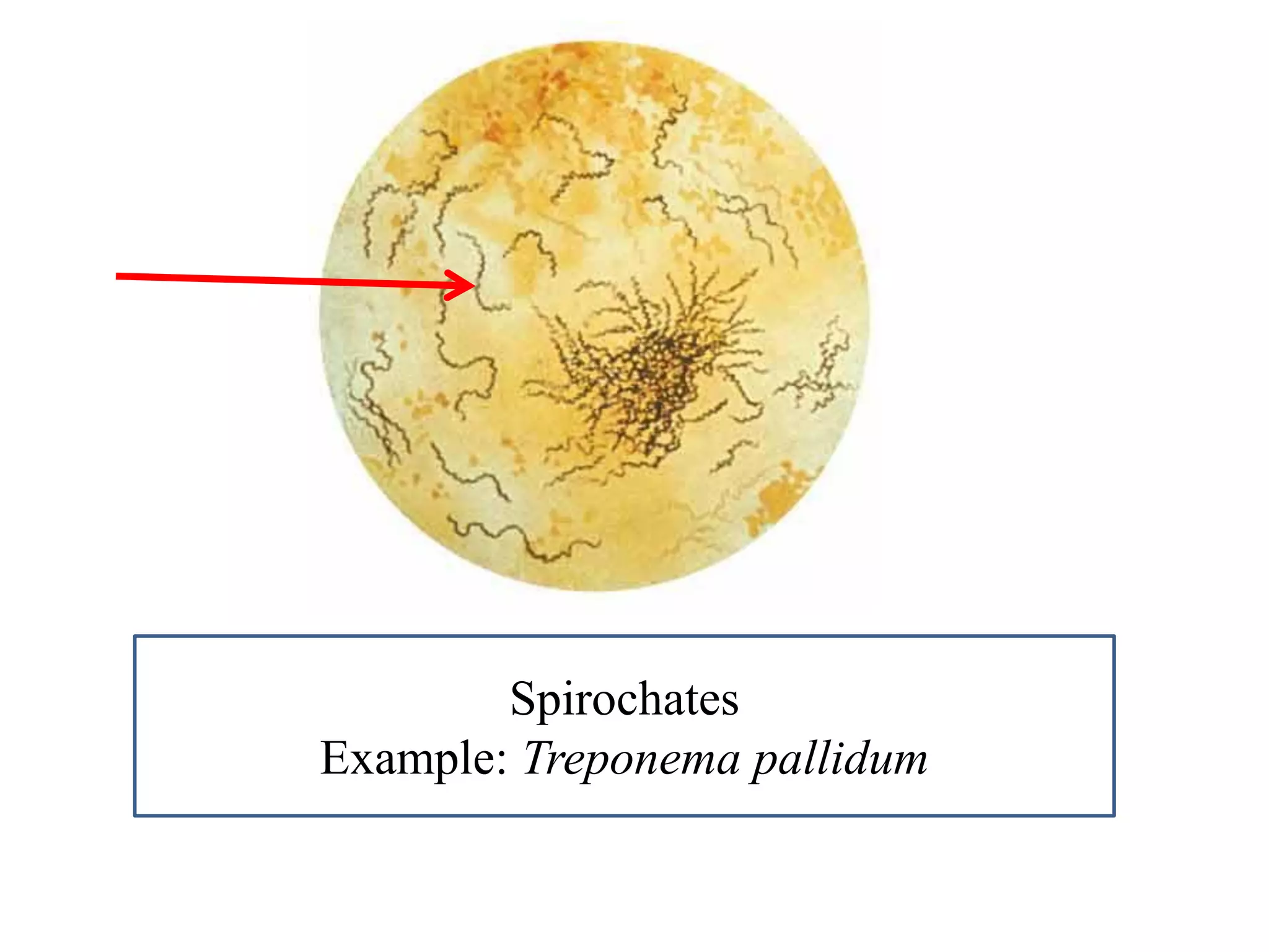
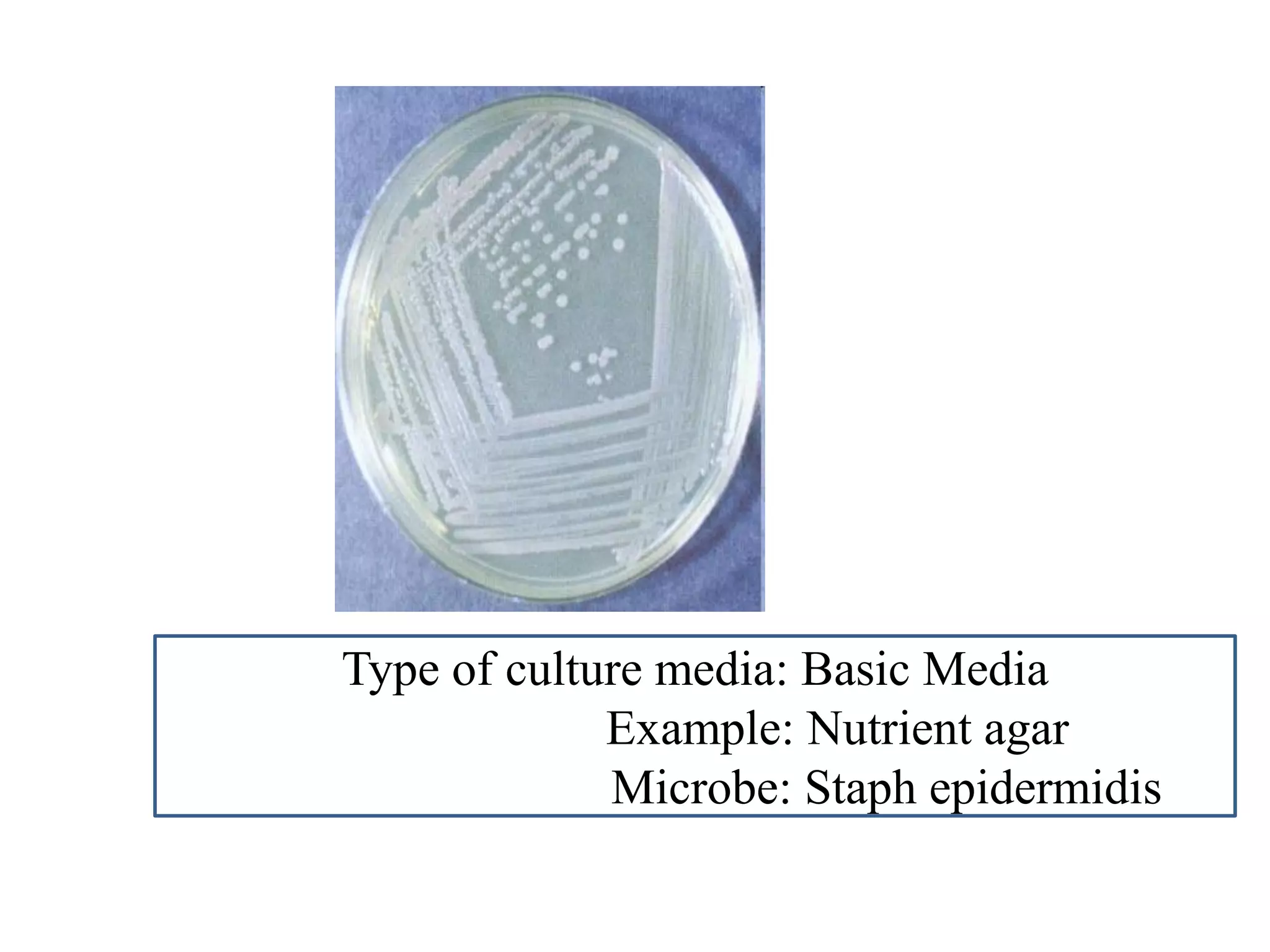
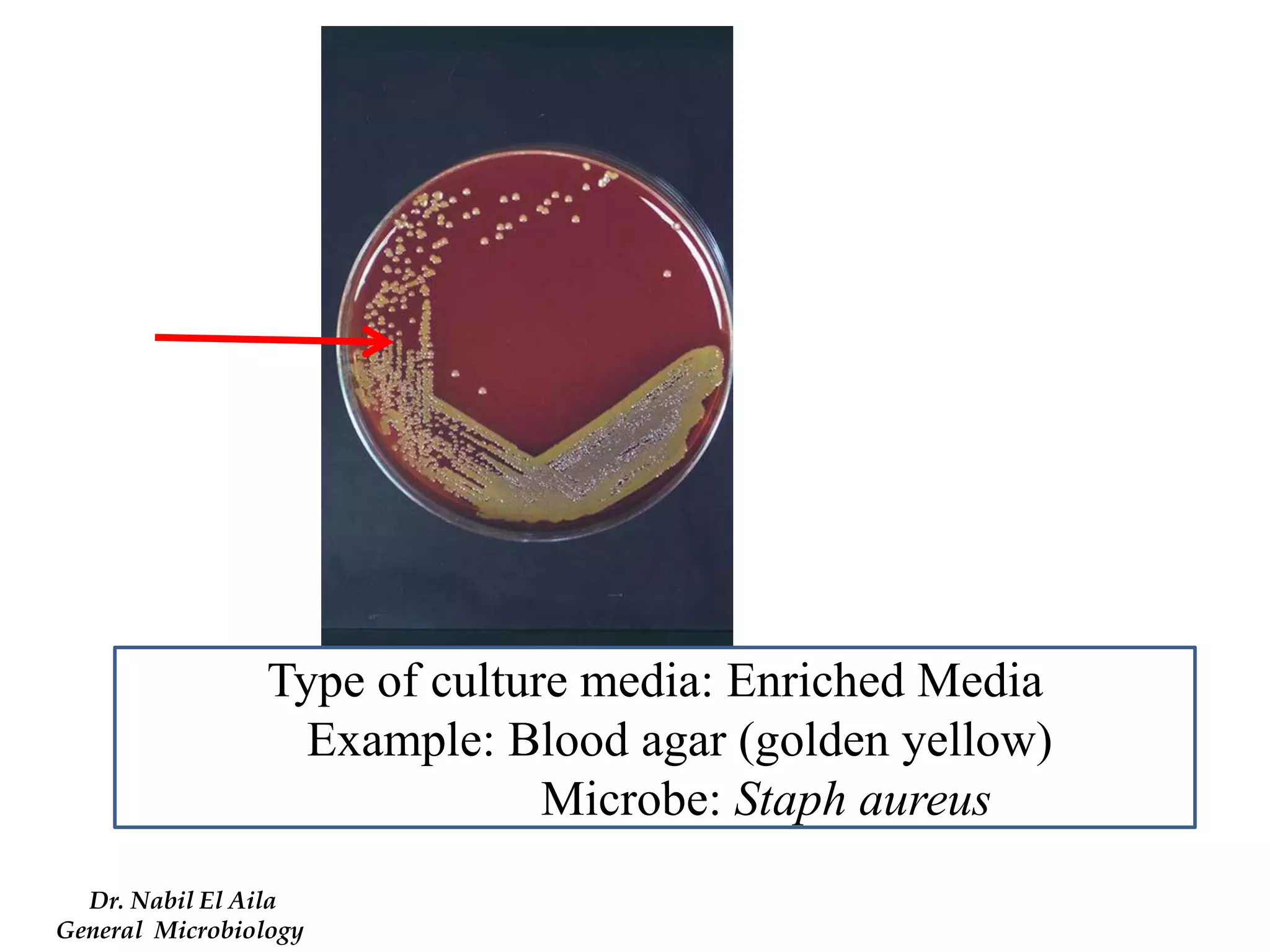
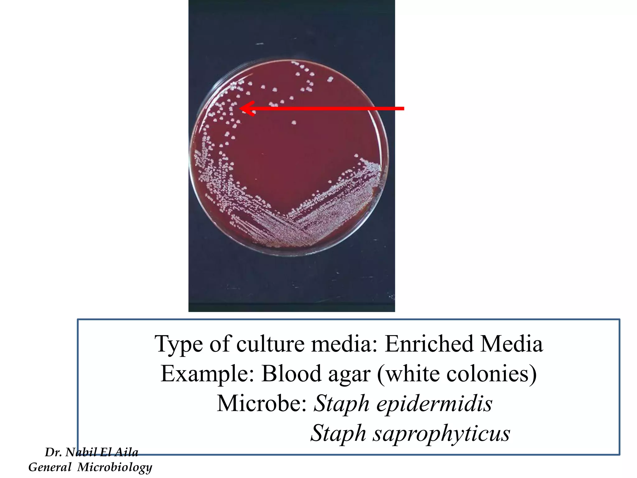
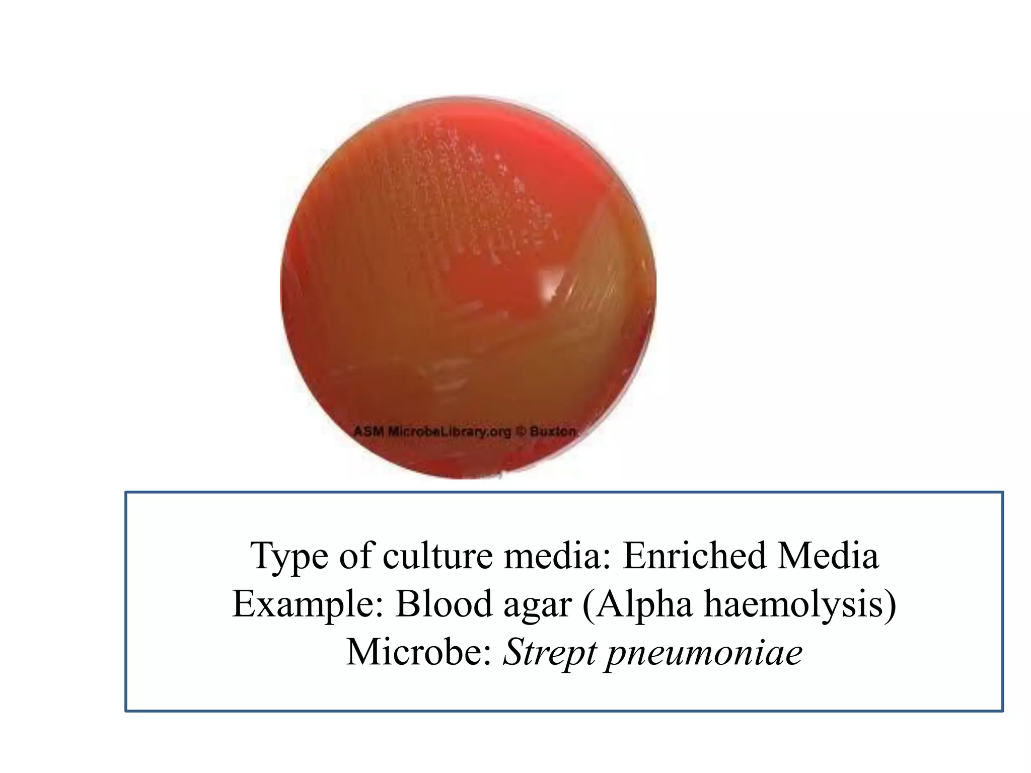
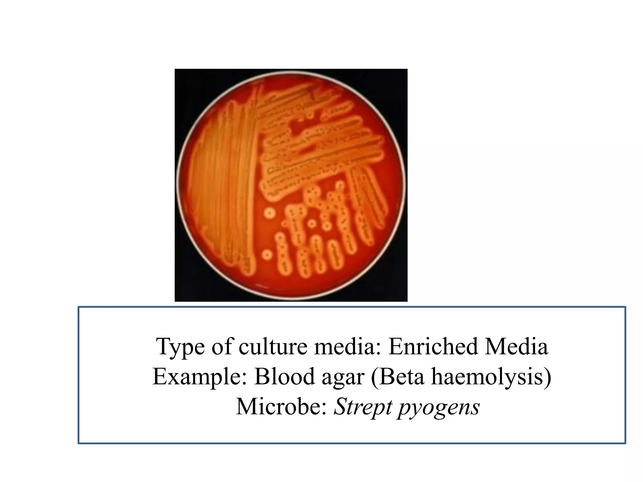
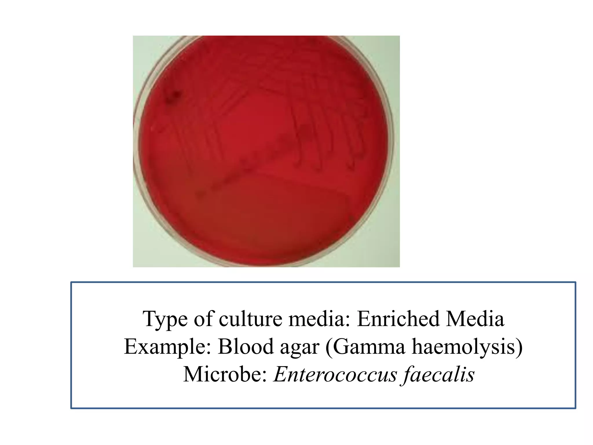
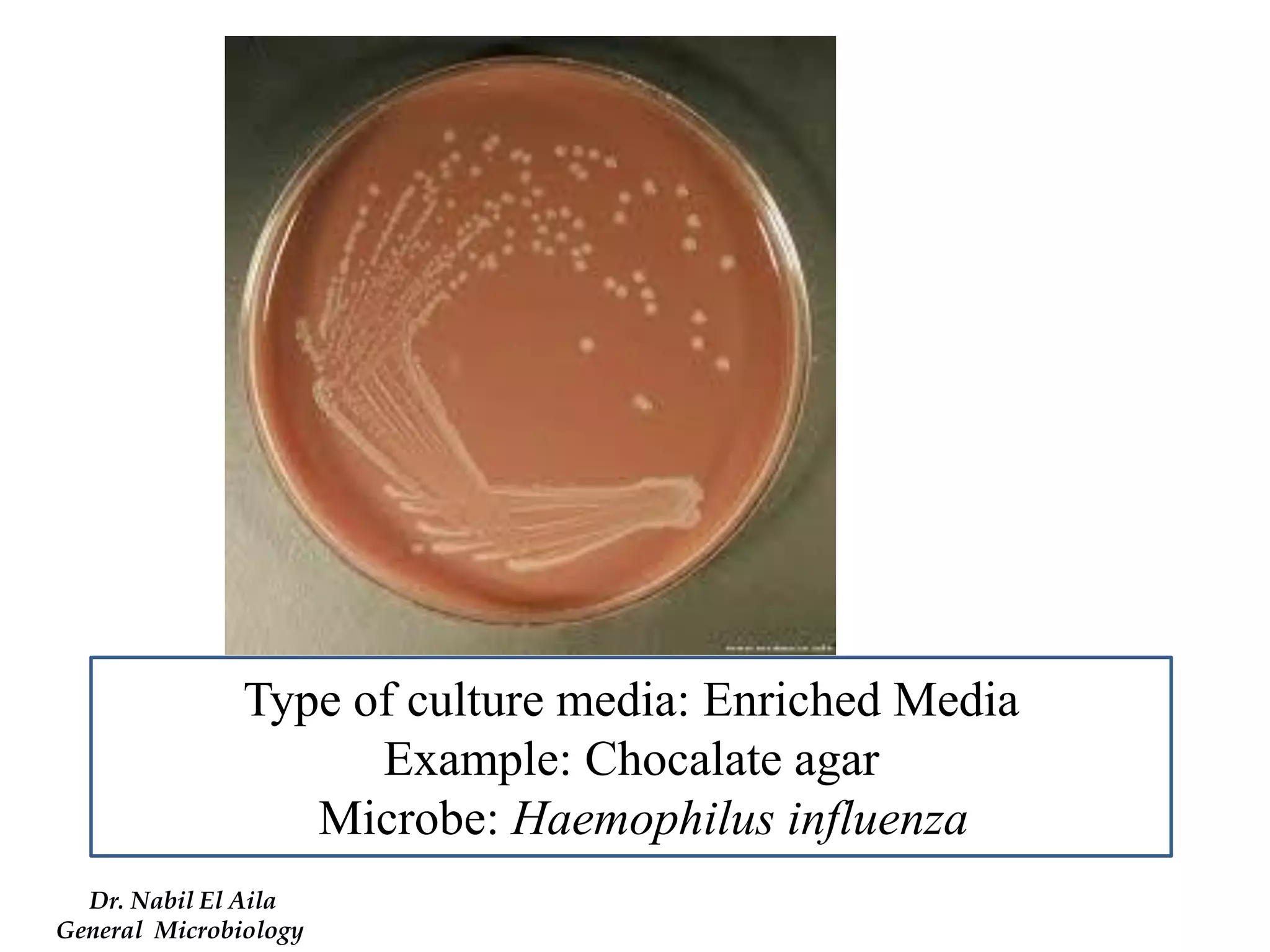
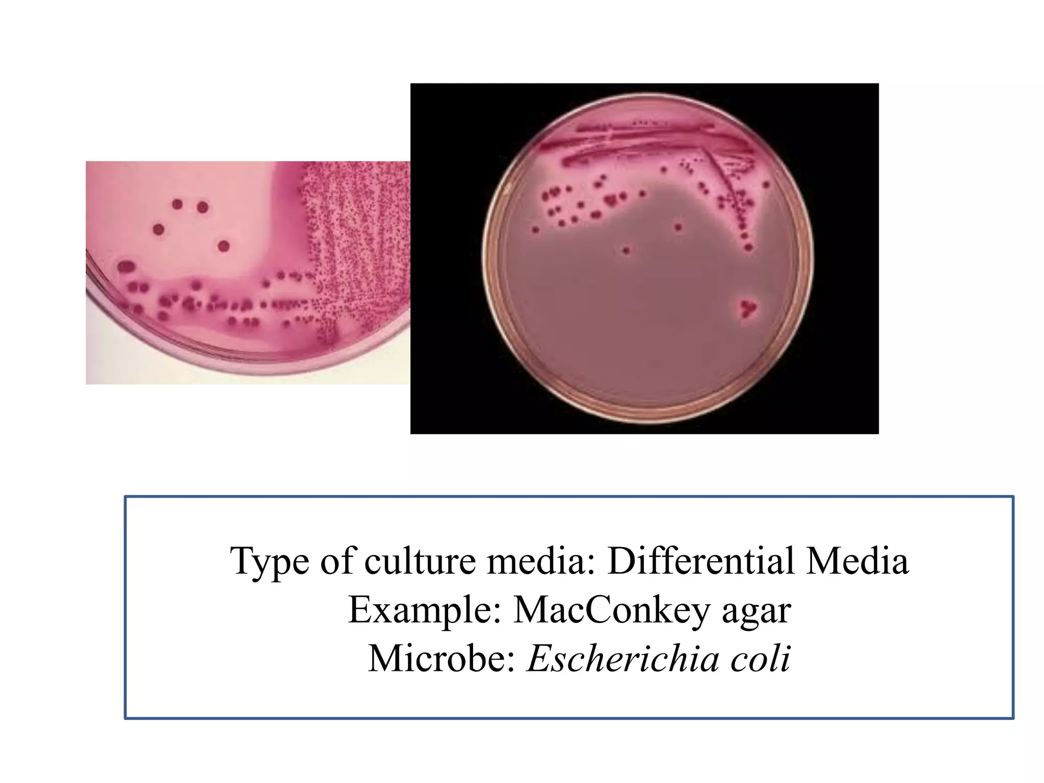
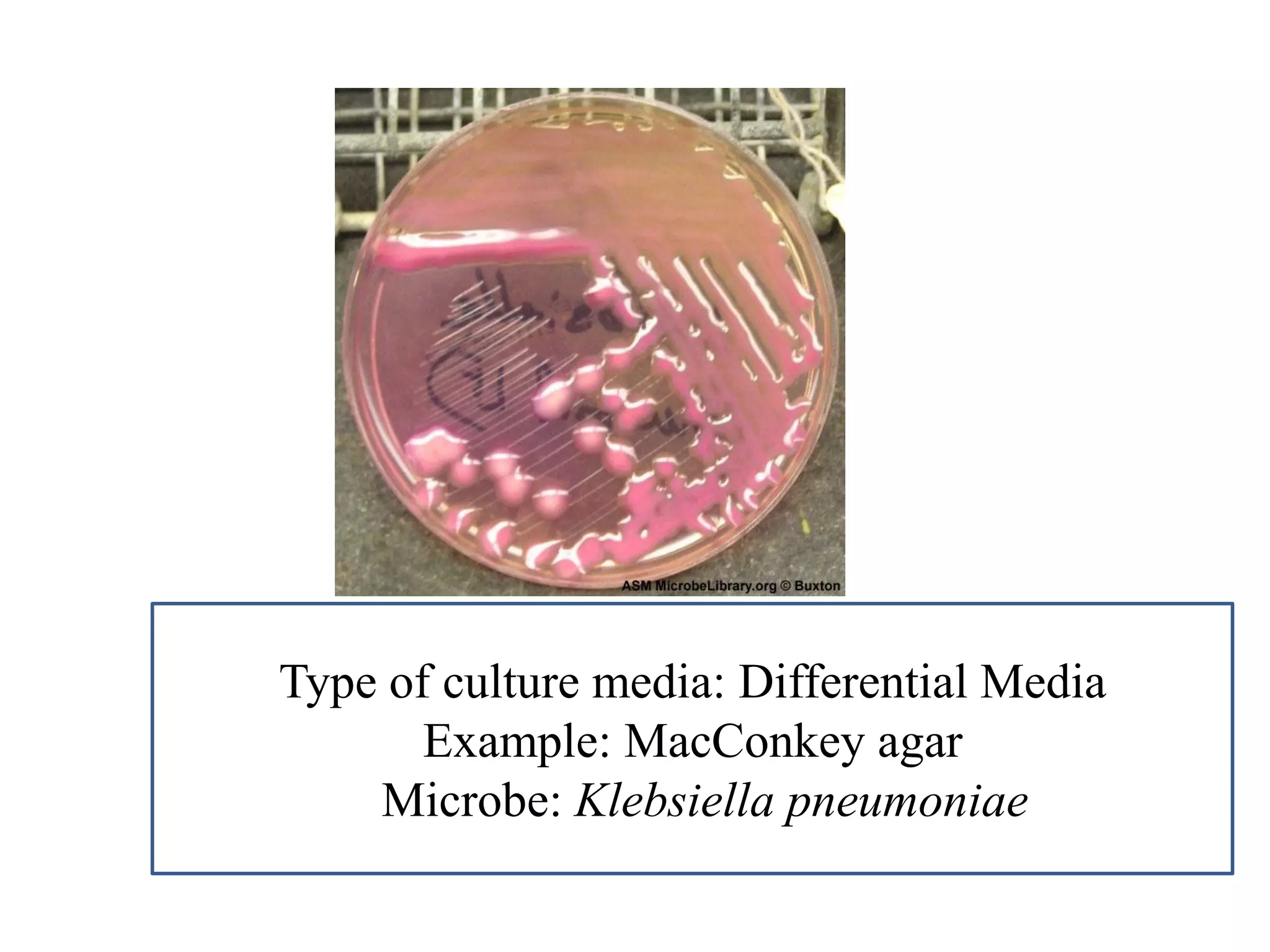
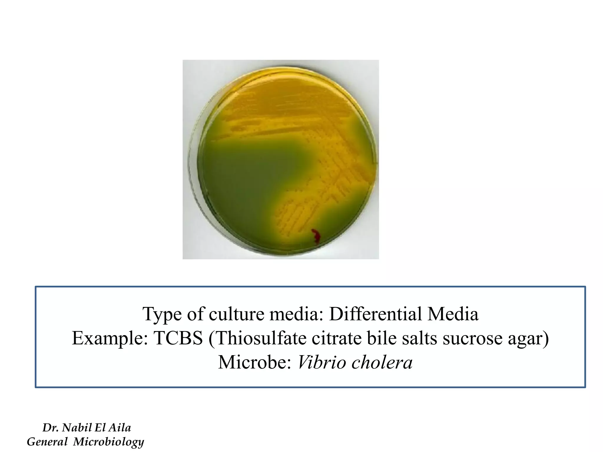
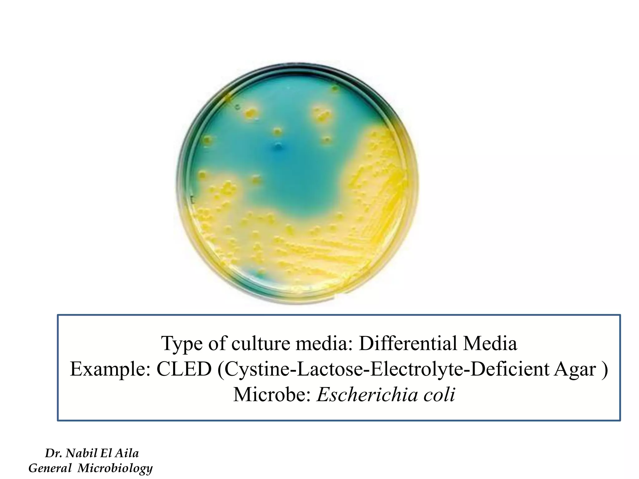
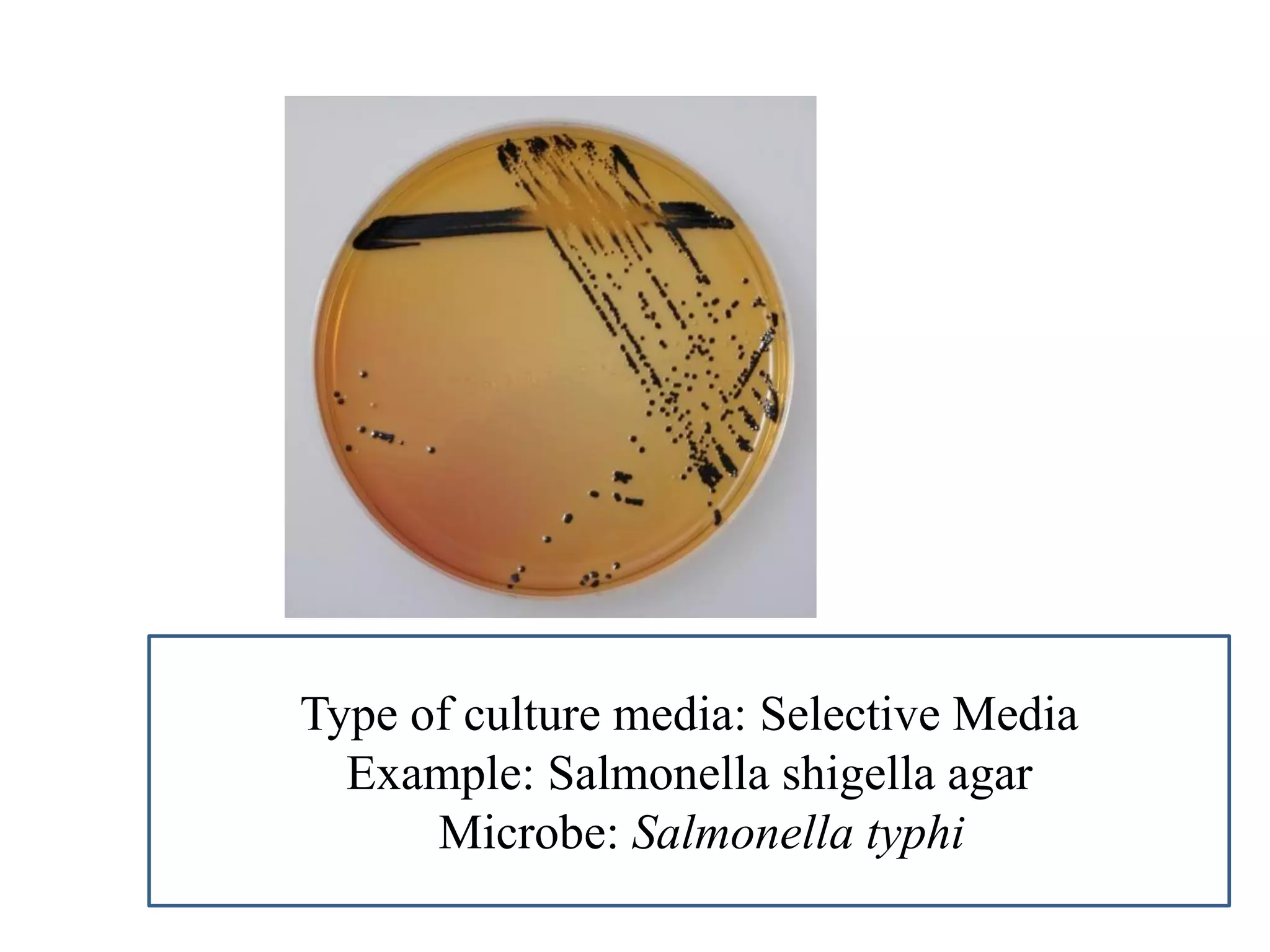
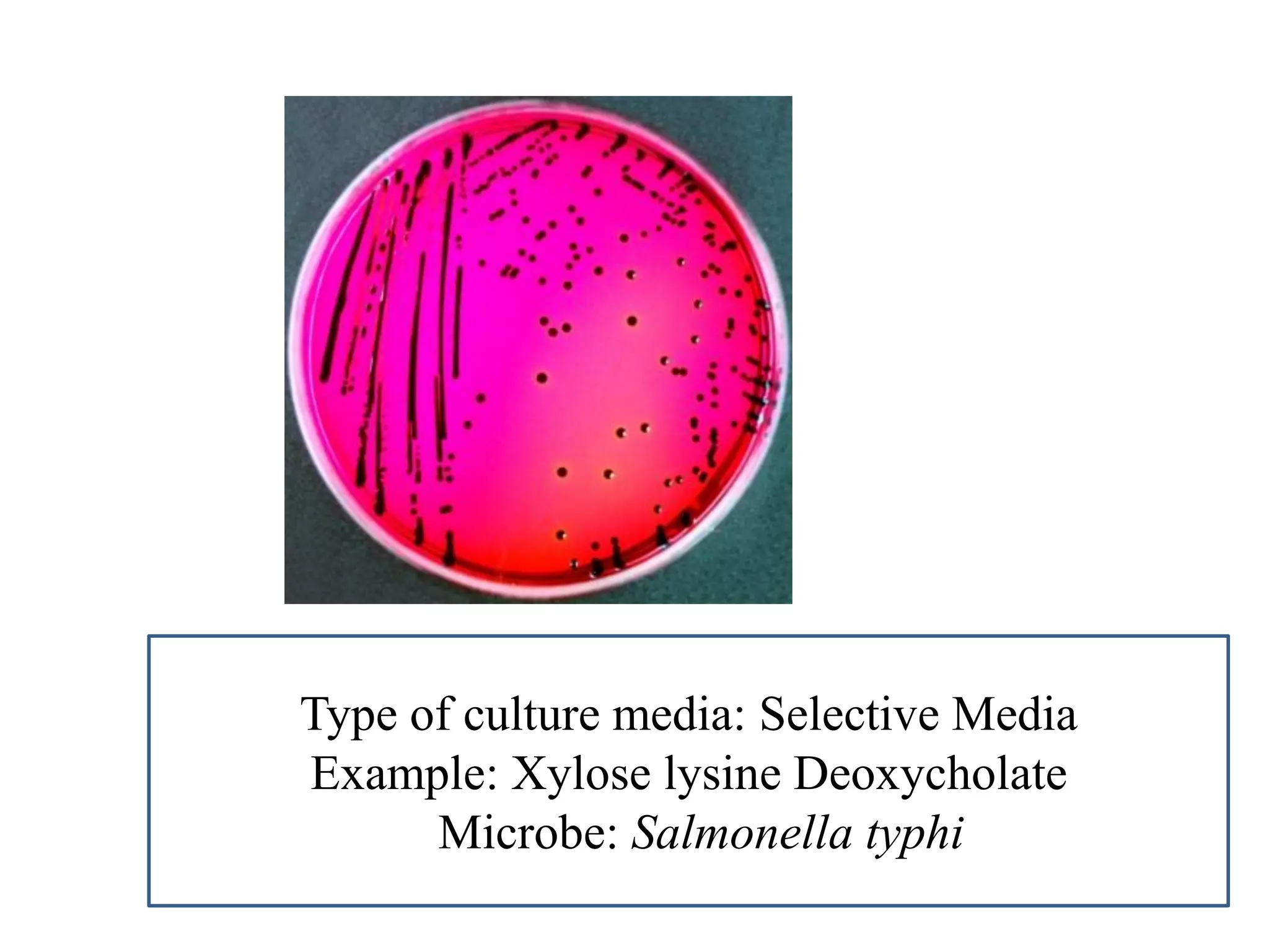
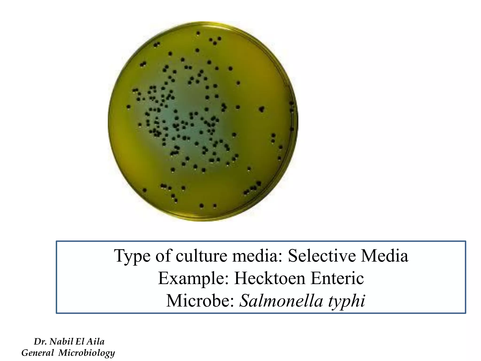
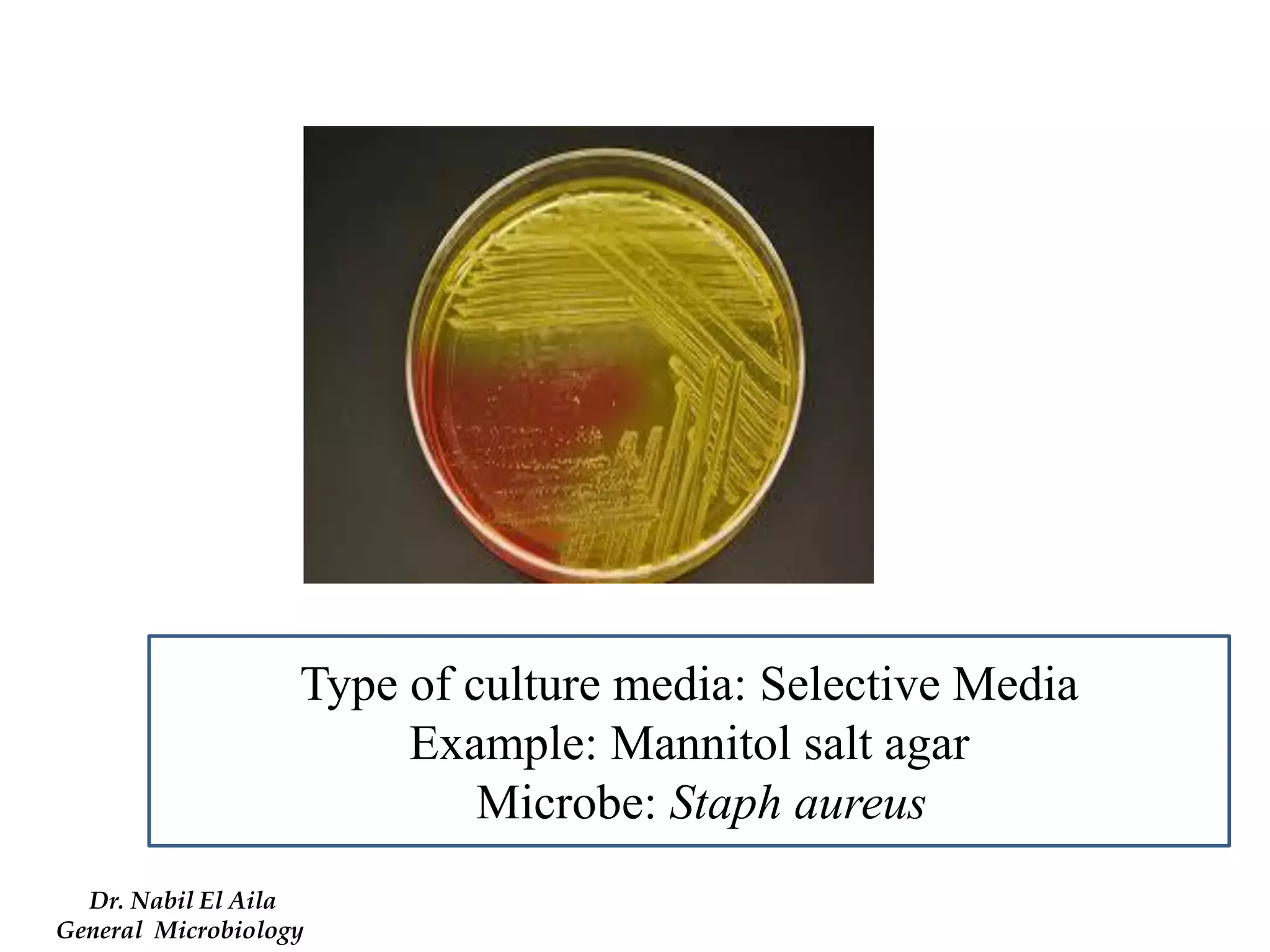
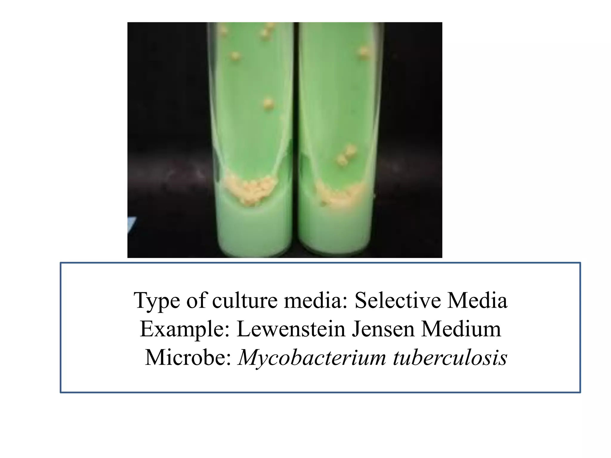
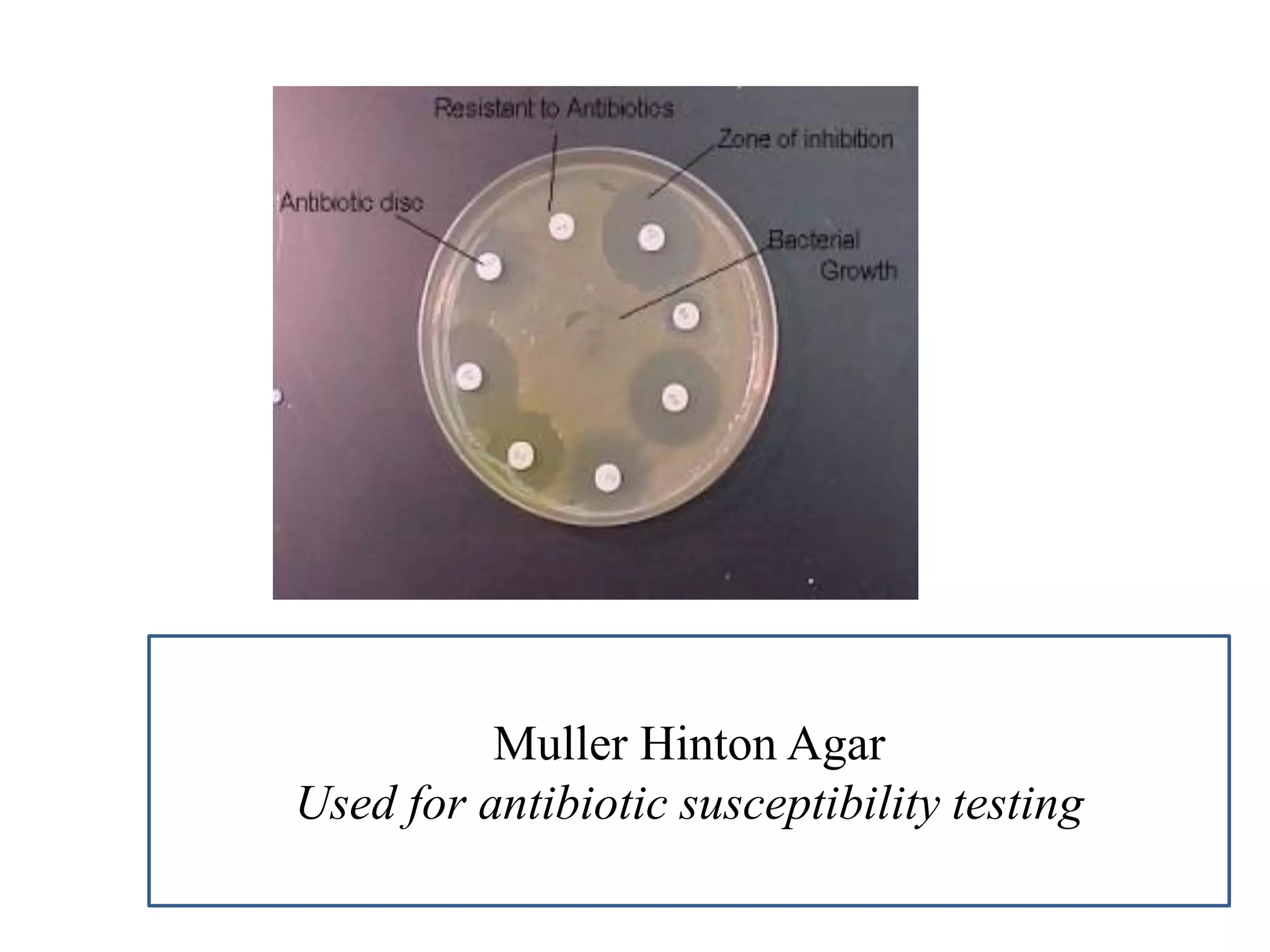
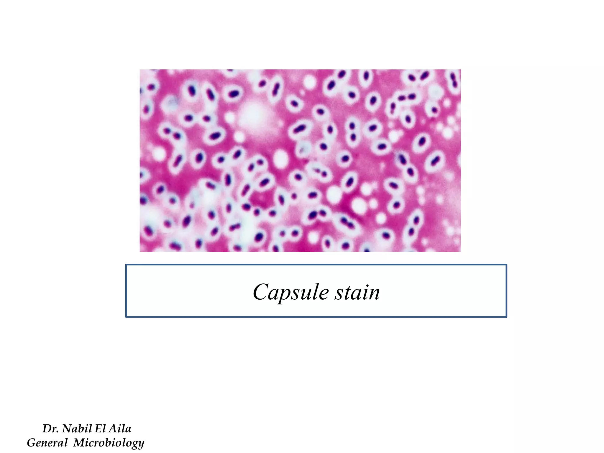
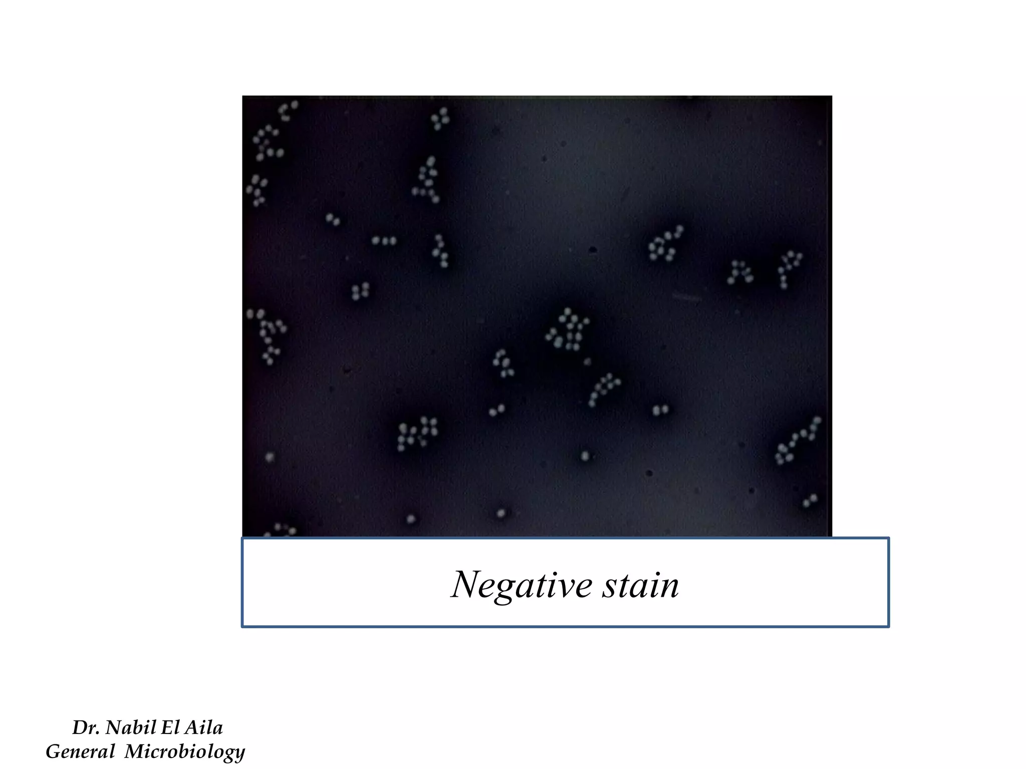
This document provides information on various microbiology tests used to identify bacterial species, including examples of positive and negative results. It describes tests such as the catalase test to distinguish Staphylococci from Streptococci, the coagulase test for identifying Staph aureus, and the DNase test to differentiate Staph aureus from Staph epidermidis. It also summarizes culture-based tests on different media types to isolate and identify bacteria such as Salmonella, E. coli, and Streptococcus species.
1. Diagnostic Microbiology Lab.
By
Dr. Nabil El Aila
Assistant Professor of Molecular Microbiology
Medical Technology Department
Al -Aqsa University
2. A
B
Name of the test: Catalase test
Example A: positive - Staphylococci
Example B: Negative - Streptococci
Principle: 2 H2O2 2 H2O + O2
Catalase
3. A
B
Name of the test: Coagulase test
Example A: Positive - Staph aureus
Example B: Negative - Staph epidermedis
Principle: Fibrinogen in plasma Fibrin clot
Coagulase
4. A
B
Name of the test: DNase test
Example A: Positive - Staph aureus
Example B: Negative - Staph epidermedis
Priciple: DNA is insoluble in acid
DNA is hydrolyzed into oligonucleotides by the action of Dnase
Nucleotides soluble in acid (After addition of 1N HCl)
5. Name of the test: Mannitol salt agar
Example A: Negative - Staph epidermidis
Example B: Positive - Staph aureus
Principle: Staph aureus ferment mannitol (sugar) and phenol red (pH indicators turns yellow in acidic pH and turns red in alkaline pH).
A
B
6. Name of the test: Novobiocin sensitivity
Example A: Staph epidermidis (Sensitive)
Example B:Staph saprophyticus (Resistant)
A
B
7. Name of the test: Bacitracin sensitivity
Example A: Strept pyogens (sensitive)
Example B: Strept agalactiae (resistant)
A
B
8. A
B
Name of the test: CAMP test
Example A: Positive - Strept agalactiae (Arrow shaped)
Example B: Negative - Strept pyogens
Principle : Strept agalactiae produce CAMP factor (a diffusible extracelluar protein) that synergistically acts with the beta-lysin of Staphylococcus aureus and enhances the lysis of red blood cells.
9. Name of the test: Optochin sensitivity
Example A: Strept pneumonia (sensitive)
Example B: Strept viridans (resistant)
A
B
10. A
B
Name of the test: Bile Esculin test
Example A: Positive - Group D streptococcus (Enterococcus species)
Example B: Negative - Group B streptococcus
Principle: The selective agent bile, inhibits most gram positive bacteria.
Esculin in the medium is hydrolyzed to esculetin and dextrose.
The esculetin reacts with ferric chloride in the media to form a black-brown color.
11. B
A
Name of the test: Bile Solubility
Example A: Strept mitis (Negative)
Example B: Strept pneumoniae (positive)
Principle: S. pneumoniae is bile soluble whereas all other alpha-hemolytic streptococci are bile resistant. Sodium deoxycholate (2% in water) will lyse the pneumococcal cell wall
12. A
B
Name of the test: Pyrrolidonyl arylamidase Test (PYR)
Example A: Strept agalactiae (Negative)
Example B: group A streptococci and Enterococcu s species (positive)
Principle: Group A streptococci and Enterococcu s species produce pyrrolidonyl arylamidase which hydrolyzes the substrate L- pyrrolidonyl -β- naphthylamide to form β-naphthylamine.
A pink to red color forms when p-dimethylaminocinnam-aldehyde (PYR reagent) is added to β-naphthylamine.
13. A
B
Tryptophane
amino acids
Tryptophanase
Indole + Pyurvic acid + NH3
Kovac’s Reagent
Red color in upper organic layer`
Name of the test: Indole test
Example A: Klebsiella pneumonia (Negative)
Example B: Escherichia coli (Positive)
Indicator: Kovac’s Reagent (4 (p) – dimethylamino benzaldehyde)
Principle:
14. A
B
Name of the test: Methyl red
Example A: Klebsiella pneumonia (Negative)
Example B: Escherichia coli (Positive)
Indicator: Methy red indicator
Principle: follow next slides
15. A
B
Name of the test: Voges Proskauer test
Example A: Escherichia coli (Negative)
Example B: Klebsiella pneumonia (Positive)
Indicator: -naphthol and 40% KOH
Principle: follow next slide
16. IMViC test Methyl Red-Voges Proskauer (MR-VP) Tests
Glucose
Acidic pathway
Mixed acids
pH less than 4.4
Methyl Red
indicator
Red color
Principle
MR positive
E. coli
Or
Neutral pathway
Acety methyl carbinol
(ACETOIN)
Barrit’s A (-naphthol)
Barrit;s B (40% KOH)
Pink color
VP positive
Klebsiella
17. Name of the test: Citrate test
Example A: Klebsiella pneumonia (Positive)
Example B: Escherichia coli (Negative)
Indicator: Bromothymol blue
Principle: follow next slide
A
B
18. Principle:
Citrate Na2CO3
Alkaline,↑pH
Bromothymol blue
Simmone’s Citrate media
Pyruvate CO2 + Na + H2O
Positive test
Contains Citrate as a sole of C source
IMViC: CITRATE TEST
Dr. Nabil El Aila
Diagnostic Microbiology
19. Name of the test: Urea test
Example A: Proteus miribalis (Positive)
Example B: Escherichia coli (Negative)
Indicator: Phenol red
Principle:
A
B
Urea
Urease
CO2 + NH3
H2O
NH4 OH
↑ in pH
Phenol Red
Pink
Positive test
20. A
B
Name of the test: Motility test
Example A: Escherichia coli (Negative)
Example B: Proteus miribalis (Positive)
21. A
B
Name of the test: H2S production
Example A: Escherichia coli (Negative)
Example B : Proteus miribalis (Positive)
Principle:
Bacteria use enzyme cysteine desulfurase to hydrolyze the amino acid cysteine, forming hydrogen sulfide as end-product.
The hydrogen sulfide will combine with the iron salt to form a visible black ferric sulfide (FeS) in the tube
22. A
B
Name of the test: Triple sugar Iron
Example A: K/A - No H2S – no gas production
Glucose fermenter - Shigella species
Example B: A/A - No H2S – gas production
All three sugars fermented – Escherichia coli
23. A
B
Name of the test: Triple sugar Iron
Example A: A/A - H2S positive –gas production
Glucose fermenter - Proteus species
Example B: K/A - H2S positive – gas production
All three sugars fermented – Salmonella species
24. A
B
Name of the test: Triple sugar Iron
Example A: K/K – No H2S – No gas production
No action on sugars - Pseudomonas species
Example B: K/A – No H2S – No gas production
Glucose fermenter - Shigella species
25. Name of the test: Lysine Decarboxylase Agar
Example A: Positive decarboxylation (butt), negative deamination (slant)
: Salmonella species
Example B: Negative decarboxylation (butt), Positive deamination (slant)
: Proteus species
A
B
26. A
B
Name of the test: Lysine Decarboxylase Agar
Example A: Positive decarboxylation (butt), negative deamination (slant)
: Salmonella species
Example B: Negative decarboxylation (butt), Negative deamination (slant)
: Citrobacter species
27. Name of the test: Oxidase test
Example A: Positive - Pseudomonas species
Example B: Negative - Proteus species
Princile: follow next slide
A
B
28. Oxidase Test: Principal
Oxidase Reagent
Cytochrome Oxidase
Indophenol
Play role in aerobic respiration
Pseudomonas
Vibrio
Alternative substrate for Cytochrome
Tetramethyl-P- Pheneylenediamine
Colorless
Purple color
Oxidize the reagent from colorless to purple color
29. A
B
Name of the test: Oxidation Fermentation test
Example A: Oxidation negative (Open tube remain green)
Example B: Fermantation negative ( Covered tube remain green)
: Oxidation negative/ Fermentation negative (Alcaligenes faecalis)
30. A
B
Name of the test: Oxidation Fermentation test
Example A: Oxidation positive (Open tube turns yellow)
Example B: Fermantation negative ( Covered tube remain green)
: Oxidation positive/ Fermentation negative (Pseudomonas species)
31. A
B
Name of the test: Oxidation Fermentation test
Example A: Oxidation positive (Open tube turns yellow)
Example B: Fermantation positive( Covered tube turns yellow)
: Oxidation positive/ Fermentation positive (Enterobacteriacea)
32. Name of the test: Gelatin Liquification test
Example A: Negative (Proteus species)
Example B: Positive ( Pseudomonas species)
Principle :
Nutrient gelatin
Protein/Polypeptides
Solid
Gelatinase
Incubation at 37/overnight
Nutrient gelatin
Amino acids
Liquid at > 25 C
Gelatinase hydrolyze the protein to aminoacids
Pseudomonas
A
B
33. A
B
C
D
Name of the test: Nitrate reduction test
Example A: Negative nitrate reduction - no change with alpha naphthol and sulf.acid
Example B: Positive nitrate reduction – turns red with alpha naphthol and sulf.acid sulf.acid
Example C: Positive nitrate reduction . No red color after addition of zinc dust ( Nitrate reduced into nitrite and further reduction to Nitrogen.
Example D: Negative nitrate reduction red color after addition of zinc dust Nitrate unreduced
Principle : follow next slide
34. Nitrate Reductase Test: Principal
Nitrate
(NO3)
Nitrate reductase
Nitrite
(NO2)
α-naphthylamine
Sulfanilic acid
Red diazonium salt
If no red color!
Further reduction
Nitrogen gas
N2
Add zinc dust (reducing agent)
Nitrate Reductase
No red color after addition of zinc dust
( Nitrate reduced into nitrite and further reduction to Nitrogen (Positive nitrate reduction )
or
red color after addition of zinc dust Nitrate unreduced (Negative nitrate reduction )
35. Name of the test: Satellitism
Principle:
- A lawn of test bacteria is plated onto a fresh blood agar plate. This provides a supply of haem.
- An inoculum of Staphylococcus aureus, which can provide NAD, is placed on the plate and the culture is incubated.
- Haemophili can be seen growing larger near to the staphylococcal colony, where the supply of NAD is greatest.
36. Name of the test: Swarming
Example: Proteus species move very actively by means of peritrichous flagella, resulting in "swarming" on solid medium
37. Name of the test: Exopigment production on Muller Hinton agar
Example: pseudomonas species produces exopigments in culture eg pyocyanin, fluorescein, pyorubrin, pyoverdin, and pyomelanin
38. Bacteria grow in the presence of X factor
Example: Haemophillus aphrophilus
39. Bacteria grow in the presence of V factor
Example: Haemophillus parainfluenza
40. A
B
A: Vaginal saline preperation: normal epithelial cells
B: Vaginal saline preperation: Clue cells
58. Type of culture media: Basic Media
Example: Nutrient agar
Microbe: Staph epidermidis
59. Type of culture media: Enriched Media
Example: Blood agar (golden yellow)
Microbe: Staph aureus
Dr. Nabil El Aila
General Microbiology
60. Type of culture media: Enriched Media
Example: Blood agar (white colonies)
Microbe: Staph epidermidis
Staph saprophyticus
Dr. Nabil El Aila
General Microbiology
61. Type of culture media: Enriched Media
Example: Blood agar (Alpha haemolysis)
Microbe: Strept pneumoniae
62. Type of culture media: Enriched Media
Example: Blood agar (Beta haemolysis)
Microbe: Strept pyogens
63. Type of culture media: Enriched Media
Example: Blood agar (Gamma haemolysis)
Microbe: Enterococcus faecalis
64. Type of culture media: Enriched Media
Example: Chocalate agar
Microbe: Haemophilus influenza
Dr. Nabil El Aila
General Microbiology
65. Type of culture media: Differential Media
Example: MacConkey agar
Microbe: Escherichia coli
66. Type of culture media: Differential Media
Example: MacConkey agar
Microbe: Klebsiella pneumoniae
67. Type of culture media: Differential Media
Example: TCBS (Thiosulfate citrate bile salts sucrose agar)
Microbe: Vibrio cholera
Dr. Nabil El Aila
General Microbiology
68. Type of culture media: Differential Media
Example: CLED (Cystine-Lactose-Electrolyte-Deficient Agar )
Microbe: Escherichia coli
Dr. Nabil El Aila
General Microbiology
69. Type of culture media: Selective Media
Example: Salmonella shigella agar
Microbe: Salmonella typhi
70. Type of culture media: Selective Media
Example: Xylose lysine Deoxycholate
Microbe: Salmonella typhi
71. Type of culture media: Selective Media
Example: Hecktoen Enteric
Microbe: Salmonella typhi
Dr. Nabil El Aila
General Microbiology
72. Type of culture media: Selective Media
Example: Mannitol salt agar
Microbe: Staph aureus
Dr. Nabil El Aila
General Microbiology
73. Type of culture media: Selective Media
Example: Lewenstein Jensen Medium
Microbe: Mycobacterium tuberculosis