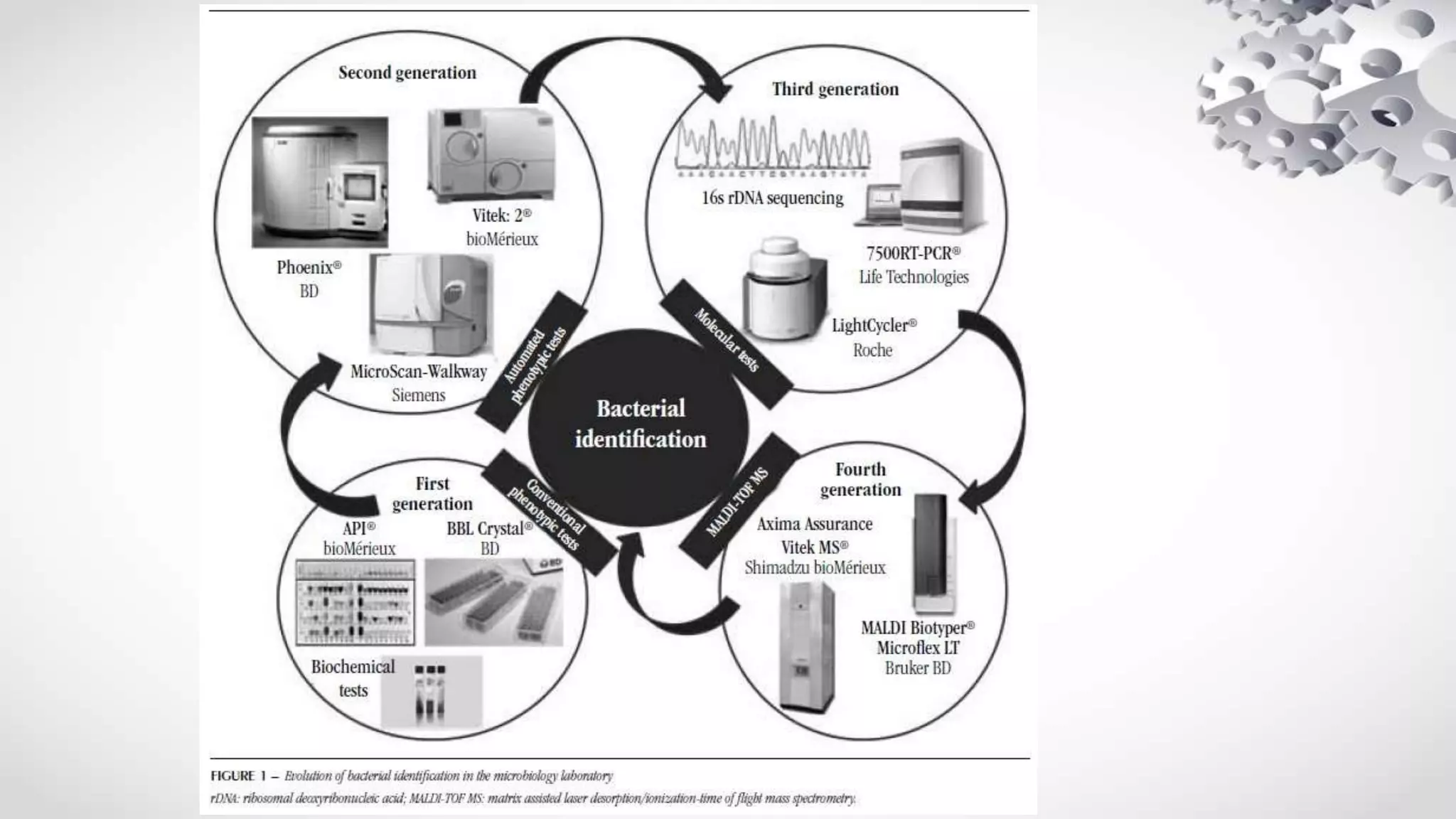
The document discusses various methods of automation used in microbiology laboratories, including automation of specimen collection, transport, processing, microscopy, identification and antimicrobial susceptibility testing. It describes several automated systems that have been developed to perform functions like automated staining, urine sediment analysis, specimen plating, bacterial identification using techniques like MALDI-TOF, VITEK and reaction-based systems. The document also provides examples of various automated equipment and their applications in microbiology laboratories.


![differences....
• Fixation 1]heat
2] alcohol 95%
• Staining methods
1]Bath stainers-chance of carry over of stain
2]Spray stainers-less wastage
3]cuvette designed staining
• Capacity- 30-120/hour
• Turn over time-3-5 min](https://image.slidesharecdn.com/automationinmicrobiology8dec-1-230605013033-532dc885/75/AUTOMATION-IN-MICROBIOLOGY-11-2048.jpg)


![UF-100 ,UF- 50,uf 1000i
• Laser based flow cytometry along with impedance detection ,forward
light scatter and fluorescence to identify cells
• two dyes are used
1] phenathridine - DNA
2]carbocyanine - -ve charged cell membrane
nuclear membrane
mitochondria](https://image.slidesharecdn.com/automationinmicrobiology8dec-1-230605013033-532dc885/75/AUTOMATION-IN-MICROBIOLOGY-14-2048.jpg)
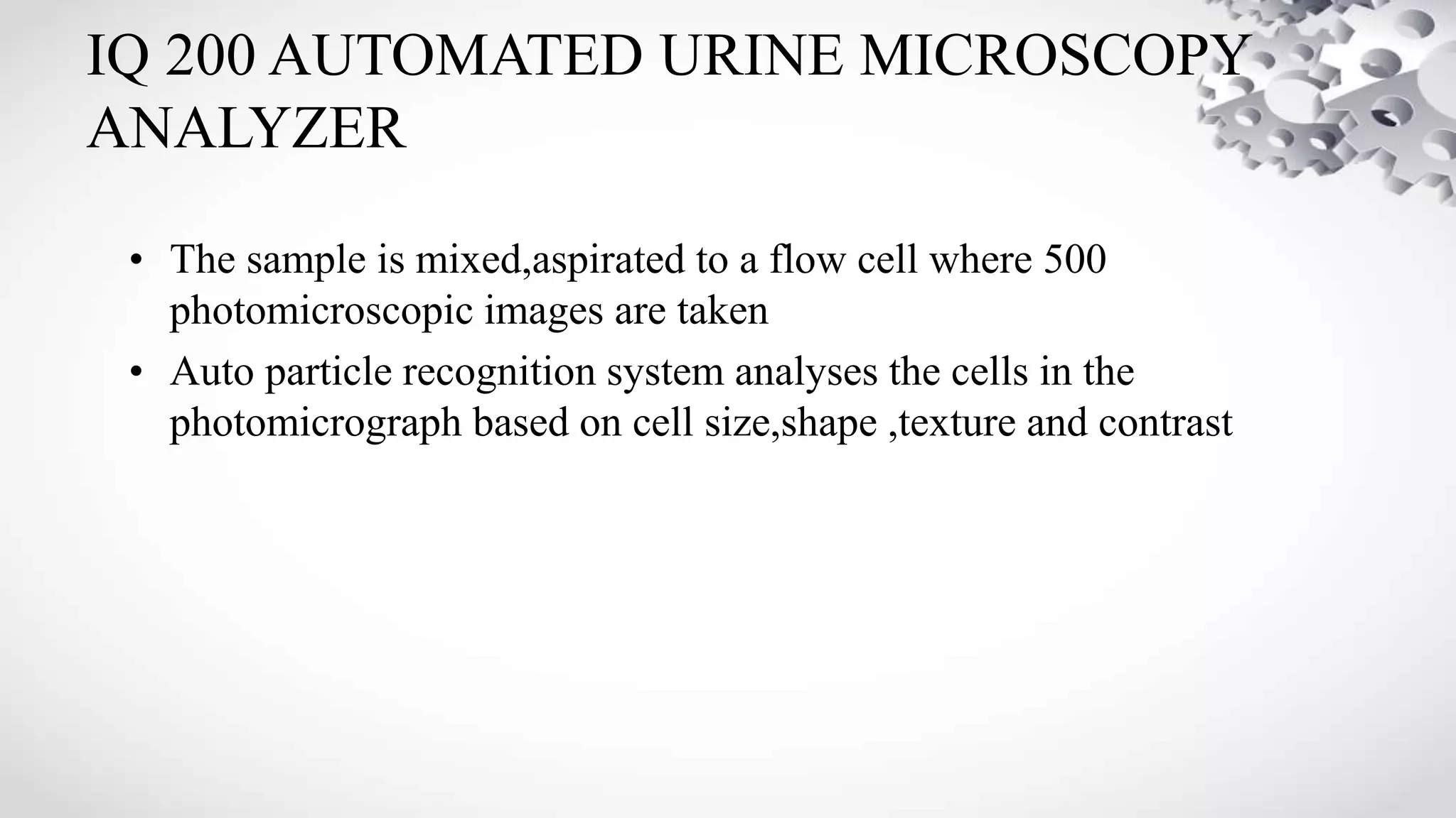
![• The system aspirates 0.8 mL of urine .
• analyse cells [erythrocyte,leukocytes (WBC) and epithelial cells],
Bacteria and casts .
• use electrical impedance for volume
• forward light scatter for size
• uses a couple of fluorescent dyes for nuclear and cytoplasmic
characteristics
• The formed elements are categorised in a two-dimensional space
(scattergrams) on the basis of their size, shape, volume,and staining
characteristics.](https://image.slidesharecdn.com/automationinmicrobiology8dec-1-230605013033-532dc885/75/AUTOMATION-IN-MICROBIOLOGY-15-2048.jpg)

![AUTOMATION IN SPECIMEN PROCESSING
• The currently available speciemen processors include:
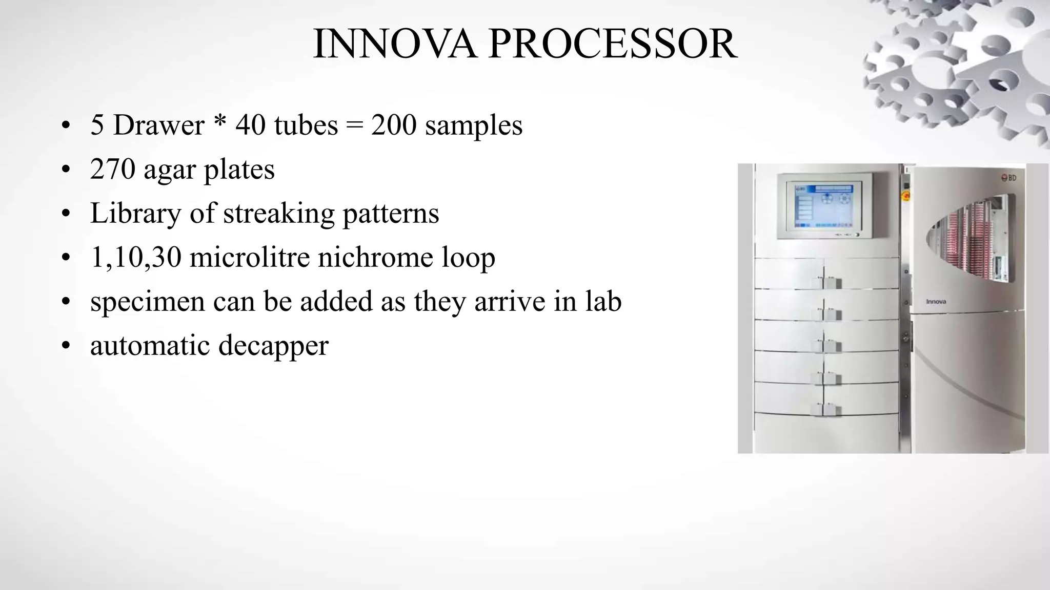
1]Innova processor -BD diagnostics
2]InoqulA fullautomation/manual automation [FA/MI] specimen
processing device - BD Diagnostics
3]Previ Isola Automatic plate streaker-Biomerieux
4] Walk Away Specimen Processor[WASP]-Cogan diagnostics
• Process LIQUID -BASED SPECIMENS](https://image.slidesharecdn.com/automationinmicrobiology8dec-1-230605013033-532dc885/75/AUTOMATION-IN-MICROBIOLOGY-19-2048.jpg)


![BActerial Rapid Detection using Optical scatter
Technology (BARDOT)
• irradiation of bacterial colonies grown in a Petri dish with a
red laser to generate light scattering patterns. The light
scattering patterns are dependent on the three-
dimensional (3D) morphology of bacterial colonies.
• distinguished Listeria, Staphylococcus, Salmonella,
Vibrio, and Escherichia with classification accuracy of 90–
99% . Five species of Listeria , three species of Vibrio [7],
and seven serogroups of E. coli have been discriminated
with the accuracy of >91%, >96%, and >81%,
respectively.](https://image.slidesharecdn.com/automationinmicrobiology8dec-1-230605013033-532dc885/75/AUTOMATION-IN-MICROBIOLOGY-29-2048.jpg)


![COMPONENTS
• Integrated modular system
1] filling - sealer unit
2]reader-incubater
3]computer control module
4]data terminal
5]multicopy printer](https://image.slidesharecdn.com/automationinmicrobiology8dec-1-230605013033-532dc885/75/AUTOMATION-IN-MICROBIOLOGY-39-2048.jpg)


![MALDI-TOF
• MATRIX ASSISTED LASER DESORPTION/IONISATION -TIME OF
FLIGHT MASS SPECTROMETRY
• Identification Based on PEPTIDE MASS FINGERPRINT
• commercially available systems include
-Vitek MS -[bioMerieux]
-MALDI Biotyper-[Bruker]](https://image.slidesharecdn.com/automationinmicrobiology8dec-1-230605013033-532dc885/75/AUTOMATION-IN-MICROBIOLOGY-48-2048.jpg)

![MECHANISM
• sample[ A] is mixed with excess
matrix[M] solution.
• laser ionises [M]→MH+
• [MH+] + A→M + AH+](https://image.slidesharecdn.com/automationinmicrobiology8dec-1-230605013033-532dc885/75/AUTOMATION-IN-MICROBIOLOGY-50-2048.jpg)


![DIRECT RAPID ANTIMICROBIAL SUSCEPTIBILITY
TESTING[DRAST]
• AST from positive blood culture bottles
• microscopic image analysis
• Growth detection and time lapse for MIC calculation](https://image.slidesharecdn.com/automationinmicrobiology8dec-1-230605013033-532dc885/75/AUTOMATION-IN-MICROBIOLOGY-75-2048.jpg)


![AUTOMATION IN BLOOD CULTURE
CULTURE METHODS
RAPID
IDENTIFICATION
METHODS
OLD NEW
[CONT. MONITORING]
OLDER METHODS
BACTEC BACTEC 9240/9120
NOVEL METHODS
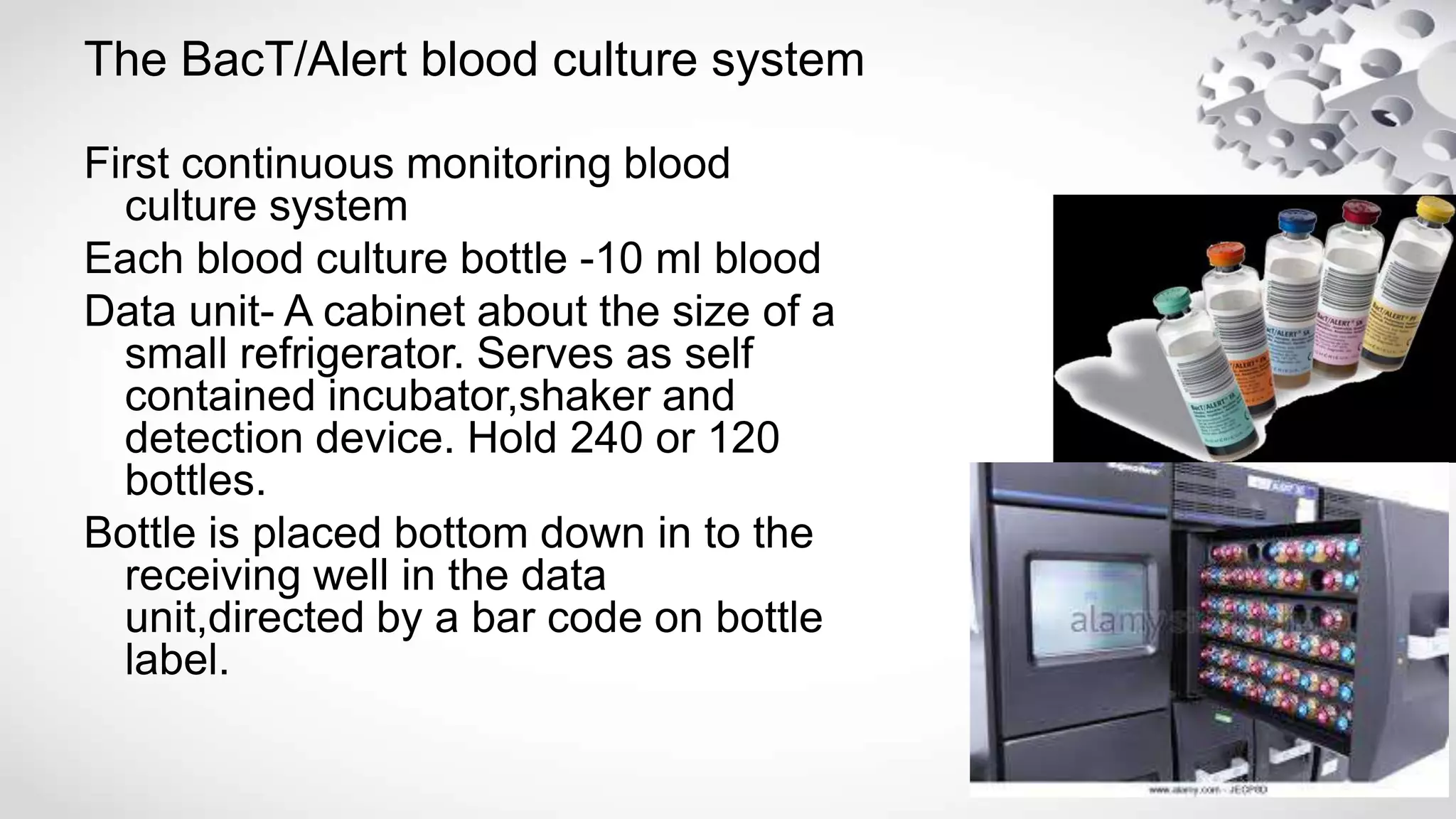
BACT/ALERT
1]Lysis centrifugation intrinsic
fluorescence
2]Integrated Comprehensive
direct droplet
TREK ESP](https://image.slidesharecdn.com/automationinmicrobiology8dec-1-230605013033-532dc885/75/AUTOMATION-IN-MICROBIOLOGY-87-2048.jpg)

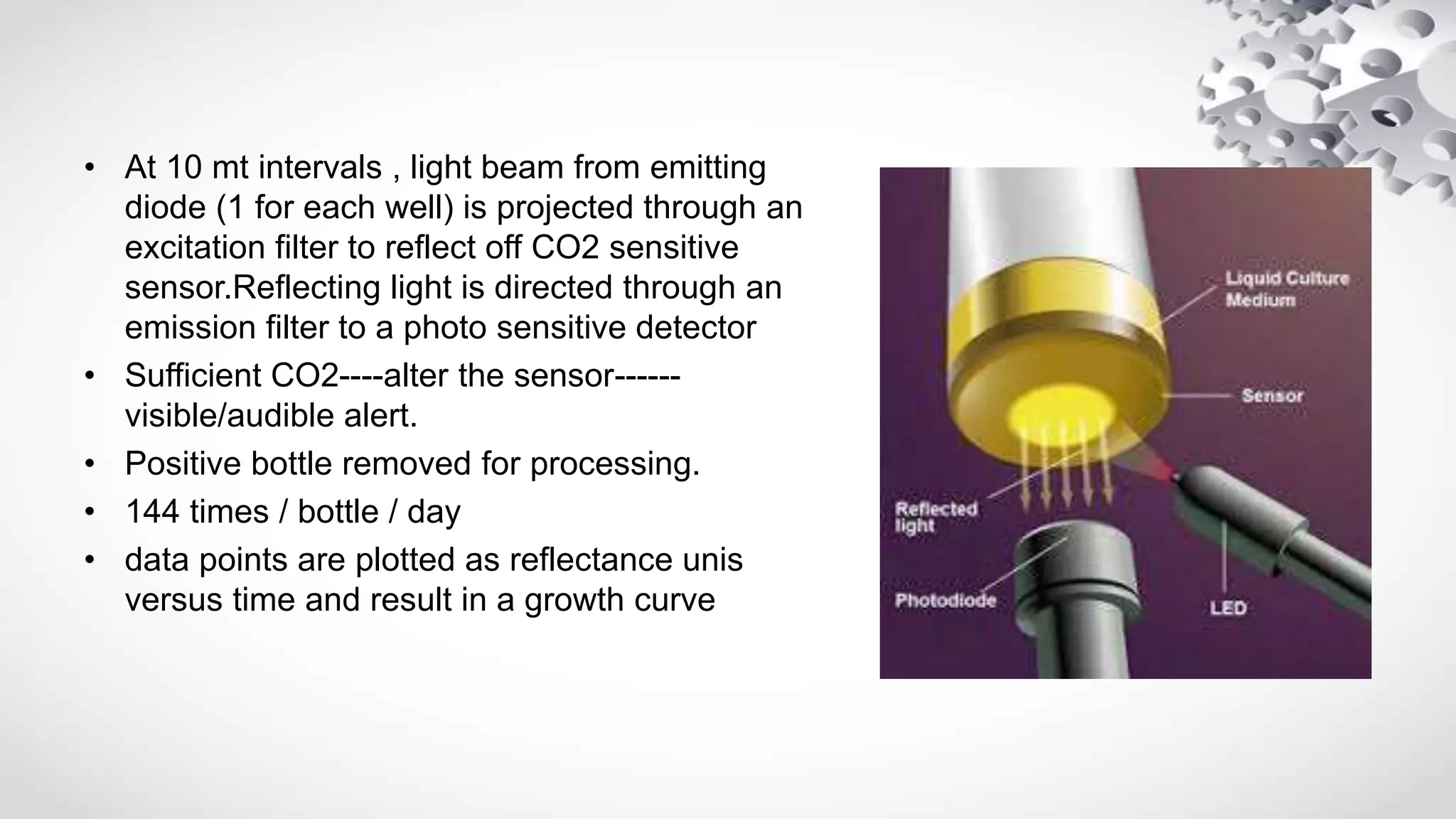
![Micro organism grow in blood broth mixture
CO2 liberated
CO2 + H20 → H2C03↔ [H+] + {HCO3-}
{ACIDIC pH]
Colour of sensor turns from green to yellow](https://image.slidesharecdn.com/automationinmicrobiology8dec-1-230605013033-532dc885/75/AUTOMATION-IN-MICROBIOLOGY-94-2048.jpg)


![Overview of Fungal Infections[1].ppt pptx](https://cdn.slidesharecdn.com/ss_thumbnails/overviewoffungalinfections1-250811070114-3c4fbad5-thumbnail.jpg?width=640&height=640&fit=bounds)


![Gas gangrene [Autosaved].pptxGas gangrene [Autosaved].pptx](https://cdn.slidesharecdn.com/ss_thumbnails/gasgangreneautosaved-250608063811-c85a18a4-thumbnail.jpg?width=640&height=640&fit=bounds)


