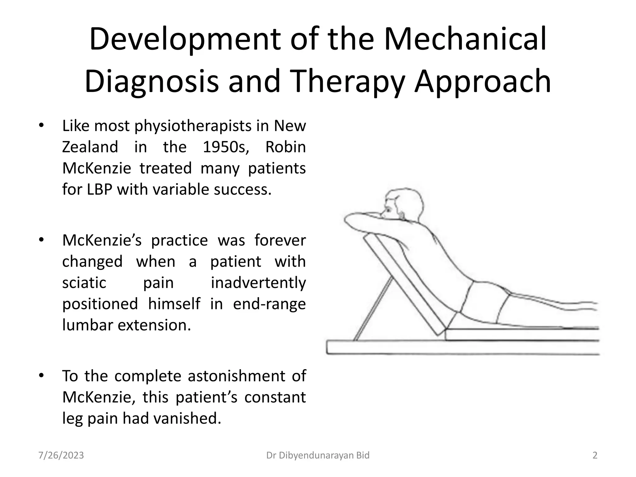
VIP Call Girls Tirunelveli Aaradhya 8250192130 Independent Escort Service Tir...
McKenzie Technique dnbid 2022.pptx
1. Dr Dibyendunarayan Bid
MPT, PGDSPT, PhD, FOMT, PGDHS (Acu)
The Sarvajanik College of Physiotherapy,
Rampura, Surat
The McKenzie Method®
of Mechanical Diagnosis
and Therapy®
2. • Like most physiotherapists in New
Zealand in the 1950s, Robin
McKenzie treated many patients
for LBP with variable success.
• McKenzie’s practice was forever
changed when a patient with
sciatic pain inadvertently
positioned himself in end-range
lumbar extension.
• To the complete astonishment of
McKenzie, this patient’s constant
leg pain had vanished.
Development of the Mechanical
Diagnosis and Therapy Approach
7/26/2023 Dr Dibyendunarayan Bid 2
3. • Over several additional visits,
McKenzie continued to use
this position with his patient,
which culminated in complete
resolution of his pain and full
restoration of lumbar range of
motion.
• At that time, the use of
extension was not considered
a beneficial practice in the
care of lumbar spine disorders
and was contrary to all that
McKenzie and his colleagues
had been taught.
7/26/2023 Dr Dibyendunarayan Bid 3
4. • McKenzie’s search for an explanation of the dramatic
changes that he had witnessed in this patient led him to
the writings of James Cyriax.
• McKenzie extrapolated from Cyriax’s work that his patient’s
recovery likely occurred “because the pressure on his sciatic
nerve was removed.”
• Operating under the premise that movement of
the lumbar spine can alter pressures on painful structures,
McKenzie continued to explore this intervention concept
with other patients.
7/26/2023 4
Dr Dibyendunarayan Bid
5. • He noticed that in some individuals, end-range extension
resolved their symptoms.
• For others, movements such as end-range flexion or lateral
movements were necessary for symptom resolution.
• Over time, McKenzie formalized his process of examining and
treating patients based on their symptomatic response to
movement and position.
• From this rather inauspicious beginning, the McKenzie
Method® of Mechanical Diagnosis and Therapy® (MDT)
was born.
•
7/26/2023 5
Dr Dibyendunarayan Bid
7. • MDT is a comprehensive approach to the
conservative management of most activity-related
spinal disorders.
• It is a system of patient examination, classification,
and intervention that is based on an individual’s
symptomatic and mechanical response to movement
and position.
7/26/2023 7
Dr Dibyendunarayan Bid
8. • Since 1990, the McKenzie Method® has evolved
to include management of mechanical disorders
of the cervical and thoracic spine, as well as the
extremities.
• The conceptual approach is the same, but the
process incorporates regional differences in
movements and positions.
7/26/2023 8
Dr Dibyendunarayan Bid
9. PHILOSOPHICAL FRAMEWORK AND FUNDAMENTAL
CONCEPTS: Philosophical Underpinnings
• Underlying the MDT approach is the belief that most
individuals with mechanical spinal disorders have the physical
capacity, intellectual wherewithal, and the self-discipline to
successfully manage their condition when provided with
appropriate education, guidance, and exercise.
• Given this, it is the responsibility of the MDT practitioner to
correctly classify responders to MDT, prescribe effective
interventions, create therapeutic alliances, educate patients
on fundamental principles of mechanical pain, and provide
strategies to control or prevent symptoms and restore
function (Box 9-1).
7/26/2023 9
Dr Dibyendunarayan Bid
14. Fundamental Concepts and Diagnostic Classification
• MDT is a systematic approach to the conservative
management of most activity-related spinal disorders that is
“diagnostic, prognostic, therapeutic, and prophylactic.”
• Perhaps one of the greatest features of this approach is the
use of a well-defined classification system (Fig. 9-1) that
categorizes patients according to their symptomatic response
to movement and position, rather than a system that is based
on a pathoanatomical diagnosis.
• The value of using impairment-based classification systems to
guide intervention in this population has been well
established.
7/26/2023 14
Dr Dibyendunarayan Bid
15. • MDT is consistent with the Quebec Task Force
classification system for activity-related spinal disorders
(Fig. 9-2) and meets the criteria for classification schemes
described in the APTA’s Guide to Physical Therapist
Practice.
• Unless contraindicated, all patients with spine-related
pain, with or without referred symptoms, are suitable for
mechanical examination.
7/26/2023 15
Dr Dibyendunarayan Bid
17. • Classification by MDT focuses on the use of repeated
movements and positions.
• At the conclusion of the examination, the individual is
classified as having a derangement syndrome, dysfunction
syndrome, postural syndrome, or “other.”
• Individuals classified within one of these mechanical
syndromes are considered to be ideal candidates for
MDT, whereas those falling into the “other” category require
additional examination or referral.
• The hallmark features for each of the three mechanical
syndromes are summarized in Table 9-1.
7/26/2023 17
Dr Dibyendunarayan Bid
18. The Derangement Syndrome
• The derangement syndrome is the most frequently observed
and best studied of the mechanical syndromes.
• The pathoanatomical model for the presence of a
derangement is an internal displacement of the intervertebral
disc, which affects the normal resting position of the joint
surfaces.
• Displacement of intradiscal material is presumed to obstruct
normal segmental spinal motion to varying degrees.
• Derangements typically develop as a result of sustained or
repetitive loading (often into flexion and/or rotation), chronic
postural stresses (often into flexion and/or rotation), or
trauma.
•
7/26/2023 18
Dr Dibyendunarayan Bid
23. The Dysfunction Syndrome
• For patients classified with a dysfunction syndrome, it is
hypothesized that the periarticular soft tissues
surrounding one or more of their spinal segments are
contracted, adhered, or adaptively shortened.
• Movement or prolonged positioning becomes painful
when restricted soft tissues are brought to the
end of their available motion.
• Pain with movement may lead to the avoidance of end-
range positions, which results in greater restrictions and
a more profound loss of movement.
7/26/2023 23
Dr Dibyendunarayan Bid
26. The Postural Syndrome
• In patients presenting with postural syndrome, it is
theorized that lumbar pain is experienced when
normal soft tissues experience abnormal stresses,
typically in response to prolonged static loading at end
range.
• The effects of a postural syndrome are most commonly
experienced after prolonged slouched sitting (Fig. 9-6)
in sedentary individuals such as students and
deskbound workers.
7/26/2023 26
Dr Dibyendunarayan Bid
27. • McKenzie states that “low back pain starts for the
same reason as pain arising in the forefinger when it
is bent backwards far enough to stimulate the free
nerve endings of periarticular structures.
• No pathology needs to exist, and no chemical
intervention will cure this form of mechanical pain.”
7/26/2023 27
Dr Dibyendunarayan Bid
28. Other Syndromes
• Individuals who fail to meet the criteria for classification into
one of the mechanical diagnostic syndromes after
examination and provisional intervention may be classified
within the other syndrome category.
• It is important to note that individuals with other syndrome
pathologies may have concomitant mechanical back pain and
should be afforded an MDT examination to determine if their
pain can be managed through mechanical intervention.
7/26/2023 28
Dr Dibyendunarayan Bid
32. PRINCIPLES OF EXAMINATION
• Examination begins with a thorough review of the patient’s history,
proceeds to a mechanical examination, and concludes in the provisional
classification of the patient’s condition.
• The classification directs the practitioner to the optimal mechanical
intervention to address the patient’s complaints.
• The McKenzie Institute® has developed examination protocols, complete
with McKenzie assessment forms (Figs. 9-7 and 9-8), which may be
accessed on the Institute’s website at www.mckenziemdt.org/forms.cfm.
• The reader is also referred to McKenzie’s The Lumbar Spine: Mechanical
Diagnosis & Therapy, Volume Two for more detailed information.
7/26/2023 32
Dr Dibyendunarayan Bid
35. The History
• The first page of the McKenzie Institute® Lumbar Spine
Assessment® (Fig. 9-7) summarizes the patient history.
• Within this approach, the process of history taking can
be referred to as empathetic interrogation owing to
the detailed manner in which this information is
obtained.
• This interrogation is undertaken to denote the patient’s
mechanical behavior throughout the course of a typical
day.
7/26/2023 35
Dr Dibyendunarayan Bid
37. • A comprehensive history is designed to efficiently
gather information about the present episode of
symptoms, including the mechanism of injury,
symptom presentation, and functional limitations.
• During the examination, the quantity of data to be
collected that relates to the impact of movement and
position on the patient’s symptoms is extensive.
• The examiner begins to formulate an idea of the
patient’s mechanical classification during the interview.
7/26/2023 37
Dr Dibyendunarayan Bid
42. Specific Questions
• Cough/Sneeze/Strain
• Symptoms that are worsened by coughing, sneezing, and or
straining suggest the presence of an active condition that is
aggravated by sudden or increased internal pressures.
• These characteristics often occur in the presence of a derangement
syndrome.
• Changes in bladder function (initiation, retention, or incontinence)
and/or saddle anesthesia that occur in conjunction with low back
pain may be indicative of a systemic problem such as bladder
pathology or cauda equina syndrome and require immediate
referral.
7/26/2023 42
Dr Dibyendunarayan Bid
43. • Gait
• A new onset of gait dysfunction should be explored to
expose the underlying cause(s).
• Antalgia, for example, may arise from a derangement
with a lateral component or from an adherent nerve
root, when the sciatic nerve is tensioned at heel strike.
• Other gait issues such as drop foot or ataxia may arise
from neuropathy or myelopathy, and additional
examination procedures are warranted.
7/26/2023 Dr Dibyendunarayan Bid 43
44. • Medications
• It is important to review all medications that are
being taken by the patient prior to examination.
• Strong analgesics taken prior to examination may
alter the patient’s pain perception during testing.
• Anticoagulants or long-term steroid use is
considered a precaution for the use of manual
techniques.
• Aspirin that provides disproportional relief of
symptoms is a “red flag for bone cancer.”
7/26/2023 Dr Dibyendunarayan Bid 44
45. • General Health
• During this portion of the examination, the therapist identifies
the patient’s perception of his or her overall health, including
a list of comorbidities.
• Constitutional symptoms such as fever, chills, or night sweats
occurring in concert with complaints of LBP suggest a systemic,
rather than musculoskeletal, origin.
• A previous history of cancer that is not being monitored
warrants referral if other warning signs are present.
7/26/2023 Dr Dibyendunarayan Bid 45
46. • Imaging Studies
• Imaging studies are essential for the purpose of ruling out
serious pathology in individuals with a history of trauma or
symptoms that suggest the presence of malignancy.
• A significant percentage of asymptomatic individuals have
been found to have substantial abnormalities, such as a
herniated nucleus pulposus, upon imaging.
• Therefore, the results of the mechanical examination
rather than the results of diagnostic imaging must
be used to determine the course of intervention.
7/26/2023 Dr Dibyendunarayan Bid 46
47. • Recent or Major Surgery
• Information regarding previous surgeries can alert the examiner to
the potential for other causes of symptoms.
• Patients with constant, unremitting pain, especially if accompanied
by fever, following a recent surgical procedure may suggest the
presence of an infection.
• Night Pain
As mentioned, unremitting night pain may be indicative of a
more serious spinal pathology such as cancer or ankylosing
spondylitis.
7/26/2023 Dr Dibyendunarayan Bid 47
48. • Accidents
• The presence of fractures, instabilities, and other injuries must be ruled
out following trauma. Modification of testing procedures is often indicated
after trauma and in the presence of active inflammation.
• The examiner should restrict range of motion and repeated movement
testing to remain within the pain-free limits.
• Unexplained Weight Loss
• While some patients lose their appetites secondary to pain or medication
side effects, more gain weight because of decreased activity levels.
• Unintentional weight loss greater than 10% of a patient’s total body
weight over a 4-week period of time is
indicative of malignancy.
7/26/2023 Dr Dibyendunarayan Bid 48
51. The Working Hypothesis:
• After completion of the first page of the Lumbar Spine
Assessment®, the examiner should attempt to develop an
initial working hypothesis regarding the origin and nature of
the patient’s condition.
• First and foremost, the examiner must determine whether or
not there are any “red flags.”
• Additionally, the examiner must determine if the history
should influence the remainder of the physical examination
and whether or not there are any psychosocial issues, such as
fear avoidance or depression.
•
7/26/2023 51
Dr Dibyendunarayan Bid
52. The Physical Examination
• The physical examination is designed to confirm or
reject the working hypothesis by testing the patient’s
symptomatic response to loading and by observing
the quality and quantity of movement.
• During the examination, testing error must be
minimized.
7/26/2023 52
Dr Dibyendunarayan Bid
53. Postures
• Sitting
• The patient’s posture is grossly assessed while the
examiner is obtaining the subjective history.
• If pain is reported in sitting, the location of the pain is
recorded. The examiner corrects any aberrant postures (Fig.
9-9a) and monitors the patient’s response to postural
correction.
• If symptoms are reduced, the patient is educated on how to
reproduce this posture, including the use of a lumbar roll
(Fig. 9-9b).
7/26/2023 53
Dr Dibyendunarayan Bid
54. • Standing
• When the patient is standing, posture is assessed in the
sagittal and frontal planes.
• The presence of a reduced or accentuated lordosis is noted. If
pain is reported in standing, the location is recorded as well as
any response to postural correction.
• Chronic deviations, such as scoliosis, may be present but have
no effect on symptoms.
• Acute deviations, such as a lateral shift, also known as an
acute lumbosacral or sciatic scoliosis, may also be present.
• This postural deviation is nonstructural and is caused by
pressure on a nerve root from a disc herniation or other
space-occupying lesion.
7/26/2023 54
Dr Dibyendunarayan Bid
55. • The lateral shift is named by the direction in which the
upper torso is displaced.
• The criteria for confirming the presence of a lateral
shift that is relevant to the current condition includes
the following:
(1) the deformity is clearly visible;
(2) the onset is concurrent with the present episode of
pain;
(3) the lateral shift cannot be voluntarily corrected or
maintained;
(4) both flexion and extension
movements are painful in weight-bearing; and
(5) the pain is worse in standing or walking than it is
when lying down.
7/26/2023 55
Dr Dibyendunarayan Bid
59. • If a lateral shift is confirmed during the examination,
lateral shift correction techniques (Fig. 9-10) should
be commenced prior to initiation of repeated
movement testing and intervention.
• Poor tolerance for repeated movements is often
noted in the presence of a lateral shift.
7/26/2023 59
Dr Dibyendunarayan Bid
60. • FIGURE 9–10. Lateral shift
correction progression.
• A. Right relevant lateral
shift.
7/26/2023 60
Dr Dibyendunarayan Bid
62. • B. Manual correction of lateral shift (shown for correction of a right
lateral shift): The patient stands with feet shoulder-width apart and
weight evenly distributed. Her right elbow is flexed 90 degrees and
placed against the trunk above the level of the iliac crest. The
clinician stands perpendicular to the patient on her right side in a
lunge position, with her right shoulder against the patient’s right
upper arm. The clinician’s hands are clasped around the patient’s
left iliac crest (B1).
• The clinician gently pulls the patient’s pelvis toward her while
simultaneously pushing the patient’s trunk away by the pressure
exerted through the clinician’s right shoulder. The patient must
remain weight-bearing symmetrically. Intermittent gentle pressure
is applied and partially released, with more pressure given with
each repetition until a slight overcorrection of the deformity is
accomplished (B2).
7/26/2023 62
Dr Dibyendunarayan Bid
63. • The patient is then asked to
perform extension while the
therapist maintains a slight
overcorrection of the shift
(B3).
7/26/2023 63
Dr Dibyendunarayan Bid
65. • C. Self-lateral shift correction against a wall. The
patient stands with feet together about shoulder’s
width from the wall with the side of the shift near the
wall (usually the shoulder contralateral to the painful
side).
• The patient’s elbow is flexed above the level of the iliac
crest, and the upper arm is placed against the wall (C1).
• The patient then uses her other hand to apply rhythmic
pressure through the pelvis toward the wall until
overcorrection is achieved (C2).
• In this position, lordosis is restored by performing
extension in standing in the overcorrected position.
7/26/2023 65
Dr Dibyendunarayan Bid
67. The Provisional Classification
• At the conclusion of the mechanical examination, the
examiner should have sufficient information to make a
provisional classification.
• Refer to the classification algorithm (see Fig. 9-1) and
characteristics (Table 9-1) to guide the decision-making
process. If the classification is a derangement, the pattern of
pain presentation should be identified, as well as the
directional preference, in order to select the appropriate
intervention.
• If the classification is a dysfunction, the direction of restriction
must be identified in order to select the proper intervention.
7/26/2023 67
Dr Dibyendunarayan Bid
68. • If there is insufficient evidence at the conclusion of
the initial examination to make a provisional classification,
patients are asked to keep a record of their symptomatic
response to movement and position.
• At the conclusion of the examination, the therapist should
have identified:
– a mechanical versus nonmechanical condition,
– the syndrome classification,
– the directional preference, and
– the subsequent principle of intervention.
7/26/2023 68
Dr Dibyendunarayan Bid
70. PRINCIPLES OF INTERVENTION
• General Principles
• As previously stated, mechanical classification drives the
intervention.
• In a derangement syndrome, the primary objectives of
intervention are to reduce the internal displacement,
maintain the reduction, and restore full movement.
• In a dysfunction syndrome, the primary objectives are to
remodel adaptively shortened tissues and reduce movement
restrictions.
• The objective of intervention for patients presenting
with a postural syndrome is to remove abnormal stresses on
normal tissues.
•
7/26/2023 70
Dr Dibyendunarayan Bid
71. • A critical principle of intervention is the confirmation
or rejection of the provisional classification.
• Upon subsequent visits, if the patient’s symptoms are
worse, or not improved, compliance and technique are
checked before assuming an incorrect mechanical
diagnosis has been made.
• Confirmation of the classification should be
determined within five sessions.
7/26/2023 71
Dr Dibyendunarayan Bid
73. • FIGURE 9–11. Sequence of repeated movement
testing.
• A. Flexion in standing (FIS): Stand with feet shoulder-
width apart. Bend forward from the waist and slide the
hands down the legs as far as possible while keeping
the knees straight.
• B. Extension in standing (EIS): Stand with feet shoulder-
width apart. Place the hands in the small of the back
and arch backward as far as possible while keeping the
knees straight.
7/26/2023 Dr Dibyendunarayan Bid 73
74. • C. Flexion in lying (FIL): From the hook-lying position, bring
the knees toward the chest as far as possible. Clasp the
hands over the knees to further flex the lumbar spine.
• D. Extension in lying (EIL): Place hands directly under the
shoulders. Extend the elbows slowly to raise the upper
body off the plinth. Keep the hips and thighs relaxed, and
allow the abdomen to sag
7/26/2023 74
Dr Dibyendunarayan Bid
76. • FIGURE 9–12. Repeated movement test movements with
lateral bias.
• A. Extension in lying (EIL) with hips off center: In prone,
translate the hips laterally, usually away from the pain.
Perform EIL: Place hands directly under the shoulders.
Extend the elbows slowly to raise the upper body off the
plinth. Keep the hips and thighs relaxed, and allow the
abdomen to sag.
• B. Side glide in standing (SGIS): Directions for right SGIS.
Stand with feet shoulder-width apart. Translate the hips to
the left while maintaining the trunk in neutral with
shoulders parallel to the ground. The clinician may initially
guide the movement at with hands at the right iliac crest
and left shoulder
7/26/2023 Dr Dibyendunarayan Bid 76
79. • A key feature of this approach is the emphasis on holding
the patient responsible for his or her own care, which begins
on the first visit, through a substantial amount of patient
education.
• Patients are educated on appropriate posture and body
mechanics, given their classification, including proper sitting
and sleeping postures, transitional movements, and postural
support.
• The use of a lumbar roll in the region of the lumbar
lordotic curve when sitting is often incorporated to improve
sitting posture by preserving lumbar lordosis and reducing the
effects of prolonged flexion forces (Fig. 9-13a and b).
7/26/2023 Dr Dibyendunarayan Bid 79
80. • Patients are provided with a specific prescription for
exercise(s) to perform until the next appointment,
usually in 1 to 2 days.
• Patients are informed about warning signs, such as
peripheralization, and are given instructions
regarding what to do if such a situation arises.
7/26/2023 Dr Dibyendunarayan Bid 80
81. • When performing exercises, force progression is
considered to be a valuable concept for two reasons:
(1) physiologically, mechanical pain may resolve using
a range of forces, and
(2) philosophically, using the least amount of force
necessary to resolve the symptoms is safer.
7/26/2023 Dr Dibyendunarayan Bid 81
82. • Therefore, only when individuals are unable to
control their own symptoms should additional forces
be introduced.
• The sequence of force progression begins with
patient-generated forces that take place in midrange
with eventual progression to end range and end
range with self-overpressure.
7/26/2023 Dr Dibyendunarayan Bid 82
83. • Once patient generated forces have been
exhausted, intervention may progress to
clinician-generated forces, which involve
assisting the patient with movement from
midrange to end range with therapist
overpressure.
• Passive nonthrust and thrust mobilization
performed by the clinician are considered when
patient-generated forces are inadequate to
produce the desired outcome.
7/26/2023 Dr Dibyendunarayan Bid 83
84. • Once the patient’s directional preference has been confirmed,
progression along the force continuum is dictated by
symptom response.
• A patient who has responded well to movement into end-
range extension but continues to display movement loss (Fig.
9-14a) should progress to end-range extension with self-
overpressure (Fig. 9-14b).
• Additionally, once improvement plateaus, modifications such
as altering the starting position (loaded or unloaded), the
direction of loading (sagittal, frontal, or combination), and
duration of the technique may be explored.
•
7/26/2023 Dr Dibyendunarayan Bid 84
85. • Lastly, this approach espouses the use of prophylactic
measures designed to equip patients with the knowledge
and activities to prevent and manage future episodes by
using “first aid” exercises.
• Patients using an MDT approach demonstrate high levels of
satisfaction in their ability to manage their current
symptoms and recurrences, demonstrate lower rates of
recurrence, have less sick leave, and seek less medical
assistance.
7/26/2023 Dr Dibyendunarayan Bid 85
86. Intervention for Derangement Syndrome
• Reduction of a derangement is achieved and maintained by
consistent application of the loading strategy that
centralized the patient’s symptoms during the examination.
• Once reduced, it is important to educate the patient to
consistently avoid the provocative positions and
movements.
• Extensive patient education regarding centralization and
peripheralization principles as well as postural education
and correction are vital to maintaining reduction and
preventing recurrence.
7/26/2023 Dr Dibyendunarayan Bid 86
89. • Ideally, exercises must be performed 10 times every 1 to
2 waking hours, or more frequently if symptoms recur.
• Assuming the mechanical classification given at examination is
correct, patients should report a decrease in symptoms with
centralization, increased mobility, and tolerance for
progression of forces.
• If centralization is not achieved within five sessions, it is
unlikely that centralization will occur.
• A common error is that patients may not have achieved end
range and then are mistakenly believed to be noncentralizers.
7/26/2023 Dr Dibyendunarayan Bid 89
90. • When the patient’s symptoms are no longer provoked or
peripheralized with movements or postures, the derangement
is considered to be fully reduced.
• During intervention, provocative motions are avoided to
reduce the risk of rederangement.
• A return to provocative motions must be done gradually, with
the goal of restoring any residual motion loss and with an
awareness that any limitation in spinal motion is a risk factor
for future derangement.
• Intervention is complete when the patient reports restoration
of normal activities and pain-free movement in all directions.
7/26/2023 Dr Dibyendunarayan Bid 90
91. • Empowering patients to intelligently manage their own pain
by providing them with the tools needed to recognize and
manage recurrences is one of the greatest virtues of the MDT
approach.
• When warning signs are present, patients should initiate self-
management measures for 48 hours, such as:
(1) avoiding positions and movements that provoke pain,
(2) sitting with the lumbar spine unsupported for no longer than 5 to 10
minutes at a time and resting in either the prone or supine position, and
(3) commencing with prior exercise 10 to 15 times every 1 to 2 waking
hours.
• Patients are instructed to continue their exercise program for
at least 6 weeks after discharge, maintain good postural
habits, reduce their biomechanical risk factors, and
incorporate general fitness activities into their lifestyle.
7/26/2023 Dr Dibyendunarayan Bid 91
92. • Four groups of derangements are typically seen with
unique directional preferences.
• The intervention approach is the same for each
group although the exact loading strategies may vary.
7/26/2023 Dr Dibyendunarayan Bid 92
93. Intervention for Posterior Derangement
Syndrome
7/26/2023
• The intervention for a posterior derangement follows the extension
principle (Fig. 9-15).
• Most patients who present with a derangement fall into this
subclassification.
• Extension in lying (EIL) is the exercise of choice for a posterior
derangement because of reduced compressive forces.
• In some individuals, the first few repetitions may provoke pain because
motion is obstructed by the posterior displacement of the disc. However,
as the patient performs more repetitions, reduction occurs, pain resolves,
and motion improves.
Dr Dibyendunarayan Bid 93
94. • Extension in standing (EIS) is performed throughout the day
when extension in lying is not possible.
• Preservation of the lumbar lordosis in sitting (see Fig. 9-13) by
using a lumbar roll for mechanical and tactile cueing is
essential in order to maintain the reduction.
• Additionally, maintenance of lumbar lordosis during
transfers and activities of daily living may require patient
education and cueing by the therapist.
• To be effective, the patient must take primary responsibility
for performing exercises and avoiding provocative movements
and positions.
7/26/2023 Dr Dibyendunarayan Bid 94
95. • Prior to discharge, intervention for a patient with a posterior
derangement must include restoration of pain-free flexion
(Fig. 9-11c).
• Once patients can reliably manage their symptoms,
movement into flexion is explored in a controlled fashion.
• While supervised, patients perform up to 10 repetitions of
flexion in lying (FIL).
• If pain is not worsened or peripheralized and extension range
of motion is not reduced, then FIL is added to the intervention
plan.
• If there is a poor response, then the derangement is unstable
and repeated flexion should be delayed.
7/26/2023 Dr Dibyendunarayan Bid 95
96. • FIGURE 9–15. Extension
Principle/progression of
forces.
• A. Lying prone. The head is
rotated to one side and the
arms relaxed by the side of the
trunk. The position may be
modified if the patient has an
acute kyphotic deformity by
placing pillows under the
abdomen to accommodate the
deformity, then sequentially
removing pillows until lying
prone is attained.
7/26/2023 Dr Dibyendunarayan Bid 96
97. • B. Lying prone in extension:
Place elbows directly under
the shoulders with the
forearms parallel and the
hips flat on the plinth.
• The lumbar spine should
sag into lumbar lordosis.
7/26/2023 Dr Dibyendunarayan Bid 97
98. • C. Extension in lying (EIL):
Place hands directly
under the shoulders (C1).
Extend the elbows slowly
to raise the upper body
off the plinth.
• Keep the hips and thighs
relaxed and allow the
abdomen to sag
(C2).
7/26/2023 Dr Dibyendunarayan Bid 98
99. • D. Extension in lying
with self-overpressure.
Perform EIL. Apply self-
overpressure: Lock the
elbows at end-range
extension, exhale fully,
and allow the abdomen
to sag prior to lowering
the chest to the table.
7/26/2023 Dr Dibyendunarayan Bid 99
100. • E. Extension in standing
(EIS): Stand with feet
shoulder-width apart.
• Place the hands in the
small of the back and
arch backward as far as
possible while keeping
the knees straight.
7/26/2023 Dr Dibyendunarayan Bid 100
101. • FIGURE 9–15. cont’d F.
Extension in lying with
clinician overpressure:
The clinician places the
heels of her hands on
the selected transverse
processes of the lumbar
segment (F1).
7/26/2023 Dr Dibyendunarayan Bid 101
102. • The patient performs EIL.
As the patient progresses
into greater
extension (F2, F3), the
clinician shifts her body
backward to maintain a
force “parallel to the
motion segment.”
• Overpressure is
maintained until the
patient fully lowers to the
start position.
7/26/2023 Dr Dibyendunarayan Bid 102
103. • The patient performs EIL.
As the patient progresses
into greater extension
(F2, F3), the clinician
shifts her body backward
to maintain a force
“parallel to the motion
segment.”
• Overpressure is
maintained until the
patient fully lowers to the
start position.
7/26/2023 Dr Dibyendunarayan Bid 103
104. • G. Extension mobilization.
• The clinician places her hands
perpendicular to each other over the
transverse processes of the spinal
segment. The clinician’s shoulders
must be directly over her hands and
her elbows extended.
• Gradual rhythmic and symmetrical
pressure is applied in a
posteroanterior direction to end
range.
• Symptom response is monitored at
each segment. Mobilization is
provided to the same segment 10
times before progressing to the next
segment.
7/26/2023 Dr Dibyendunarayan Bid 104
105. Intervention for Posterior Derangement
Syndrome With Lateral Component
• Some patients with a posterior derangement require the
application of either frontal or transverse plane directed
forces along with sagittal plane-directed forces (Fig. 9-16) in
order for a complete reduction to occur.
• These patients are said to have a lateral component to their
derangements.
• In this syndrome, the patient’s symptoms are unilateral or
asymmetrical, and during examination the symptoms do not
respond to or are worsened by pure sagittal plane
movements.
7/26/2023 Dr Dibyendunarayan Bid 105
106. • Intervention begins with EIL, with the hips shifted laterally
(Fig. 9-16a), and progresses by adding clinician overpressure
(Fig. 9-16b).
• To reduce or centralize symptoms, the hips are most
frequently shifted away from, but occasionally toward, the
painful side.
• Other options for reduction of symptoms using lateral forces
include side gliding in standing (SGIS) (Fig. 9-16c), rotation
mobilization in extension bilaterally (Fig. 9-16d1) or
unilaterally (Fig. 9-16d2), rotation in flexion (Fig. 9-16e), and
rotation mobilization in flexion (Fig. 9-16f).
7/26/2023 Dr Dibyendunarayan Bid 106
107. • When symptoms have centralized or become
symmetrical, EIL in the pure sagittal plane should be
retested to see if it is safe to perform, after which the
treatment plan is progressed to pure extension-
biased exercises.
• If pain is worsened or peripheralized, then the
derangement is not sufficiently stable to
cease lateral forces.
7/26/2023 Dr Dibyendunarayan Bid 107
108. FIGURE 9–16. Treatment principle for posterior derangement
with lateral component/progression of forces. When sagittal
plane movements do not reduce a posterior derangement (Fig.
9-15) lateral forces may be necessary.
• A. Extension in lying
(EIL) with hips off center: In
prone, translate the hips
laterally, usually away from
the pain. Perform EIL: Place
hands directly under the
shoulders.
• Extend the elbows slowly to
raise the upper body off the
plinth. Keep the hips and
thighs relaxed and
allow the abdomen to sag.
7/26/2023 Dr Dibyendunarayan Bid 108
109. • B. EIL with hips off center
with lateral overpressure
by clinician: With the
patient positioned in
prone with hips off
center, the clinician
applies and maintains
overpressure at the iliac
crests to further enhance
lateral forces as EIL is
performed.
7/26/2023 Dr Dibyendunarayan Bid 109
110. • C. Side glide in standing
(SGIS): Directions for right
SGIS. Stand with feet
shoulder-width apart.
• Translate the hips to the left
while maintaining the trunk
in neutral with shoulders
parallel to the ground.
• The clinician may initially
guide the movement with
hands at the right iliac crest
and left shoulder.
7/26/2023 Dr Dibyendunarayan Bid 110
111. • D1. Rotation mobilization in
extension: The clinician’s hands are
placed perpendicular to each other
with hypothenar eminences over the
area of the transverse processes of
the spinal segment to be mobilized
and shoulders over the hands. The
mobilization is performed by
alternating forces from one side of
the spinal segment to the other.
• First, an anteromedially directed
force is applied by shifting the
shoulders forward over the extended
arm. The shoulders are then shifted
back to apply force through the
opposite hypothenar eminence.
Rhythmical application of forces is
continued.
7/26/2023 Dr Dibyendunarayan Bid 111
112. • If application of force on
one side centralizes the
symptoms, a unilateral
rotation mobilization is
performed (D2) by
placing one hand on top
of the other.
• Rotation in flexion: In
hook-lying position, the
patient rotates the knees
to the side (usually
toward the side of the
pain).
7/26/2023 Dr Dibyendunarayan Bid 112
113. • FIGURE 9–16. cont’d E1.
Rotation mobilization in
flexion: The patient lies supine
with hips and knees extended.
The clinician stands to the side
of the patient, facing
proximally.
• The clinician passively flexes
the patient’s hips and knees to
90 degrees and presets the
pelvis by rotating it prior to
bringing the knees to one side
(usually toward the side of
pain).
7/26/2023 Dr Dibyendunarayan Bid 113
114. • E2. The patient’s ankles
rest on the clinician’s
hips or pelvis.
• The hand closest to the
patient fixes either the
far shoulder or rib cage
through the patient’s
clasped hands.
7/26/2023 Dr Dibyendunarayan Bid 114
115. • E3.The therapist’s far
hand pushes the knees
downward, either
sustaining the force or by
applying intermittent
pressure.
• Symptom response is
monitored. The lower
extremities are passively
returned to the starting
position.
7/26/2023 Dr Dibyendunarayan Bid 115
117. Intervention for Lateral Shift
• A lateral shift is defined as a frontal plane postural deviation
in which the upper trunk is displaced laterally relative to the
lower trunk and the upper trunk is unable to move past the
midline.
• The majority of patients with a relevant lateral shift will
deviate away from the painful side.
7/26/2023 Dr Dibyendunarayan Bid 117
118. • Correction of the lateral shift must be achieved before
attempting to restore extension range of motion.
• Attempts to apply sagittal forces in the presence of a true
lateral shift will worsen the patient’s symptoms.
• Manual correction of a lateral shift is one of the more
common manual therapy procedures used within the MDT
approach (Fig. 9-10).
7/26/2023 Dr Dibyendunarayan Bid 118
119. • Correction, or overcorrection, in which the patient’s shifted
trunk is brought to midline and then slightly beyond, may be
painful and may produce vasovagal syncope.
• Once overcorrection is achieved, patients must immediately
perform extension in standing while overpressure is
maintained by the clinician to maintain the correction (Fig. 9-
17).
7/26/2023 Dr Dibyendunarayan Bid 119
120. • Following lateral shift correction in the clinic, it is imperative
that the patient maintain lordosis at all times to prevent
recurrence.
• Additionally, patients are instructed in self-correction
techniques, including SGIS (Fig. 9-10c).
• The patient stands with the shoulder (on the side to which the
shift has occurred) against the wall and the feet
together, approximately 12 inches away from the wall.
• The opposite hand is placed on the outside hip as the patient
moves the pelvis toward the wall; the position is held for
several seconds and repeated until the shift is corrected.
7/26/2023 Dr Dibyendunarayan Bid 120
121. • After self-correction, the patient immediately performs EIS or
EIL with hips offset.
• The shift correction is maintained by avoiding trunk flexion
and strict adherence to good posture.
• Once a shift is corrected, exercise proceeds as for a posterior
derangement (Fig. 9-15).
7/26/2023 Dr Dibyendunarayan Bid 121
122. Intervention for Anterior Derangement
Syndrome
• The intervention of an anterior derangement follows the
flexion principle (Fig. 9-18), which increases compressive
forces anteriorly.
• Ten repetitions of the selected exercise must be performed
every 1 to 2 hours.
• A progression of force is made when the patient’s symptoms
have improved, but are no longer progressing with the current
exercise regimen.
• It is important to note that less than 7% of all derangements
are anterior.
7/26/2023 Dr Dibyendunarayan Bid 122
123. Intervention for Dysfunction
Syndrome
• There are four types of dysfunction syndromes, which are
named by the restricted direction or the direction in which
symptoms are reproduced.
• They are:
– flexion dysfunction,
– extension dysfunction,
– side gliding dysfunction, and
– adherent nerve root dysfunction (Box 9-5).
7/26/2023 Dr Dibyendunarayan Bid 123
125. • As described, with the exception of an adherent nerve root
dysfunction syndrome, a dysfunction produces only local
symptoms without peripheralization.
• The progression of intervention for an ANR is displayed in
Figure 9-19.
• Progressions are made when the prescribed exercises are no
longer producing symptoms, but full range of motion has not
yet been achieved.
• Many ANRs occur as complications of a previous posterior
derangement; therefore, flexion exercises should be avoided
during the first 4 hours of the day and should always be
immediately followed by extension procedures.
7/26/2023 Dr Dibyendunarayan Bid 125
126. • The appropriate intervention for a patient presenting with a
dysfunction syndrome is progressive movement in the
direction of restriction.
• The main goal of intervention is to improve motion by
gradually eliminating the barriers to full motion.
• Patient education regarding the warning signs of
overstretching, such as pain lasting more than 20 minutes
after the completion of exercises, as well as postural
education and correction, are an integral part of the plan of
care for a patient with a dysfunction syndrome.
7/26/2023 Dr Dibyendunarayan Bid 126
127. • Exercises must be performed 10 times every 2 to 3 waking
hours.
• Patients should move into the range of motion where
symptoms are reproduced; however, the increase in
symptoms should only be temporary and localized to the
spine, except for the case of an ANR, as mentioned above.
• The symptoms should abate rapidly when the position is
released, and there must be no lasting or residual increase
in pain.
7/26/2023 Dr Dibyendunarayan Bid 127
128. • Progressions are made when exercises no longer produce
end-range pain or when additional increases in range have
ceased, but full range of motion has not yet been achieved.
• A response to intervention is expected within 4 to 6 weeks.
• As in the management of the other syndromes, patients are
provided with a structured home exercise program and
instructed to see their physical therapist as necessary to
monitor pain and range of motion and to progress their
program.
7/26/2023 Dr Dibyendunarayan Bid 128
129. • Manual techniques are rarely required in the case of
a dysfunction; however, they may be indicated when
patient-generated forces have been exhausted and
progress has plateaued.
• The progression for intervention of an extension
dysfunction is presented in Figure 9-15 and the
progression for a flexion dysfunction is displayed in
Figure 9-18.
7/26/2023 Dr Dibyendunarayan Bid 129
130. FIGURE 9–18. Flexion principle/progression of forces.
• A. Flexion in lying (FIL):
From the hook-lying
position, bring the knees
toward the chest as far as
possible.
• Clasp the hands over the
knees to further flex the
lumbar spine.
7/26/2023 Dr Dibyendunarayan Bid 130
131. • B. Flexion in sitting (FISit):
The patient sits in a straight-
back chair and bends
forward, bringing the head
between the knees, and
then returns to the full
upright position.
• C. Flexion in standing (FIS):
Stand with feet shoulder-
width apart. Bend forward
from the waist and slide the
hands down the legs as far
as possible while keeping
the knees straight.
7/26/2023 Dr Dibyendunarayan Bid 131
132. • D. Flexion in step standing
(FISS): This procedure is used
when there is a deviation in
flexion. The patient stands
with one leg on a chair so that
the knee is flexed
90 degrees and the other leg
extended (D1).
• The leg on the chair is on the
side contralateral to the side
of deviation. The patient
forward flexes her lumbar
spine by grasping her ankle
and bringing her shoulder to
the raised knee (D2).
• Between each repetition, the
patient must return to
standing and restore the
lordosis.
7/26/2023 Dr Dibyendunarayan Bid 132
133. • E. Flexion in lying with
clinician overpressure:
The patient performs FIL.
The clinician evenly applies
overpressure through the
patient’s knees.
7/26/2023 Dr Dibyendunarayan Bid 133
134. FIGURE 9–19. Adherent nerve root progression.
• A. Flexion in lying (FIL):
From the hook-lying
position, bring the
knees toward the chest
as far as possible.
Clasp the hands over
the knees to further flex
the lumbar spine.
7/26/2023 Dr Dibyendunarayan Bid 134
135. • B. Extension in lying (EIL):
Place hands directly
under the shoulders.
Extend the elbows slowly
to raise the upper body
off the plinth.
Keep the hips and thighs
relaxed and allow the
abdomen to sag.
7/26/2023 Dr Dibyendunarayan Bid 135
136. • C. Flexion in step standing
(FISS): In ANR, stand with
the asymptomatic leg on
a chair so that the knee is
flexed 90 degrees and the
symptomatic leg
extended (C1).
• The patient forward flexes
her lumbar spine by
grasping her ankle and
bringing her shoulder to
the raised knee (C2).
• Between each repetition,
the patient must return to
standing between each
repetition and restore the
lordosis.
7/26/2023 Dr Dibyendunarayan Bid 136
137. • D. Flexion in standing (FIS):
Stand with feet shoulder-
width apart.
• Flex the lumbar spine by
sliding the hands down the
legs as far as possible while
keeping the knees straight.
• Between each repetition
the patient must return to
standing and restore the
lordosis.
7/26/2023 Dr Dibyendunarayan Bid 137
138. Intervention for Postural Syndrome
• Education is the key to the management of a postural
syndrome.
• Spinal joint capsules, spinal ligaments, and muscles are
strained at the end of their range of motion owing to
prolonged static loading.
• It is vital to discuss how this concept translates into
patients’ lives and to instruct patients in good postural
habits for sitting, standing, and sleeping, as well as
proper body mechanics for functional activities.
7/26/2023 Dr Dibyendunarayan Bid 138
139. • The slouch-overcorrect exercise (Fig. 9-20) is
given to teach patients how to find good posture
in sitting.
• Intervention for postural syndrome must include
a discussion of the longterm consequences of
poor posture, including the increased risk for
future back and neck pain.
7/26/2023 Dr Dibyendunarayan Bid 139
140. FIGURE 9–20. Slouch-overcorrect exercise.
• A. The patient is
instructed
to sit slouched,
7/26/2023 Dr Dibyendunarayan Bid 140
141. • B. then sit erect in
exaggerated good
posture with the
lumbar spine arched,
chest up, and cervical
spine in retraction.
• C. The patient is then
instructed to relax 10%
into good sitting
posture.
7/26/2023 Dr Dibyendunarayan Bid 141
142. DIFFERENTIATING CHARACTERISTICS
• In summary, the McKenzie Method® of Mechanical
Diagnosis and Therapy® is a systematic approach to
conservative management of activity-related spinal
disorders.
• As defined, McKenzie’s system may not be
considered a manual therapy approach in the truest
sense.
• MDT encourages a more active approach that places
the responsibility of care in the hands of
the patient.
7/26/2023 Dr Dibyendunarayan Bid 142
143. • Considered a comprehensive approach, MDT includes a full
continuum of procedures ranging from examination to
intervention to prevention of recurrence.
• The progression through each phase of management is
logical and sequential and guided by the results of the
examination.
• This system is unique in that it clearly identifies patients
who present with mechanical versus nonmechanical
conditions and then specifically assigns individuals into one
of the three mechanical syndrome classifications.
7/26/2023 Dr Dibyendunarayan Bid 143
144. • The use of diagnostic classification systems in the care of
spinal conditions has been deemed as an important initiative.
• An impairment-based system of classification is preferred over
a pathoanatomical tissue-based system that attempts to
identify the specific anatomical origin of an individual’s
reported back pain.
• The MDT approach uses a classification system that is based
on an individual’s mechanical response to movement and
position.
7/26/2023 Dr Dibyendunarayan Bid 144
145. • Another unique feature of this approach is the manner in
which active movements are tested.
• To classify patients, examination includes the use of repeated
movements.
• Typically, 8 to 10 repetitions are performed as patients report
their pain levels, the specific location of their symptoms, the
point within the movement at which symptoms occur, and if
the symptoms are provoked during the movement or at end
range.
7/26/2023 Dr Dibyendunarayan Bid 145
146. • Side gliding in standing, replaces the typically performed
rotational and side-bending movements.
• Repeated movements are tested both in weight-bearing (i.e.
EIS, FIS, SGIS) and non-weight-bearing positions (i.e. EIL, FIL).
7/26/2023 Dr Dibyendunarayan Bid 146
147. • The concept of centralization and peripheralization and the
manner in which intervention is guided by these features are
unique to this approach.
• Centralization and peripheralization of symptoms in the
presence of a derangement syndrome is used to gauge a
patient’s progress in response to intervention.
• Centralization is associated with favorable outcomes, whereas
noncentralization is associated with poorer outcomes.
• Using these principles to guide intervention has been shown
in the literature to be effective and predictive.
7/26/2023 Dr Dibyendunarayan Bid 147
148. • The MDT approach espouses a criterion-based progression of
forces.
• The value of this approach lies in its simplicity.
• The examination findings of greatest relevance involve an
appreciation of the patient’s symptomatic and mechanical
response to active movement and/or position.
• The movements used for examination become the
intervention.
7/26/2023 Dr Dibyendunarayan Bid 148
149. • This approach is preoccupied with empowering patients to
take personal responsibility for their own care.
• Significant emphasis is placed on patient education and self-
care procedures.
7/26/2023 Dr Dibyendunarayan Bid 149
150. • Each patient is instructed in a specific course of exercises
based on his or her syndrome classification that are to be
performed routinely throughout the day along with following
specific guidelines related to posture and body mechanics.
• OMPT procedures are not enlisted until self-management
measures have reached their maximal benefit.
7/26/2023 Dr Dibyendunarayan Bid 150