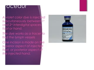
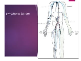
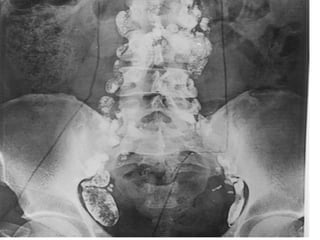
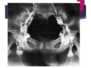
Lymphography
 Lymphography: Lymph/o: Lymph /
-graphy:………
 Invasive procedure
 Oil base contrast (the radiographic one)
 Uses gross-visualized dye contrast and
radiopaque contrast
 Lymphogram is the term used to cover
both lymph vessels and lymph nodes’
studies: lymphangiography and
lymphadenography
Basics of the Lymphatic System
 Returns proteins and fluid that has leaked out of the
bloodstream back into the blood
 Relates to the immune system. Lymphocytes and
monocytes
 The Lymphatic system is divided in lymph nodes,
lymph vessels and lymph capillaries
Lymph Nodes
 Most common and largest clusters of lymph nodes:
 Cervical
 Axillary
 Mediastinal
 And inguinal (groin)
Note: tonsils are masses of lymph
tissue.
Note: adenoids are enlarged lymph
tissue in the nasopharynx.
Procedur
e
A violet color dye is injected
subcutaneously between 1st
and 2nd
interdigital spaces,
foot or hand.
The dye works as a tracer to
find the lymph vessels
One incision is made on the
superior aspect of injected
foot, or posterior aspect of
the injected hand.
Procedure
 The incision should be just big enough to access the
tinted lymph vessels (visualized thanks to the violet
dye)
 The injection of radiographic oil-base contrast is
done trough the uncovered lymph vessels
 The injection takes about 30 min. using a 27 or 30
gauge needle
Procedure
 Confirmation of injection in the lymphatic system
could be done with fluoroscopy
 The first radiograph is taken 1 hr after injection, to
visualize only lymph vessels
 Lymph Nodes radiographs are taken 24 hrs after
injection.
 Lymph nodes retain contrast for 3 to 4 weeks
Procedure
 Abnormal physiology may retain contrast for months
 Delayed films may be required
 The violet coloration can be observed on skin and
urine and it usually disappear within a few hrs after
injection.
Lymphatic System
Alternative Exams and
Indications
 MRI and CT have replaced this procedure almost
completely. However, lymphography is useful
evaluating extent of lymphomas and staging of
radiation. Also, demonstrating obstructions of lymph
vessels and others.

Lymphography rad 205-2013

  • 1.
    Lymphography  Lymphography: Lymph/o:Lymph / -graphy:………  Invasive procedure  Oil base contrast (the radiographic one)  Uses gross-visualized dye contrast and radiopaque contrast  Lymphogram is the term used to cover both lymph vessels and lymph nodes’ studies: lymphangiography and lymphadenography
  • 2.
    Basics of theLymphatic System  Returns proteins and fluid that has leaked out of the bloodstream back into the blood  Relates to the immune system. Lymphocytes and monocytes  The Lymphatic system is divided in lymph nodes, lymph vessels and lymph capillaries
  • 3.
    Lymph Nodes  Mostcommon and largest clusters of lymph nodes:  Cervical  Axillary  Mediastinal  And inguinal (groin) Note: tonsils are masses of lymph tissue. Note: adenoids are enlarged lymph tissue in the nasopharynx.
  • 4.
    Procedur e A violet colordye is injected subcutaneously between 1st and 2nd interdigital spaces, foot or hand. The dye works as a tracer to find the lymph vessels One incision is made on the superior aspect of injected foot, or posterior aspect of the injected hand.
  • 5.
    Procedure  The incisionshould be just big enough to access the tinted lymph vessels (visualized thanks to the violet dye)  The injection of radiographic oil-base contrast is done trough the uncovered lymph vessels  The injection takes about 30 min. using a 27 or 30 gauge needle
  • 6.
    Procedure  Confirmation ofinjection in the lymphatic system could be done with fluoroscopy  The first radiograph is taken 1 hr after injection, to visualize only lymph vessels  Lymph Nodes radiographs are taken 24 hrs after injection.  Lymph nodes retain contrast for 3 to 4 weeks
  • 7.
    Procedure  Abnormal physiologymay retain contrast for months  Delayed films may be required  The violet coloration can be observed on skin and urine and it usually disappear within a few hrs after injection.
  • 8.
  • 11.
    Alternative Exams and Indications MRI and CT have replaced this procedure almost completely. However, lymphography is useful evaluating extent of lymphomas and staging of radiation. Also, demonstrating obstructions of lymph vessels and others.