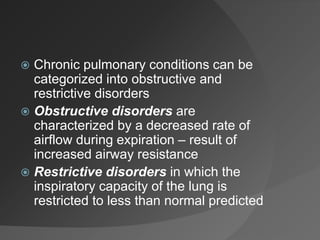
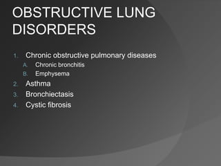
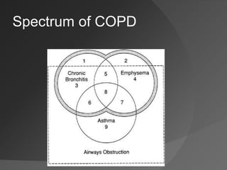
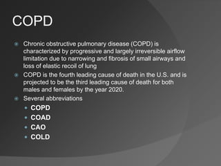
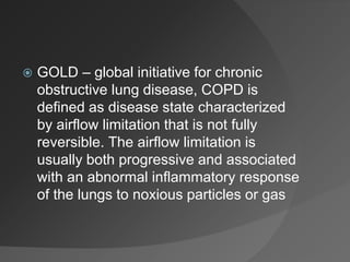
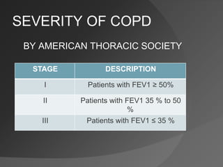
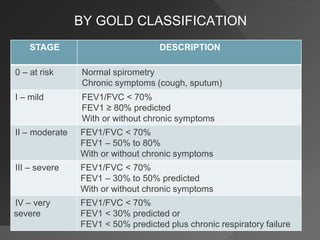
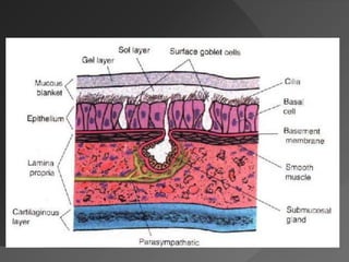
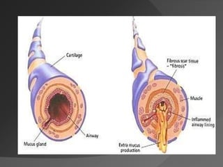
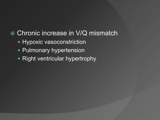
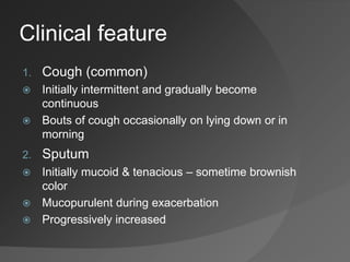
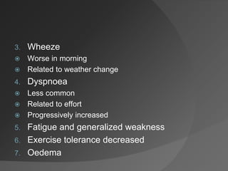
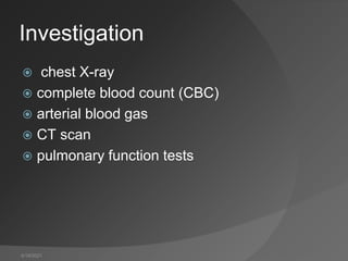
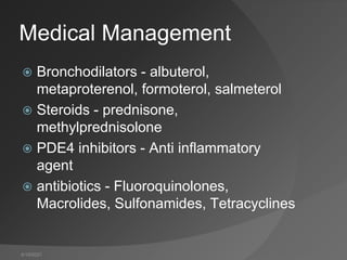
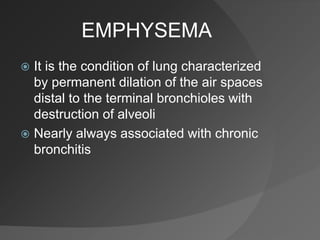
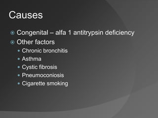
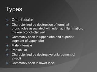
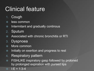
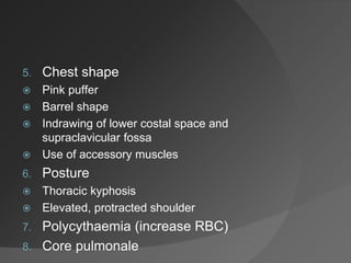
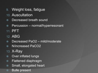
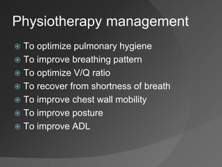
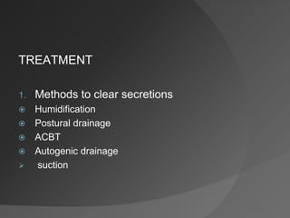
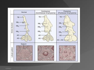
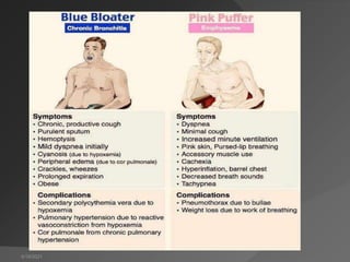
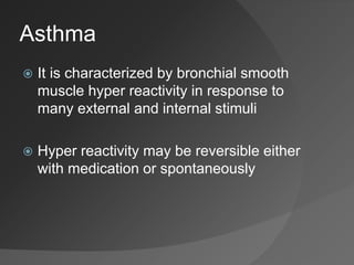
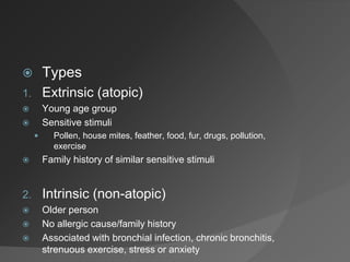
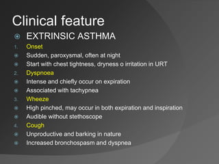
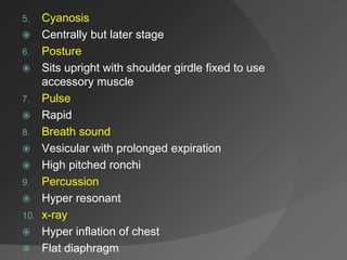
RESPIRATORY
DISORDERS
Dr. Arjun Patel
 Chronic pulmonary conditions can be
categorized into obstructive and
restrictive disorders
 Obstructive disorders are
characterized by a decreased rate of
airflow during expiration – result of
increased airway resistance
 Restrictive disorders in which the
inspiratory capacity of the lung is
restricted to less than normal predicted
OBSTRUCTIVE LUNG
DISORDERS
1. Chronic obstructive pulmonary diseases
A. Chronic bronchitis
B. Emphysema
2. Asthma
3. Bronchiectasis
4. Cystic fibrosis
Spectrum of COPD
COPD
 Chronic obstructive pulmonary disease (COPD) is
characterized by progressive and largely irreversible airflow
limitation due to narrowing and fibrosis of small airways and
loss of elastic recoil of lung
 COPD is the fourth leading cause of death in the U.S. and is
projected to be the third leading cause of death for both
males and females by the year 2020.
 Several abbreviations
 COPD
 COAD
 CAO
 COLD
 GOLD – global initiative for chronic
obstructive lung disease, COPD is
defined as disease state characterized
by airflow limitation that is not fully
reversible. The airflow limitation is
usually both progressive and associated
with an abnormal inflammatory response
of the lungs to noxious particles or gas
SEVERITY OF COPD
BY AMERICAN THORACIC SOCIETY
STAGE DESCRIPTION
I Patients with FEV1 ≥ 50%
II Patients with FEV1 35 % to 50
%
III Patients with FEV1 ≤ 35 %
BY GOLD CLASSIFICATION
STAGE DESCRIPTION
0 – at risk Normal spirometry
Chronic symptoms (cough, sputum)
I – mild FEV1/FVC < 70%
FEV1 ≥ 80% predicted
With or without chronic symptoms
II – moderate FEV1/FVC < 70%
FEV1 – 50% to 80%
With or without chronic symptoms
III – severe FEV1/FVC < 70%
FEV1 – 30% to 50% predicted
With or without chronic symptoms
IV – very
severe
FEV1/FVC < 70%
FEV1 < 30% predicted or
FEV1 < 50% predicted plus chronic respiratory failure
Chronic Bronchitis
 Chronic bronchitis is a disease
characterized by a cough producing
sputum for at least 3 months and for 2
consecutive years
 American thoracic society
 Etiology
 Smoking
 Quantity of tobacco consumption
 Cigarettes
 Pipe and Cigar
 Infection
 Viral
 Bacterial
 Air pollution
 Occupational exposure
 Pathophysiology
 Increase in mucus production
 Hypertrophy bronchial mucous glands
 Goblet cells increase in number
 Damaged cilia
 Bronchial mucosa inflammation and narrowing of airway
 Bronchial smooth muscle hyperreactivity
 Bronchospasm
 Increase V/Q mismatch
 Mucus plugging
 Airway obstruction
 Hypoxemia
 Chronic increase in V/Q mismatch
 Hypoxic vasoconstriction
 Pulmonary hypertension
 Right ventricular hypertrophy
Clinical feature
1. Cough (common)
 Initially intermittent and gradually become
continuous
 Bouts of cough occasionally on lying down or in
morning
2. Sputum
 Initially mucoid & tenacious – sometime brownish
color
 Mucopurulent during exacerbation
 Progressively increased
3. Wheeze
 Worse in morning
 Related to weather change
4. Dyspnoea
 Less common
 Related to effort
 Progressively increased
5. Fatigue and generalized weakness
6. Exercise tolerance decreased
7. Oedema
8. Chest shape
 Blue bloater
 Barrel chest
 Thoracic movt gradually diminished
 Use of accessory muscles
 Paradoxical indrowing in intercostals space
9. Cynosis
 Due to complication of hypoxia, polycythaemia, rt
ventricular failure
10. Cor pulmonale
 In later stage
11. PFT
 FVC, FEV1 reduced
 RV, FRC, TLC increased
12. ABG
 PaO2 decrease, PaCO2 increased
13. Auscultation
 Wheezing, diminished breath sound
 Percussion – hyperresonant
14. X-Ray
 Initial no abnormality
 Hyperinflation of lung
 Hypertrophy of RT ventricle
 Flatten diaphram
4/19/2021
Investigation
 chest X-ray
 complete blood count (CBC)
 arterial blood gas
 CT scan
 pulmonary function tests
4/19/2021
Medical Management
 Bronchodilators - albuterol,
metaproterenol, formoterol, salmeterol
 Steroids - prednisone,
methylprednisolone
 PDE4 inhibitors - Anti inflammatory
agent
 antibiotics - Fluoroquinolones,
Macrolides, Sulfonamides, Tetracyclines
EMPHYSEMA
 It is the condition of lung characterized
by permanent dilation of the air spaces
distal to the terminal bronchioles with
destruction of alveoli
 Nearly always associated with chronic
bronchitis
Causes
 Congenital – alfa 1 antitrypsin deficiency
 Other factors
 Chronic bronchitis
 Asthma
 Cystic fibrosis
 Pneumoconiosis
 Cigarette smoking
Types
1. Centrilobular
 Characterized by destruction of terminal
bronchioles associated with edema, inflammation,
thicken bronchiolar wall
 Commonly seen in upper lobe and superior
segment of upper lobe
 Male > female
2. Panlobular
 Characterized by destructive enlargement of
alveoli
 Commonly seen in lower lobe
Clinical feature
1. Cough
 less common
 Intermitant and gradually continous
2. Sputum
 Associated with chronic bronchitis or RTI
3. Dyspnoea
 More common
 Initially on exertion and progress to rest
4. Respiratory pattern
 FISHLIKE inspiratory gasp followed by proloned
by prolonged expiration with pursed lips
 I:E = 1:3-4
5. Chest shape
 Pink puffer
 Barrel shape
 Indrawing of lower costal space and
supraclavicular fossa
 Use of accessory muscles
6. Posture
 Thoracic kyphosis
 Elevated, protracted shoulder
7. Polycythaemia (increase RBC)
8. Core pulmonale
9. Weight loss, fatigue
10. Auscultation
 Decreased breath sound
 Percussion – normal/hyperresonant
11. PFT
12. ABG
 Decreased PaO2 – mild/moderate
 N/increased PaCO2
13. X-Ray
 Over inflated lungs
 Flattened diaphragm
 Small, elongated heart
 Bulle present
Physiotherapy management
 To optimize pulmonary hygiene
 To improve breathing pattern
 To optimize V/Q ratio
 To recover from shortness of breath
 To improve chest wall mobility
 To improve posture
 To improve ADL
TREATMENT
1. Methods to clear secretions
 Humidification
 Postural drainage
 ACBT
 Autogenic drainage
 suction
2. Methods to decrease work of breathing
 Positioning
 Breathing exercise
3. Methods to increase lung volumes
 Controlled mobilization
 Breathing exercise
 Non-invasive positive pressure ventilation (NIPPV)
4. Methods to increase strength of
respiratory muscle
 Breathing exercise
 Inspiratory muscle trainer
5. Methods to improve ADL
 Flexibility, stretching and posture
 Strengthening
4/19/2021
4/19/2021
Asthma
 It is characterized by bronchial smooth
muscle hyper reactivity in response to
many external and internal stimuli
 Hyper reactivity may be reversible either
with medication or spontaneously
 Types
1. Extrinsic (atopic)
 Young age group
 Sensitive stimuli
 Pollen, house mites, feather, food, fur, drugs, pollution,
exercise
 Family history of similar sensitive stimuli
2. Intrinsic (non-atopic)
 Older person
 No allergic cause/family history
 Associated with bronchial infection, chronic bronchitis,
strenuous exercise, stress or anxiety
Clinical feature
 EXTRINSIC ASTHMA
1. Onset
 Sudden, paroxysmal, often at night
 Start with chest tightness, dryness o irritation in URT
2. Dyspnoea
 Intense and chiefly occur on expiration
 Associated with tachypnea
3. Wheeze
 High pinched, may occur in both expiration and inspiration
 Audible without stethoscope
4. Cough
 Unproductive and barking in nature
 Increased bronchospasm and dyspnea
5. Cyanosis
 Centrally but later stage
6. Posture
 Sits upright with shoulder girdle fixed to use
accessory muscle
7. Pulse
 Rapid
8. Breath sound
 Vesicular with prolonged expiration
 High pitched ronchi
9. Percussion
 Hyper resonant
10. x-ray
 Hyper inflation of chest
 Flat diaphragm
11. PFT
 Increased TLC, FRC, RV
 Decreased FEV1, FVC
12. ABG
 Decreased PaCO2 – hyperventilation of alveoli
 Decreased PaO2 – V/Q mismatch
 INTRINSIC ASTHMA
 Same as above
 Wheeze and dyspnea more sever
Physiotherapy management
 Aims
1. To provide psychological support
2. To relieve bronchospasm
3. To clear secretion
4. To improve breathing control
5. To teach relaxation and improve
posture
6. To improve ADL
 Psychological support
 Methods to clear secretions
1. Nebulizer
2. Postural drainage
3. ACBT
4. FET
5. Breathing exercise
 To decrease work of breathing
1. Relaxation Positioning
2. Breathing exercise
 To improve ADL
1. Strengthen respiratory muscle
2. General mobility exercise
3. Short distance walking
During acute attack
 Removal of secretion
1. ACBT
2. Postural drainage
3. Breathing exercise
 Breathing control
 Relaxation position
 Oxygen therapy
 Inhalation therapy
1. Pressurized inhaler
2. Dry powder inhaler
3. Large volume spacers
 Nabulizer
BRONCHIECTASIS
 It is defined as an abnormal dilatation of
medium size of the bronchi associated with
obstruction and infection
Aetiology
 Occurs following
 Bacterial infection
 Aspiration
 Neoplasm
 Foreign bodies in bronchi
 Abnormality in immune system
 Several genetic disorders are almost always
associated with bronchiectasis
 Cystic fibrosis
 Kartagener’s syndrome
○ bronchiectasis
○ Frontal sinusitis
○ visceral transposition
 Immotile cilia syndrome
Pathology
Clinical feature
 Onset
 Symptoms begin in childhood, but diagnosis not made
until adult life
 Cough and sputum
 Persistent cough with purulent sputum since childhood
 Sputum – greenish color, foul smelling
 Cough – early in morning, changing position
 Sputum is collected and allowed to stand than 3 layers
seen
○ Upper – frothy
○ Mid – mucopurulent
○ Lower – purulent with grayish or yellowish plug
 Severity of disease
○ 24 hours sputum volume collected and measure
 < 10 ml – mild
 10 – 150 ml – moderate
 > 150 ml – severe
 Dyspnea
 If disease is severe and wide spread
 Haemoptysis
 Quite common, associated with acute infection
 Bronchial arterioles anastomosis with pulmonary capillary
 Life-threatening if severe
 Recurrent pneumonia
 Chronic sinusitis
 General ill health
 Pyrexia
 Night sweats
 Anorexia
 Weight loss
 Clubbing
 50% of patient affected
 Thoracic mobility
 Gradually decreased, use of accessory muscles
 Radiography
 X-Ray -- Initially normal, later increased multiple
cysts with fluid level
 Bronchography – accurate localization of lung,
dilation of bronchi
 CT scan – bronchial wall thickening, dilatation of
bronchi, cysts
Physiotherapy management
 Aims
 To counseling the patient
 To remove secretions
 To promote general good health
 To improve exercise tolerance
 To improve ADL activity
Treatment
 counseling the patient
 To clear secretions
 Postural drainage
 ACBT
 FET
 Breathing exercise
 To increase exercise tolerance
 Aerobic exercise
 Thoracic mobility exercise
 To improve ADL activity
Cystic fibrosis
 It is a hereditary disorder of exocrine glands, with
sodium chloride content in sweat and pancreatic
insufficiency, resulting in malabsorption
 It affects mostly lungs, pancreas, liver, intestines,
sinuses, and sex organs
 Normally, mucus is watery. It keeps the linings of
certain organs moist and prevents them from
drying out or getting infected. But in CF, an
abnormal gene causes mucus to become thick
and sticky
 The abnormal gene also causes sweat to
become extremely salty. As a result, when
you perspire, your body loses large
amounts of salt. This can upset the
balance of minerals in your blood
 Respiratory failure is the most common
cause of death in people with CF
 Cystic fibrosis (CF) is caused by a defect in
a gene called the cystic fibrosis
transmembrane conductance regulator
(CFTR) gene.
 This gene makes a protein that controls the
movement of salt and water in and out of
the cells in body. In people with CF, the
gene does not work effectively.
 This causes the thick, sticky mucus and
very salty sweat that are the main features
of CF
Signs and symptoms
 General (any age)
 Family history of cystic fibrosis
 Salty-tasting skin
 Clubbing of fingers and toes
 Cough with sputum production
 Mucoid Pseudomonas aeruginosa isolated
from airway secretions
 Hypochloraemic metabolic alkalosis
 Neonatal
 jaundice
 Abdominal or scrotal calcifications
 Intestinal obstruction
 Infancy
 Failure to thrive
 hypoproteinaemia
 Chronic diarrhoea
 Abdominal distention
 Staphylococcus aureus pneumonia
 Idiopathic intracranial hypertension
 Haemolytic anaemia (vitamin E deficiency
causes anaemia by increasing fragility and
reducing lifespan of red blood cells)
 Childhood
 Mechonium ileus
 Chronic pansinusitis
 Rectal prolapse
 Idiopathic recurrent or chronic pancreatitis
 Liver disease
 Dyspnoea
 Wheeze
 Foul smelling stool
 Adolescence and adulthood
 Allergic bronchopulmonary aspergillosis
 Chronic pansinusitis
 Bronchiectasis
 Haemoptysis
 Idiopathic recurrent pancreatitis
 Portal hypertension
 Delayed puberty
 Finger clubbing
 Azoospermia secondary to congenital
bilateral absence of the vas deferens
Complications
 Sinus disease
 Haemoptysis
 Pneumothorax
 Biliary cirrhosis
 Diabetes
 Infertility
 Bone disease
 Chest Deformity
 incontinence
Physiotherapy management
 Aims
1. To reduce bronchospasm and to clear lung
field
2. To maintain physical fitness
3. To correct posture and provide relaxation
4. To educate patient in self management
Treatment
 To clear secretions
 Humidification
 Nebulizer
 Postural drainage
 FET
 ACBT
 AD
 To correct posture and provide
relaxation
 To maintain physical fitness
 Aerobic exercise
 Thoracic mobility exercise
THANK YOU
4/19/2021

Chronic Obstructive Pulmonary Disease

  • 1.
  • 2.
     Chronic pulmonaryconditions can be categorized into obstructive and restrictive disorders  Obstructive disorders are characterized by a decreased rate of airflow during expiration – result of increased airway resistance  Restrictive disorders in which the inspiratory capacity of the lung is restricted to less than normal predicted
  • 3.
    OBSTRUCTIVE LUNG DISORDERS 1. Chronicobstructive pulmonary diseases A. Chronic bronchitis B. Emphysema 2. Asthma 3. Bronchiectasis 4. Cystic fibrosis
  • 4.
  • 5.
    COPD  Chronic obstructivepulmonary disease (COPD) is characterized by progressive and largely irreversible airflow limitation due to narrowing and fibrosis of small airways and loss of elastic recoil of lung  COPD is the fourth leading cause of death in the U.S. and is projected to be the third leading cause of death for both males and females by the year 2020.  Several abbreviations  COPD  COAD  CAO  COLD
  • 6.
     GOLD –global initiative for chronic obstructive lung disease, COPD is defined as disease state characterized by airflow limitation that is not fully reversible. The airflow limitation is usually both progressive and associated with an abnormal inflammatory response of the lungs to noxious particles or gas
  • 7.
    SEVERITY OF COPD BYAMERICAN THORACIC SOCIETY STAGE DESCRIPTION I Patients with FEV1 ≥ 50% II Patients with FEV1 35 % to 50 % III Patients with FEV1 ≤ 35 %
  • 8.
    BY GOLD CLASSIFICATION STAGEDESCRIPTION 0 – at risk Normal spirometry Chronic symptoms (cough, sputum) I – mild FEV1/FVC < 70% FEV1 ≥ 80% predicted With or without chronic symptoms II – moderate FEV1/FVC < 70% FEV1 – 50% to 80% With or without chronic symptoms III – severe FEV1/FVC < 70% FEV1 – 30% to 50% predicted With or without chronic symptoms IV – very severe FEV1/FVC < 70% FEV1 < 30% predicted or FEV1 < 50% predicted plus chronic respiratory failure
  • 9.
    Chronic Bronchitis  Chronicbronchitis is a disease characterized by a cough producing sputum for at least 3 months and for 2 consecutive years  American thoracic society
  • 10.
     Etiology  Smoking Quantity of tobacco consumption  Cigarettes  Pipe and Cigar  Infection  Viral  Bacterial  Air pollution  Occupational exposure
  • 11.
     Pathophysiology  Increasein mucus production  Hypertrophy bronchial mucous glands  Goblet cells increase in number  Damaged cilia  Bronchial mucosa inflammation and narrowing of airway  Bronchial smooth muscle hyperreactivity  Bronchospasm  Increase V/Q mismatch  Mucus plugging  Airway obstruction  Hypoxemia
  • 14.
     Chronic increasein V/Q mismatch  Hypoxic vasoconstriction  Pulmonary hypertension  Right ventricular hypertrophy
  • 15.
    Clinical feature 1. Cough(common)  Initially intermittent and gradually become continuous  Bouts of cough occasionally on lying down or in morning 2. Sputum  Initially mucoid & tenacious – sometime brownish color  Mucopurulent during exacerbation  Progressively increased
  • 16.
    3. Wheeze  Worsein morning  Related to weather change 4. Dyspnoea  Less common  Related to effort  Progressively increased 5. Fatigue and generalized weakness 6. Exercise tolerance decreased 7. Oedema
  • 17.
    8. Chest shape Blue bloater  Barrel chest  Thoracic movt gradually diminished  Use of accessory muscles  Paradoxical indrowing in intercostals space 9. Cynosis  Due to complication of hypoxia, polycythaemia, rt ventricular failure 10. Cor pulmonale  In later stage
  • 18.
    11. PFT  FVC,FEV1 reduced  RV, FRC, TLC increased 12. ABG  PaO2 decrease, PaCO2 increased 13. Auscultation  Wheezing, diminished breath sound  Percussion – hyperresonant 14. X-Ray  Initial no abnormality  Hyperinflation of lung  Hypertrophy of RT ventricle  Flatten diaphram
  • 19.
    4/19/2021 Investigation  chest X-ray complete blood count (CBC)  arterial blood gas  CT scan  pulmonary function tests
  • 20.
    4/19/2021 Medical Management  Bronchodilators- albuterol, metaproterenol, formoterol, salmeterol  Steroids - prednisone, methylprednisolone  PDE4 inhibitors - Anti inflammatory agent  antibiotics - Fluoroquinolones, Macrolides, Sulfonamides, Tetracyclines
  • 21.
    EMPHYSEMA  It isthe condition of lung characterized by permanent dilation of the air spaces distal to the terminal bronchioles with destruction of alveoli  Nearly always associated with chronic bronchitis
  • 22.
    Causes  Congenital –alfa 1 antitrypsin deficiency  Other factors  Chronic bronchitis  Asthma  Cystic fibrosis  Pneumoconiosis  Cigarette smoking
  • 23.
    Types 1. Centrilobular  Characterizedby destruction of terminal bronchioles associated with edema, inflammation, thicken bronchiolar wall  Commonly seen in upper lobe and superior segment of upper lobe  Male > female 2. Panlobular  Characterized by destructive enlargement of alveoli  Commonly seen in lower lobe
  • 24.
    Clinical feature 1. Cough less common  Intermitant and gradually continous 2. Sputum  Associated with chronic bronchitis or RTI 3. Dyspnoea  More common  Initially on exertion and progress to rest 4. Respiratory pattern  FISHLIKE inspiratory gasp followed by proloned by prolonged expiration with pursed lips  I:E = 1:3-4
  • 25.
    5. Chest shape Pink puffer  Barrel shape  Indrawing of lower costal space and supraclavicular fossa  Use of accessory muscles 6. Posture  Thoracic kyphosis  Elevated, protracted shoulder 7. Polycythaemia (increase RBC) 8. Core pulmonale
  • 26.
    9. Weight loss,fatigue 10. Auscultation  Decreased breath sound  Percussion – normal/hyperresonant 11. PFT 12. ABG  Decreased PaO2 – mild/moderate  N/increased PaCO2 13. X-Ray  Over inflated lungs  Flattened diaphragm  Small, elongated heart  Bulle present
  • 27.
    Physiotherapy management  Tooptimize pulmonary hygiene  To improve breathing pattern  To optimize V/Q ratio  To recover from shortness of breath  To improve chest wall mobility  To improve posture  To improve ADL
  • 28.
    TREATMENT 1. Methods toclear secretions  Humidification  Postural drainage  ACBT  Autogenic drainage  suction
  • 29.
    2. Methods todecrease work of breathing  Positioning  Breathing exercise 3. Methods to increase lung volumes  Controlled mobilization  Breathing exercise  Non-invasive positive pressure ventilation (NIPPV)
  • 30.
    4. Methods toincrease strength of respiratory muscle  Breathing exercise  Inspiratory muscle trainer 5. Methods to improve ADL  Flexibility, stretching and posture  Strengthening
  • 31.
  • 32.
  • 33.
    Asthma  It ischaracterized by bronchial smooth muscle hyper reactivity in response to many external and internal stimuli  Hyper reactivity may be reversible either with medication or spontaneously
  • 34.
     Types 1. Extrinsic(atopic)  Young age group  Sensitive stimuli  Pollen, house mites, feather, food, fur, drugs, pollution, exercise  Family history of similar sensitive stimuli 2. Intrinsic (non-atopic)  Older person  No allergic cause/family history  Associated with bronchial infection, chronic bronchitis, strenuous exercise, stress or anxiety
  • 35.
    Clinical feature  EXTRINSICASTHMA 1. Onset  Sudden, paroxysmal, often at night  Start with chest tightness, dryness o irritation in URT 2. Dyspnoea  Intense and chiefly occur on expiration  Associated with tachypnea 3. Wheeze  High pinched, may occur in both expiration and inspiration  Audible without stethoscope 4. Cough  Unproductive and barking in nature  Increased bronchospasm and dyspnea
  • 36.
    5. Cyanosis  Centrallybut later stage 6. Posture  Sits upright with shoulder girdle fixed to use accessory muscle 7. Pulse  Rapid 8. Breath sound  Vesicular with prolonged expiration  High pitched ronchi 9. Percussion  Hyper resonant 10. x-ray  Hyper inflation of chest  Flat diaphragm
  • 37.
    11. PFT  IncreasedTLC, FRC, RV  Decreased FEV1, FVC 12. ABG  Decreased PaCO2 – hyperventilation of alveoli  Decreased PaO2 – V/Q mismatch  INTRINSIC ASTHMA  Same as above  Wheeze and dyspnea more sever
  • 38.
    Physiotherapy management  Aims 1.To provide psychological support 2. To relieve bronchospasm 3. To clear secretion 4. To improve breathing control 5. To teach relaxation and improve posture 6. To improve ADL
  • 39.
     Psychological support Methods to clear secretions 1. Nebulizer 2. Postural drainage 3. ACBT 4. FET 5. Breathing exercise
  • 40.
     To decreasework of breathing 1. Relaxation Positioning 2. Breathing exercise  To improve ADL 1. Strengthen respiratory muscle 2. General mobility exercise 3. Short distance walking
  • 41.
    During acute attack Removal of secretion 1. ACBT 2. Postural drainage 3. Breathing exercise  Breathing control  Relaxation position
  • 42.
     Oxygen therapy Inhalation therapy 1. Pressurized inhaler 2. Dry powder inhaler 3. Large volume spacers  Nabulizer
  • 43.
    BRONCHIECTASIS  It isdefined as an abnormal dilatation of medium size of the bronchi associated with obstruction and infection
  • 44.
    Aetiology  Occurs following Bacterial infection  Aspiration  Neoplasm  Foreign bodies in bronchi  Abnormality in immune system  Several genetic disorders are almost always associated with bronchiectasis  Cystic fibrosis  Kartagener’s syndrome ○ bronchiectasis ○ Frontal sinusitis ○ visceral transposition  Immotile cilia syndrome
  • 45.
  • 46.
    Clinical feature  Onset Symptoms begin in childhood, but diagnosis not made until adult life  Cough and sputum  Persistent cough with purulent sputum since childhood  Sputum – greenish color, foul smelling  Cough – early in morning, changing position  Sputum is collected and allowed to stand than 3 layers seen ○ Upper – frothy ○ Mid – mucopurulent ○ Lower – purulent with grayish or yellowish plug  Severity of disease ○ 24 hours sputum volume collected and measure  < 10 ml – mild  10 – 150 ml – moderate  > 150 ml – severe
  • 47.
     Dyspnea  Ifdisease is severe and wide spread  Haemoptysis  Quite common, associated with acute infection  Bronchial arterioles anastomosis with pulmonary capillary  Life-threatening if severe  Recurrent pneumonia  Chronic sinusitis  General ill health  Pyrexia  Night sweats  Anorexia  Weight loss
  • 48.
     Clubbing  50%of patient affected  Thoracic mobility  Gradually decreased, use of accessory muscles  Radiography  X-Ray -- Initially normal, later increased multiple cysts with fluid level  Bronchography – accurate localization of lung, dilation of bronchi  CT scan – bronchial wall thickening, dilatation of bronchi, cysts
  • 49.
    Physiotherapy management  Aims To counseling the patient  To remove secretions  To promote general good health  To improve exercise tolerance  To improve ADL activity
  • 50.
    Treatment  counseling thepatient  To clear secretions  Postural drainage  ACBT  FET  Breathing exercise
  • 51.
     To increaseexercise tolerance  Aerobic exercise  Thoracic mobility exercise  To improve ADL activity
  • 52.
    Cystic fibrosis  Itis a hereditary disorder of exocrine glands, with sodium chloride content in sweat and pancreatic insufficiency, resulting in malabsorption  It affects mostly lungs, pancreas, liver, intestines, sinuses, and sex organs  Normally, mucus is watery. It keeps the linings of certain organs moist and prevents them from drying out or getting infected. But in CF, an abnormal gene causes mucus to become thick and sticky
  • 53.
     The abnormalgene also causes sweat to become extremely salty. As a result, when you perspire, your body loses large amounts of salt. This can upset the balance of minerals in your blood  Respiratory failure is the most common cause of death in people with CF
  • 54.
     Cystic fibrosis(CF) is caused by a defect in a gene called the cystic fibrosis transmembrane conductance regulator (CFTR) gene.  This gene makes a protein that controls the movement of salt and water in and out of the cells in body. In people with CF, the gene does not work effectively.  This causes the thick, sticky mucus and very salty sweat that are the main features of CF
  • 55.
    Signs and symptoms General (any age)  Family history of cystic fibrosis  Salty-tasting skin  Clubbing of fingers and toes  Cough with sputum production  Mucoid Pseudomonas aeruginosa isolated from airway secretions  Hypochloraemic metabolic alkalosis
  • 56.
     Neonatal  jaundice Abdominal or scrotal calcifications  Intestinal obstruction
  • 57.
     Infancy  Failureto thrive  hypoproteinaemia  Chronic diarrhoea  Abdominal distention  Staphylococcus aureus pneumonia  Idiopathic intracranial hypertension  Haemolytic anaemia (vitamin E deficiency causes anaemia by increasing fragility and reducing lifespan of red blood cells)
  • 58.
     Childhood  Mechoniumileus  Chronic pansinusitis  Rectal prolapse  Idiopathic recurrent or chronic pancreatitis  Liver disease  Dyspnoea  Wheeze  Foul smelling stool
  • 59.
     Adolescence andadulthood  Allergic bronchopulmonary aspergillosis  Chronic pansinusitis  Bronchiectasis  Haemoptysis  Idiopathic recurrent pancreatitis  Portal hypertension  Delayed puberty  Finger clubbing  Azoospermia secondary to congenital bilateral absence of the vas deferens
  • 60.
    Complications  Sinus disease Haemoptysis  Pneumothorax  Biliary cirrhosis  Diabetes  Infertility  Bone disease  Chest Deformity  incontinence
  • 61.
    Physiotherapy management  Aims 1.To reduce bronchospasm and to clear lung field 2. To maintain physical fitness 3. To correct posture and provide relaxation 4. To educate patient in self management
  • 62.
    Treatment  To clearsecretions  Humidification  Nebulizer  Postural drainage  FET  ACBT  AD  To correct posture and provide relaxation
  • 63.
     To maintainphysical fitness  Aerobic exercise  Thoracic mobility exercise
  • 64.