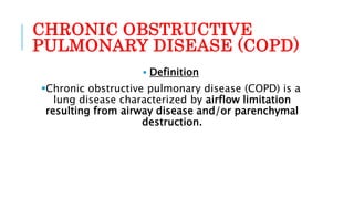
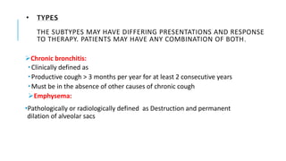
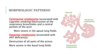
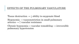
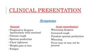
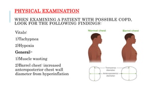
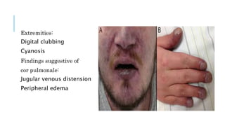
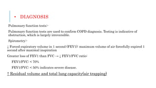
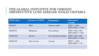
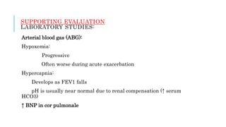
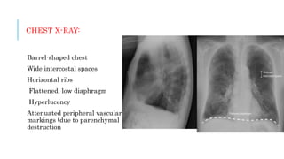
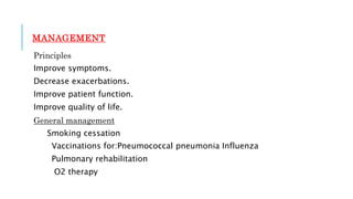
COPD By:Dr Mayur Patel
CHRONIC OBSTRUCTIVE
PULMONARY DISEASE (COPD)
 Definition
Chronic obstructive pulmonary disease (COPD) is a
lung disease characterized by airflow limitation
resulting from airway disease and/or parenchymal
destruction.
• TYPES
THE SUBTYPES MAY HAVE DIFFERING PRESENTATIONS AND RESPONSE
TO THERAPY. PATIENTS MAY HAVE ANY COMBINATION OF BOTH.
Chronic bronchitis:
 Clinically defined as
 Productive cough > 3 months per year for at least 2 consecutive years
 Must be in the absence of other causes of chronic cough
Emphysema:
•Pathologically or radiologically defined as Destruction and permanent
dilation of alveolar sacs
PATHOPHYSIOLOGY
Inflammation,
smooth
muscle hypertrop
hy, and excess
mucus
production lead
to
progressive airw
ay obstruction.
Chronic bronchitis
Inhaled agents cause chronic inflammation in the airways,
which lead to progressive airway obstruction through:
 Damage to endothelial cells → ↓ mucocilliary clearance
 Mucous gland hyperplasia → mucous hypersecretion and
plugging
 Airway edema and smooth muscle hyperplasia → luminal
narrowing
 Peribronchial fibrosis → bronchial distortion
EMPHYSEMA
In normal lungs, there is a balance between:
Proteases → break down elastin and connective tissue as part of normal tissue repair:Neutrophil
elastase
Matrix metalloproteinase (MMP)
Catharses
Antiproteases → balance protease activity:
AAT
Secretory leukoprotease inhibitor derived from airway epithelium
Elafin
MMP tissue inhibitor
IN EMPHYSEMA:
Inflammatory response → activated neutrophils release proteases
Protease activity exceeds antiprotease activity → tissue destruction
Alveolar destruction leads to:Enlarged alveoli
↓ Elastic recoil
↑ Compliance
Consequences:-Airway closure during expiration → obstruction
Air trapping → lung hyperinflation
MORPHOLOGIC PATTERNS:
Centriacinar emphysema (associated with
cigarette smoking):Destruction of the
respiratory bronchioles and a central
portion of the acini
More severe in the apical lung fields
Panacinar emphysema (associated with
AAT deficiency):
Destruction of all parts of the acinus
More severe in the basal lung fields
EFFECTS OF THE PULMONARY VASCULATURE
Tissue destruction → ↓ ability to oxygenate blood
Hypoxemia → vasoconstriction in small pulmonary
arteries → ↑ vascular resistance
Chronic hypoxemia→ vascular remodeling → irreversible
pulmonary hypertension
CLINICAL PRESENTATION
Symptoms
General
 Progressive dyspnea
(particularly with exertion)
 Chronic cough
 Sputum production
 Chest tightness
 Weight gain or loss
 Fatigue
Acute exacerbation:
Worsening dyspnea
Increased cough
Purulent sputum production
Wheezing
Fever may or may not be
present
PHYSICAL EXAMINATION
WHEN EXAMINING A PATIENT WITH POSSIBLE COPD,
LOOK FOR THE FOLLOWING FINDINGS:
Vitals:
1)Tachypnea
2)Hypoxia
General:-
1)Muscle wasting
2)Barrel chest: increased
anteroposterior chest wall
diameter from hyperinflation
Extremities:
Digital clubbing
Cyanosis
Findings suggestive of
cor pulmonale:
Jugular venous distension
Peripheral edema
CLINICAL PHENOTYPES
SIGNS AND SYMPTOMS ARE ASSOCIATED MORE FREQUENTLY
WITH EITHER CHRONIC BRONCHITIS OR EMPHYSEMA. HOWEVER,
PATIENTS OFTEN PRESENT WITH A MIXTURE OF FEATURES.
Chronic bronchitis (“blue
bloater”):
Patients are generally
overweight.
Frequent, productive cough
Peripheral edema
Cyanosis
Emphysema (“pink puffer”):
Patients are generally thin.
Barrel chest
Infrequent cough
Pursed lip breathing
Accessory muscle use Tripod
positioning
Hyperresonant chest
• DIAGNOSIS
Pulmonary function tests:-
Pulmonary function tests are used to confirm COPD diagnosis. Testing is indicative of
obstruction, which is largely irreversible.
Spirometry:-
↓ Forced expiratory volume in 1 second (FEV1): maximum volume of air forcefully expired 1
second after maximal inspiration
Greater loss of FEV1 than FVC → ↓ FEV1/FVC ratio:
FEV1/FVC: < 70%
FEV1/FVC: < 50% indicates severe disease.
↑ Residual volume and total lung capacity(air trapping)
Emphysema:-
↓ Diffusing capacity for CO:
Also known as transfer factor
Due to loss of surface area for
gas exchange
Rapid fall in expiratory flow (dynamic
airway collapse) → produces a concave
pattern
Post-bronchodilator test:
Used to assess the reversibility of the
obstructive condition
Minimal reversibility in COPD
THE GLOBAL INITIATIVE FOR CHRONIC
OBSTRUCTIVE LUNG DISEASE (GOLD) CRITERIA
GOLD class Severity of COPD Symptoms Spirometry
results
GOLD I Mild None or mild FEV1: ≥ 80%
FEV1/FVC: < 70%
GOLD II Moderate On exertion FEV1: 50%–79%
FEV1/FVC: < 70%
GOLD III Severe On minimal exertion FEV1: 30%–49%
FEV1/FVC: < 70%
GOLD IV Very severe At rest FEV1: < 30%
FEV1/FVC: < 70
SUPPORTING EVALUATION
LABORATORY STUDIES:
Arterial blood gas (ABG):
Hypoxemia:
Progressive
Often worse during acute exacerbation
Hypercapnia:
Develops as FEV1 falls
pH is usually near normal due to renal compensation (↑ serum
HCO3)
↑ BNP in cor pulmonale
CHEST X-RAY:
Barrel-shaped chest
Wide intercostal spaces
Horizontal ribs
Flattened, low diaphragm
Hyperlucency
Attenuated peripheral vascular
markings (due to parenchymal
destruction
MANAGEMENT
Principles
Improve symptoms.
Decrease exacerbations.
Improve patient function.
Improve quality of life.
General management
Smoking cessation
Vaccinations for:Pneumococcal pneumonia Influenza
Pulmonary rehabilitation
O2 therapy
MEDICAL THERAPY
Bronchodilators
Short acting (used as needed for rescue):
Beta-2 adrenergic agonists(e.g., albuterol)
Anticholinergics (e.g., ipratropium bromide
Long acting:
Beta-2 adrenergic agonists(e.g., salmeterol,
formoterol,indacaterol)
Anticholinergics (e.g., tiotropium, aclidinium, umeclidinium)
Phosphodiesterase-4 inhibitors
Reduces inflammation Can increase FEV1 and reduce
exacerbations
Inhalbronchodilatoroids:
Can produce both marginal improvements and adverse effects
Theophylline(oral bronchodilator)
Mucolytics
Surgical intervention:-
Bullectomy: removal of giant bullae to relieve local compression
Lung volume reduction
Lung transplant:-indicated in end-stage lung disease
MANAGEMENT OF ACUTE EXACERBATIONS
Short-acting bronchodilators:
Scheduled every 4–6 hours
Continuous nebulization may be needed for severe bronchospasm.
Systemic steroids
Antibiotics are indicated for:
Purulent sputum Evidence of pneumonia
Patients requiring hospitalization
Controlled O2 therapy for acute respiratory failure:
Nasal cannula
Noninvasive ventilation
Invasive ventilation
DIFFERENTIAL DIAGNOSIS
Asthma
A chronic, inflammatory condition
characterized by reversible airflow
obstruction in the lower airways.
Patients present with intermittent or
persistent wheezing, cough, and
dyspnea. Diagnosis is usually
confirmed with a pulmonary function
test showing a reversible, obstructive
pattern. Management varies based on
severity and includes bronchodilators
and inhaled corticosteroids for
inflammation control.
Bronchiectasis
A chronic condition with bronchial
dilation and destruction as a result of
inflammation and infection. Symptoms
include dyspnea, chronic cough, and
purulent sputum.
The diagnosis is made with imaging
(X-ray and CT).
Management includes bronchodilators
and antibiotics for acute exacerbations.
Pulmonary embolism:
Obstruction of the pulmonary
arteries most often due to
thrombus migration from the deep
venous system. Signs and
symptoms include pleuritic chest
pain, dyspnea, tachypnea, and
tachycardia.Severe cases can
result in hemodynamic instability
or cardiopulmonary arrest. Chest
CTA is the primary method of
diagnosis. Management includes
oxygenation, anticoagulation, and
thrombolytic therapy for unstable
patients.
Cystic fibrosis:-
Autosomal recessive disorder leading
to dysfunction of chloride channels,
which results in hyperviscous mucus
and the accumulation of
secretions.Patients often have chronic
respiratory infections, failure to
thrive, and pancreatic insufficiency.
The gold standard for diagnosis is the
sweat chloride test, which can be
complemented by genetic testing.
COPD.pptx

COPD.pptx

  • 1.
  • 2.
    CHRONIC OBSTRUCTIVE PULMONARY DISEASE(COPD)  Definition Chronic obstructive pulmonary disease (COPD) is a lung disease characterized by airflow limitation resulting from airway disease and/or parenchymal destruction.
  • 3.
    • TYPES THE SUBTYPESMAY HAVE DIFFERING PRESENTATIONS AND RESPONSE TO THERAPY. PATIENTS MAY HAVE ANY COMBINATION OF BOTH. Chronic bronchitis:  Clinically defined as  Productive cough > 3 months per year for at least 2 consecutive years  Must be in the absence of other causes of chronic cough Emphysema: •Pathologically or radiologically defined as Destruction and permanent dilation of alveolar sacs
  • 4.
    PATHOPHYSIOLOGY Inflammation, smooth muscle hypertrop hy, andexcess mucus production lead to progressive airw ay obstruction.
  • 5.
    Chronic bronchitis Inhaled agentscause chronic inflammation in the airways, which lead to progressive airway obstruction through:  Damage to endothelial cells → ↓ mucocilliary clearance  Mucous gland hyperplasia → mucous hypersecretion and plugging  Airway edema and smooth muscle hyperplasia → luminal narrowing  Peribronchial fibrosis → bronchial distortion
  • 6.
    EMPHYSEMA In normal lungs,there is a balance between: Proteases → break down elastin and connective tissue as part of normal tissue repair:Neutrophil elastase Matrix metalloproteinase (MMP) Catharses Antiproteases → balance protease activity: AAT Secretory leukoprotease inhibitor derived from airway epithelium Elafin MMP tissue inhibitor
  • 7.
    IN EMPHYSEMA: Inflammatory response→ activated neutrophils release proteases Protease activity exceeds antiprotease activity → tissue destruction Alveolar destruction leads to:Enlarged alveoli ↓ Elastic recoil ↑ Compliance Consequences:-Airway closure during expiration → obstruction Air trapping → lung hyperinflation
  • 8.
    MORPHOLOGIC PATTERNS: Centriacinar emphysema(associated with cigarette smoking):Destruction of the respiratory bronchioles and a central portion of the acini More severe in the apical lung fields Panacinar emphysema (associated with AAT deficiency): Destruction of all parts of the acinus More severe in the basal lung fields
  • 9.
    EFFECTS OF THEPULMONARY VASCULATURE Tissue destruction → ↓ ability to oxygenate blood Hypoxemia → vasoconstriction in small pulmonary arteries → ↑ vascular resistance Chronic hypoxemia→ vascular remodeling → irreversible pulmonary hypertension
  • 10.
    CLINICAL PRESENTATION Symptoms General  Progressivedyspnea (particularly with exertion)  Chronic cough  Sputum production  Chest tightness  Weight gain or loss  Fatigue Acute exacerbation: Worsening dyspnea Increased cough Purulent sputum production Wheezing Fever may or may not be present
  • 11.
    PHYSICAL EXAMINATION WHEN EXAMININGA PATIENT WITH POSSIBLE COPD, LOOK FOR THE FOLLOWING FINDINGS: Vitals: 1)Tachypnea 2)Hypoxia General:- 1)Muscle wasting 2)Barrel chest: increased anteroposterior chest wall diameter from hyperinflation
  • 12.
    Extremities: Digital clubbing Cyanosis Findings suggestiveof cor pulmonale: Jugular venous distension Peripheral edema
  • 13.
    CLINICAL PHENOTYPES SIGNS ANDSYMPTOMS ARE ASSOCIATED MORE FREQUENTLY WITH EITHER CHRONIC BRONCHITIS OR EMPHYSEMA. HOWEVER, PATIENTS OFTEN PRESENT WITH A MIXTURE OF FEATURES. Chronic bronchitis (“blue bloater”): Patients are generally overweight. Frequent, productive cough Peripheral edema Cyanosis Emphysema (“pink puffer”): Patients are generally thin. Barrel chest Infrequent cough Pursed lip breathing Accessory muscle use Tripod positioning Hyperresonant chest
  • 14.
    • DIAGNOSIS Pulmonary functiontests:- Pulmonary function tests are used to confirm COPD diagnosis. Testing is indicative of obstruction, which is largely irreversible. Spirometry:- ↓ Forced expiratory volume in 1 second (FEV1): maximum volume of air forcefully expired 1 second after maximal inspiration Greater loss of FEV1 than FVC → ↓ FEV1/FVC ratio: FEV1/FVC: < 70% FEV1/FVC: < 50% indicates severe disease. ↑ Residual volume and total lung capacity(air trapping)
  • 16.
    Emphysema:- ↓ Diffusing capacityfor CO: Also known as transfer factor Due to loss of surface area for gas exchange Rapid fall in expiratory flow (dynamic airway collapse) → produces a concave pattern Post-bronchodilator test: Used to assess the reversibility of the obstructive condition Minimal reversibility in COPD
  • 17.
    THE GLOBAL INITIATIVEFOR CHRONIC OBSTRUCTIVE LUNG DISEASE (GOLD) CRITERIA GOLD class Severity of COPD Symptoms Spirometry results GOLD I Mild None or mild FEV1: ≥ 80% FEV1/FVC: < 70% GOLD II Moderate On exertion FEV1: 50%–79% FEV1/FVC: < 70% GOLD III Severe On minimal exertion FEV1: 30%–49% FEV1/FVC: < 70% GOLD IV Very severe At rest FEV1: < 30% FEV1/FVC: < 70
  • 18.
    SUPPORTING EVALUATION LABORATORY STUDIES: Arterialblood gas (ABG): Hypoxemia: Progressive Often worse during acute exacerbation Hypercapnia: Develops as FEV1 falls pH is usually near normal due to renal compensation (↑ serum HCO3) ↑ BNP in cor pulmonale
  • 19.
    CHEST X-RAY: Barrel-shaped chest Wideintercostal spaces Horizontal ribs Flattened, low diaphragm Hyperlucency Attenuated peripheral vascular markings (due to parenchymal destruction
  • 20.
    MANAGEMENT Principles Improve symptoms. Decrease exacerbations. Improvepatient function. Improve quality of life. General management Smoking cessation Vaccinations for:Pneumococcal pneumonia Influenza Pulmonary rehabilitation O2 therapy
  • 21.
    MEDICAL THERAPY Bronchodilators Short acting(used as needed for rescue): Beta-2 adrenergic agonists(e.g., albuterol) Anticholinergics (e.g., ipratropium bromide Long acting: Beta-2 adrenergic agonists(e.g., salmeterol, formoterol,indacaterol) Anticholinergics (e.g., tiotropium, aclidinium, umeclidinium) Phosphodiesterase-4 inhibitors Reduces inflammation Can increase FEV1 and reduce exacerbations
  • 22.
    Inhalbronchodilatoroids: Can produce bothmarginal improvements and adverse effects Theophylline(oral bronchodilator) Mucolytics Surgical intervention:- Bullectomy: removal of giant bullae to relieve local compression Lung volume reduction Lung transplant:-indicated in end-stage lung disease
  • 23.
    MANAGEMENT OF ACUTEEXACERBATIONS Short-acting bronchodilators: Scheduled every 4–6 hours Continuous nebulization may be needed for severe bronchospasm. Systemic steroids Antibiotics are indicated for: Purulent sputum Evidence of pneumonia Patients requiring hospitalization Controlled O2 therapy for acute respiratory failure: Nasal cannula Noninvasive ventilation Invasive ventilation
  • 24.
    DIFFERENTIAL DIAGNOSIS Asthma A chronic,inflammatory condition characterized by reversible airflow obstruction in the lower airways. Patients present with intermittent or persistent wheezing, cough, and dyspnea. Diagnosis is usually confirmed with a pulmonary function test showing a reversible, obstructive pattern. Management varies based on severity and includes bronchodilators and inhaled corticosteroids for inflammation control. Bronchiectasis A chronic condition with bronchial dilation and destruction as a result of inflammation and infection. Symptoms include dyspnea, chronic cough, and purulent sputum. The diagnosis is made with imaging (X-ray and CT). Management includes bronchodilators and antibiotics for acute exacerbations.
  • 25.
    Pulmonary embolism: Obstruction ofthe pulmonary arteries most often due to thrombus migration from the deep venous system. Signs and symptoms include pleuritic chest pain, dyspnea, tachypnea, and tachycardia.Severe cases can result in hemodynamic instability or cardiopulmonary arrest. Chest CTA is the primary method of diagnosis. Management includes oxygenation, anticoagulation, and thrombolytic therapy for unstable patients. Cystic fibrosis:- Autosomal recessive disorder leading to dysfunction of chloride channels, which results in hyperviscous mucus and the accumulation of secretions.Patients often have chronic respiratory infections, failure to thrive, and pancreatic insufficiency. The gold standard for diagnosis is the sweat chloride test, which can be complemented by genetic testing.