CHRONIC INFLAMMATION
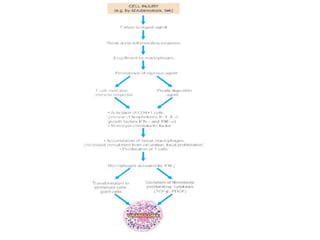
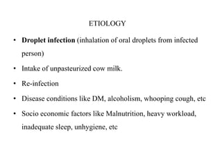
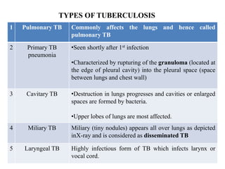
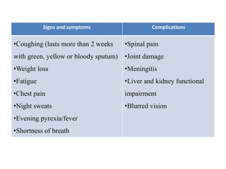
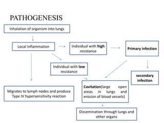
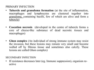
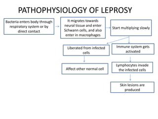
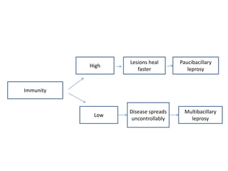
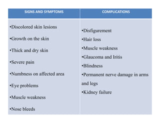
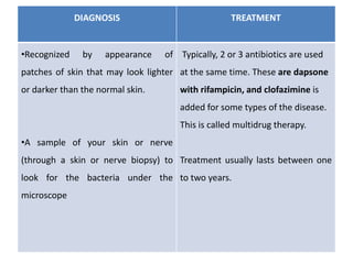
•Chronic inflammation is defined as prolonged process in which tissue
destruction and inflammation occur at the same time.
•Chronic inflammation can be caused by one of the following 3 ways:
1. Chronic inflammation following acute inflammation.
When the tissue destruction is extensive, or the bacteria survive and persist in
small numbers at the site of acute inflammation e.g. in osteomyelitis,
pneumonia terminating in lung abscess.
2. Recurrent attacks of acute inflammation.
When repeated bouts of acute inflammation culminate in chronicity of the
process e.g. in recurrent urinary tract infection leading to chronic pyelonephritis,
repeated acute infection of gallbladder leading to chronic cholecystitis.
3. Chronic inflammation starting de novo.
When the infection with organisms of low pathogenicity is chronic from the
beginning e.g. infection with Mycobacterium tuberculosis.
GENERAL FEATURES OF CHRONIC INFLAMMATION
1. MONONUCLEAR CELL INFILTRATION. Chronic inflammatory lesions are
infiltrated by mononuclear inflammatory cells like phagocytes and
lymphoid cells.
2. TISSUE DESTRUCTION OR NECROSIS. Tissue destruction and necrosis are
central features of most forms of chronic inflammatory lesions. This is
brought about by activated macrophages which release a variety of
biologically active substances.
3. PROLIFERATIVE CHANGES. As a result of necrosis, proliferation of small
blood vessels and fibroblasts is stimulated resulting in formation of
inflammatory granulation tissue. Eventually, healing by fibrosis and
collagen laying takes place.
SYSTEMIC EFFECTS OF CHRONIC INFLAMMATION
 Fever
 Anaemia
 Leucocytosis
 ESR
 Amyloidosis
TYPES OF CHRONIC INFLAMMATION
1. Non-specific, when the irritant substance produces a nonspecific chronic
inflammatory reaction with formation of granulation tissue and healing by
fibrosis e.g. chronic osteomyelitis, chronic ulcer.
2. Specific, when the injurious agent causes a characteristic histologic tissue
response e.g. tuberculosis, leprosy, syphilis.
Histological features are used for classifying chronic inflammation into 2
corresponding types:
1. Chronic non-specific inflammation. It is characterised by non-specific
inflammatory cell infiltration e.g. chronic osteomyelitis, lung abscess.
A variant of this type of chronic inflammatory response is chronic
suppurative inflammation in which infiltration by polymorphs and abscess
formation are additional features e.g. actinomycosis.
2. Chronic granulomatous inflammation. It is characterised by formation of
granulomas e.g. tuberculosis, leprosy, syphilis, actinomycosis, sarcoidosis
etc.
GRANULOMATOUS INFLAMMATION
• Granuloma is defined as a circumscribed, tiny lesion, about 1 mm in
diameter, composed predominantly of collection of modified
macrophages called epithelioid cells, and rimmed at the periphery by
lymphoid cells.
PATHOGENESIS OF GRANULOMA
1. Engulfment by macrophages. Macrophages and monocytes engulf the
antigen and try to destroy it. But since the antigen is poorly degradable,
these cells fail to digest and degrade the antigen, and instead undergo
morphologic changes to epithelioid cells.
2. CD4+ T cells. Macrophages, being antigen-presenting cells, having failed to
deal with the antigen, present it to CD4+ T lymphocytes. These
lymphocytes get activated and elaborate lymphokines (IL-1, IL-2,
interferon-γ, TNF-α).
3. Cytokines. Various cytokines formed by activated CD4+ T cells and also by
activated macrophages perform the following roles:
i) IL-1 and IL-2 stimulate proliferation of more T cells.
ii) Interferon-γ activates macrophages.
iii) TNF-α promotes fibroblast proliferation and activates endothelium to secrete
prostaglandins which have role in vascular response in inflammation.
iv) Growth factors (transforming growth factor-β, platelet derived growth factor)
elaborated by activated macrophages stimulate fibroblast growth.
Thus, a granuloma is formed of macrophages modified as epithelioid cells in the
centre, with some interspersed multinucleate giant cells, surrounded
peripherally by lymphocytes (mainly T cells), and healing by fibroblasts or
collagen depending upon the age of granuloma.
COMPOSITION OF GRANULOMA. In general, a granuloma has the following
structural composition:
1. Epithelioid cells. These are so called because of their epithelial cell-like
appearance, are modified macrophages/ histiocytes which are somewhat
elongated, having vesicular and lightly-staining slipper-shaped nucleus, and
pale- staining abundant cytoplasm with hazy outlines so that the cell
membrane of adjacent epithelioid cells is closely apposed.
Epithelioid cells are weakly phagocytic.
2. Multinucleate giant cells. Multinucleate giant cells are formed by fusion of
adjacent epithelioid cells and may have 20 or more nuclei. These nuclei may be
arranged at the periphery like horseshoe or ring, or are clustered at the two
poles (Langhans’ giant cells), or they may be present centrally (foreign body
giant cells).
3. Lymphoid cells. As a cell mediated immune reaction to antigen, the host
response by lymphocytes is integral to composition of a granuloma.
Plasma cells indicative of accelerated humoral immune response are
present in some types of granulomas.
4. Necrosis. Necrosis may be a feature of some granulomatous conditions e.g.
central caseation necrosis of tuberculosis, so called because of cheese-like
appearance and consistency of necrosis.
5. Fibrosis. Fibrosis is a feature of healing by proliferating fibroblasts at the
periphery of granuloma. The classical example of granulomatous
inflammation
EXAMPLES OF GRANULOMATOUS INFLAMMATION
ACTINOMYCOSIS
• Actinomycosis is a chronic suppurative disease caused by anaerobic bacteria,
Actinomycetes israelii.
• The organisms are commensals in the oral cavity, alimentary tract and vagina.
• The organisms invade, proliferate and disseminate in favourable conditions like break
in mucocutaneous continuity, some underlying disease etc.
MORPHOLOGIC FEATURES.
Depending upon the anatomic location of lesions, actinomycosis is of 4 types:
• 1. Cervicofacial actinomycosis. This is the commonest form (60%) and has the best
prognosis. The infection enters from tonsils, carious teeth, periodontal disease or
trauma following tooth extraction.
• Initially, a firm swelling develops in the lower jaw (‘lumpy jaw’). In time, the mass
breaks down and abscesses and sinuses are formed. The discharging pus contains
typical tiny yellow sulphur.
granules. The infection may extend into adjoining soft tissues as well as
may destroy the bone.
2. Thoracic actinomycosis. The infection in the lungs is due to aspiration of
the organism from oral cavity or extension of infection from abdominal or
hepatic lesions. Initially, the disease resembles pneumonia but
subsequently the infection spreads to the whole of lung, pleura, ribs and
vertebrae.
3. Abdominal actinomycosis. This type is common in appendix, caecum and
liver. The abdominal infection results from swallowing of organisms from
oral cavity or extension from thoracic cavity.
4. Pelvic actinomycosis. Infection in the pelvis occurs as a complication of
intrauterine contraceptive devices (IUCD’s).
Microscopically, irrespective of the location of actinomycosis,
the following features are seen
i) The inflammatory reaction is a granuloma with central suppuration. There is
formation of abscesses in the centre of lesions and at the periphery
chronic inflammatory cells, giant cells and fibroblasts are seen.
ii) The centre of each abscess contains the bacterial colony, ‘sulphur granule’,
characterised by radiating filaments (hence previously known as ray
fungus) with hyaline, eosinophilic, club-like ends representative of
secreted immunoglobulins.
iii) Bacterial stains reveal the organisms as gram-positive filaments, nonacid-
fast, which stain positively with Gomori’s methenamine silver (GMS)
staining.
SARCOIDOSIS (BOECK’S SARCOID)
• Sarcoidosis is a systemic disease of unknown etiology.
• It is worldwide in distribution and affects adults from 20-40 years of age.
• The disease is characterised by the presence of noncaseating epithelioid
cell granulomas (‘sarcoid granuloma’) in the affected tissues and organs,
notably lymph nodes and lungs.
• Other sites are the skin, spleen, uvea of the eyes, salivary glands, liver and
bones of hands and feet.
ETIOLOGY AND PATHOGENESIS
• The cause of sarcoidosis remains unknown.
• Currently, possible etiology is an infectious or noninfectious environmental
agent in a genetically susceptible individual.
• Likely infectious agents include Propionibacter acnes, atypical
mycobacteria and mycobacterial protein of M. tuberculosis.
The following observations point towards a possible immune origin of sarcoidosis:
1. poorly degradable Antigen
2. high levels of CD4+T cells
3. activated alveolar macrophages
MORPHOLOGIC FEATURES.
• The lesions in sarcoidosis are generalised and may affect various organs and
tissues at sometime in the course of disease, but severity of the disease is borne
by the lungs and lymph nodes.
Microscopically, the following features are present
1. The diagnostic feature in sarcoidosis of any organ or tissue is the non-caseating
sarcoid granuloma, composed of epithelioid cells, Langhans’ and foreign body
giant cells and surrounded peripherally by fibroblasts.
2. Typically, granulomas of sarcoidosis are ‘naked’ i.e. either devoid of
peripheral rim of lymphocytes or there is paucity of lymphocytes.
3. In late stage, the granuloma is either enclosed by hyalinised fibrous tissue
or is replaced by hyalinised fibrous mass.
4. The giant cells in sarcoid granulomas contain certain cytoplasmic inclusions
as follows:
i) Asteroid bodies which are eosinophilic and stellate shaped structures.
ii) Schaumann’s bodies or conchoid (conch like) bodies which are concentric
laminations of calcium and of iron salts, complexed with proteins.
iii) Birefringent cytoplasmic crystals which are colourless.
• Tuberculosis is an infective disease commonly affecting lungs.
• It is caused by
 Mycobacterium tuberculosis
 Mycobacterium bovis
• It is an airborne disease, spreads through air in form of small
droplets.
• Transmitted by sneezing, coughing, singing or even talking
TUBERCULOSIS
ETIOLOGY
• Droplet infection (inhalation of oral droplets from infected
person)
• Intake of unpasteurized cow milk.
• Re-infection
• Disease conditions like DM, alcoholism, whooping cough, etc
• Socio economic factors like Malnutrition, heavy workload,
inadequate sleep, unhygiene, etc
TYPES OF TUBERCULOSIS
1 Pulmonary TB Commonly affects the lungs and hence called
pulmonary TB
2 Primary TB
pneumonia
•Seen shortly after 1st infection
•Characterized by rupturing of the granuloma (located at
the edge of pleural cavity) into the pleural space (space
between lungs and chest wall)
3 Cavitary TB •Destruction in lungs progresses and cavities or enlarged
spaces are formed by bacteria.
•Upper lobes of lungs are most affected.
4 Miliary TB Miliary (tiny nodules) appears all over lungs as depicted
inX-ray and is considered as disseminated TB
5 Laryngeal TB Highly infectious form of TB which infects larynx or
vocal cord.
Signs and symptoms Complications
•Coughing (lasts more than 2 weeks
with green, yellow or bloody sputum)
•Weight loss
•Fatigue
•Chest pain
•Night sweats
•Evening pyrexia/fever
•Shortness of breath
•Spinal pain
•Joint damage
•Meningitis
•Liver and kidney functional
impairment
•Blurred vision
PATHOGENESIS
Inhalation of organism into lungs
secondary
infection
Individual with high
resistance
Migrates to lymph nodes and produce
Type IV hypersensitivity reaction
Local inflammation
Individual with low
resistance
Primary infection
Cavitation(large open
areas in lungs and
erosion of blood vessels)
Dissemination through lungs and
other organs
PRIMARY INFECTION
• Tubercle and granuloma formation (at the site of inflammation,
macrophages and lymphocytes are clustered together into
granuloma, containing bacilli, few of which are alive and form a
tubercle)
• Caseation necrosis (developed in the centre of tubercle forms a
core of cheese-like substance of dead necrotic tissues and
macrophages)
• Ghon complex (An individual of strong immune system may resist
the invasion, but these lesions may remain very small and become
walled off by fibrous tissue and sometimes also calcify. These
lesions are called Ghon complex)
SECONDARY INFECTION
• If resistance decreases later (eg. Immune suppression), organism re-
active
DIAGNOSIS TREATMENT
Mantoux test : A small amount of a
substance called PPD tuberculin is
injected just below the skin of your
inside forearm. Within 48 to 72 hours,
if a hard, raised red bump appears on
the injection site, confirms TB
Sputum (the mucus that comes up
when you cough) are tested for TB
bacteria.
Chest X-ray or a CT scan may show
white spots in your lungs where your
immune system has walled off TB
bacteria
The most common medications used
to treat tuberculosis include:
Isoniazid
Rifampin
Ethambutol
Pyrazinamide
If you have drug-resistant TB, a
combination of antibiotics called
fluoroquinolones and injectable
medications, such as amikacin,
kanamycin or capreomycin, are
generally used for 20 to 30 months.
• A chronic infectious disease caused by Mycobacterium leprae and mainly
affects the skin, peripheral nerves, mucosal surfaces of the upper
respiratory tract, and eyes is termed leprosy or Hansen’s disease
ETIOLOGY
• Mycobacterium leprae OR Mycobacterium lepromatosis
• Transmitted through direct contact
• Incubation period: Tuberculoid- 5 yrs, Lepromatous form- 12 yrs
TYPES
BASIS OF CLINICAL MANIFESTATIONS AND SKIN SMEAR RESULTS
Paucibacillary leprosy Show -ve smear test at all sites
Multibacillary leprosy Show +ve smear test at all sites
LEPROSY
REDLEY AND JOPLING CLASSIFICATION
Intermediate leprosy Few flat lesions – heal by their own
Borderline Tuberculoid Lesions small and numerous – may remain in this stage
or return to tuberculoid
Tuberculoid Large, painless lesion – left untreated, develop to
lepromatous
Borderline lepromatous Numerous painful lesions- contain papules, plaques and
nodules– may remain in this stage or progresses
Lepromatus leprosy At first, small, diffuse and cutaneous lesions develop and
later it changes to larger and deeper form
PATHOPHYSIOLOGY OF LEPROSY
Bacteria enters body through
respiratory system or by
direct contact
Skin lesions are
produced
Start multiplying slowly
It migrates towards
neural tissue and enter
Schwann cells, and also
enter in macrophages
Affect other normal cell
Immune system gets
activated
Liberated from infected
cells
Lymphocytes invade
the infected cells
Immunity
High
Low
Multibacillary
leprosy
Disease spreads
uncontrollably
Paucibacillary
leprosy
Lesions heal
faster
SIGNS AND SYMPTOMS COMPLICATIONS
•Discolored skin lesions
•Growth on the skin
•Thick and dry skin
•Severe pain
•Numbness on affected area
•Eye problems
•Muscle weakness
•Nose bleeds
•Disfigurement
•Hair loss
•Muscle weakness
•Glaucoma and Iritis
•Blindness
•Permanent nerve damage in arms
and legs
•Kidney failure
DIAGNOSIS TREATMENT
•Recognized by appearance of
patches of skin that may look lighter
or darker than the normal skin.
•A sample of your skin or nerve
(through a skin or nerve biopsy) to
look for the bacteria under the
microscope
Typically, 2 or 3 antibiotics are used
at the same time. These are dapsone
with rifampicin, and clofazimine is
added for some types of the disease.
This is called multidrug therapy.
Treatment usually lasts between one
to two years.
INFLAMMATORY CELLS
Chronic inflammation

Chronic inflammation

  • 1.
    CHRONIC INFLAMMATION •Chronic inflammationis defined as prolonged process in which tissue destruction and inflammation occur at the same time. •Chronic inflammation can be caused by one of the following 3 ways: 1. Chronic inflammation following acute inflammation. When the tissue destruction is extensive, or the bacteria survive and persist in small numbers at the site of acute inflammation e.g. in osteomyelitis, pneumonia terminating in lung abscess. 2. Recurrent attacks of acute inflammation. When repeated bouts of acute inflammation culminate in chronicity of the process e.g. in recurrent urinary tract infection leading to chronic pyelonephritis, repeated acute infection of gallbladder leading to chronic cholecystitis. 3. Chronic inflammation starting de novo. When the infection with organisms of low pathogenicity is chronic from the beginning e.g. infection with Mycobacterium tuberculosis.
  • 2.
    GENERAL FEATURES OFCHRONIC INFLAMMATION 1. MONONUCLEAR CELL INFILTRATION. Chronic inflammatory lesions are infiltrated by mononuclear inflammatory cells like phagocytes and lymphoid cells. 2. TISSUE DESTRUCTION OR NECROSIS. Tissue destruction and necrosis are central features of most forms of chronic inflammatory lesions. This is brought about by activated macrophages which release a variety of biologically active substances. 3. PROLIFERATIVE CHANGES. As a result of necrosis, proliferation of small blood vessels and fibroblasts is stimulated resulting in formation of inflammatory granulation tissue. Eventually, healing by fibrosis and collagen laying takes place.
  • 3.
    SYSTEMIC EFFECTS OFCHRONIC INFLAMMATION  Fever  Anaemia  Leucocytosis  ESR  Amyloidosis TYPES OF CHRONIC INFLAMMATION 1. Non-specific, when the irritant substance produces a nonspecific chronic inflammatory reaction with formation of granulation tissue and healing by fibrosis e.g. chronic osteomyelitis, chronic ulcer. 2. Specific, when the injurious agent causes a characteristic histologic tissue response e.g. tuberculosis, leprosy, syphilis.
  • 4.
    Histological features areused for classifying chronic inflammation into 2 corresponding types: 1. Chronic non-specific inflammation. It is characterised by non-specific inflammatory cell infiltration e.g. chronic osteomyelitis, lung abscess. A variant of this type of chronic inflammatory response is chronic suppurative inflammation in which infiltration by polymorphs and abscess formation are additional features e.g. actinomycosis. 2. Chronic granulomatous inflammation. It is characterised by formation of granulomas e.g. tuberculosis, leprosy, syphilis, actinomycosis, sarcoidosis etc.
  • 5.
    GRANULOMATOUS INFLAMMATION • Granulomais defined as a circumscribed, tiny lesion, about 1 mm in diameter, composed predominantly of collection of modified macrophages called epithelioid cells, and rimmed at the periphery by lymphoid cells. PATHOGENESIS OF GRANULOMA 1. Engulfment by macrophages. Macrophages and monocytes engulf the antigen and try to destroy it. But since the antigen is poorly degradable, these cells fail to digest and degrade the antigen, and instead undergo morphologic changes to epithelioid cells. 2. CD4+ T cells. Macrophages, being antigen-presenting cells, having failed to deal with the antigen, present it to CD4+ T lymphocytes. These lymphocytes get activated and elaborate lymphokines (IL-1, IL-2, interferon-γ, TNF-α).
  • 6.
    3. Cytokines. Variouscytokines formed by activated CD4+ T cells and also by activated macrophages perform the following roles: i) IL-1 and IL-2 stimulate proliferation of more T cells. ii) Interferon-γ activates macrophages. iii) TNF-α promotes fibroblast proliferation and activates endothelium to secrete prostaglandins which have role in vascular response in inflammation. iv) Growth factors (transforming growth factor-β, platelet derived growth factor) elaborated by activated macrophages stimulate fibroblast growth. Thus, a granuloma is formed of macrophages modified as epithelioid cells in the centre, with some interspersed multinucleate giant cells, surrounded peripherally by lymphocytes (mainly T cells), and healing by fibroblasts or collagen depending upon the age of granuloma.
  • 8.
    COMPOSITION OF GRANULOMA.In general, a granuloma has the following structural composition: 1. Epithelioid cells. These are so called because of their epithelial cell-like appearance, are modified macrophages/ histiocytes which are somewhat elongated, having vesicular and lightly-staining slipper-shaped nucleus, and pale- staining abundant cytoplasm with hazy outlines so that the cell membrane of adjacent epithelioid cells is closely apposed. Epithelioid cells are weakly phagocytic. 2. Multinucleate giant cells. Multinucleate giant cells are formed by fusion of adjacent epithelioid cells and may have 20 or more nuclei. These nuclei may be arranged at the periphery like horseshoe or ring, or are clustered at the two poles (Langhans’ giant cells), or they may be present centrally (foreign body giant cells).
  • 9.
    3. Lymphoid cells.As a cell mediated immune reaction to antigen, the host response by lymphocytes is integral to composition of a granuloma. Plasma cells indicative of accelerated humoral immune response are present in some types of granulomas. 4. Necrosis. Necrosis may be a feature of some granulomatous conditions e.g. central caseation necrosis of tuberculosis, so called because of cheese-like appearance and consistency of necrosis. 5. Fibrosis. Fibrosis is a feature of healing by proliferating fibroblasts at the periphery of granuloma. The classical example of granulomatous inflammation
  • 10.
    EXAMPLES OF GRANULOMATOUSINFLAMMATION ACTINOMYCOSIS • Actinomycosis is a chronic suppurative disease caused by anaerobic bacteria, Actinomycetes israelii. • The organisms are commensals in the oral cavity, alimentary tract and vagina. • The organisms invade, proliferate and disseminate in favourable conditions like break in mucocutaneous continuity, some underlying disease etc. MORPHOLOGIC FEATURES. Depending upon the anatomic location of lesions, actinomycosis is of 4 types: • 1. Cervicofacial actinomycosis. This is the commonest form (60%) and has the best prognosis. The infection enters from tonsils, carious teeth, periodontal disease or trauma following tooth extraction. • Initially, a firm swelling develops in the lower jaw (‘lumpy jaw’). In time, the mass breaks down and abscesses and sinuses are formed. The discharging pus contains typical tiny yellow sulphur.
  • 11.
    granules. The infectionmay extend into adjoining soft tissues as well as may destroy the bone. 2. Thoracic actinomycosis. The infection in the lungs is due to aspiration of the organism from oral cavity or extension of infection from abdominal or hepatic lesions. Initially, the disease resembles pneumonia but subsequently the infection spreads to the whole of lung, pleura, ribs and vertebrae. 3. Abdominal actinomycosis. This type is common in appendix, caecum and liver. The abdominal infection results from swallowing of organisms from oral cavity or extension from thoracic cavity. 4. Pelvic actinomycosis. Infection in the pelvis occurs as a complication of intrauterine contraceptive devices (IUCD’s).
  • 12.
    Microscopically, irrespective ofthe location of actinomycosis, the following features are seen i) The inflammatory reaction is a granuloma with central suppuration. There is formation of abscesses in the centre of lesions and at the periphery chronic inflammatory cells, giant cells and fibroblasts are seen. ii) The centre of each abscess contains the bacterial colony, ‘sulphur granule’, characterised by radiating filaments (hence previously known as ray fungus) with hyaline, eosinophilic, club-like ends representative of secreted immunoglobulins. iii) Bacterial stains reveal the organisms as gram-positive filaments, nonacid- fast, which stain positively with Gomori’s methenamine silver (GMS) staining.
  • 13.
    SARCOIDOSIS (BOECK’S SARCOID) •Sarcoidosis is a systemic disease of unknown etiology. • It is worldwide in distribution and affects adults from 20-40 years of age. • The disease is characterised by the presence of noncaseating epithelioid cell granulomas (‘sarcoid granuloma’) in the affected tissues and organs, notably lymph nodes and lungs. • Other sites are the skin, spleen, uvea of the eyes, salivary glands, liver and bones of hands and feet. ETIOLOGY AND PATHOGENESIS • The cause of sarcoidosis remains unknown. • Currently, possible etiology is an infectious or noninfectious environmental agent in a genetically susceptible individual. • Likely infectious agents include Propionibacter acnes, atypical mycobacteria and mycobacterial protein of M. tuberculosis.
  • 14.
    The following observationspoint towards a possible immune origin of sarcoidosis: 1. poorly degradable Antigen 2. high levels of CD4+T cells 3. activated alveolar macrophages MORPHOLOGIC FEATURES. • The lesions in sarcoidosis are generalised and may affect various organs and tissues at sometime in the course of disease, but severity of the disease is borne by the lungs and lymph nodes. Microscopically, the following features are present 1. The diagnostic feature in sarcoidosis of any organ or tissue is the non-caseating sarcoid granuloma, composed of epithelioid cells, Langhans’ and foreign body giant cells and surrounded peripherally by fibroblasts.
  • 15.
    2. Typically, granulomasof sarcoidosis are ‘naked’ i.e. either devoid of peripheral rim of lymphocytes or there is paucity of lymphocytes. 3. In late stage, the granuloma is either enclosed by hyalinised fibrous tissue or is replaced by hyalinised fibrous mass. 4. The giant cells in sarcoid granulomas contain certain cytoplasmic inclusions as follows: i) Asteroid bodies which are eosinophilic and stellate shaped structures. ii) Schaumann’s bodies or conchoid (conch like) bodies which are concentric laminations of calcium and of iron salts, complexed with proteins. iii) Birefringent cytoplasmic crystals which are colourless.
  • 16.
    • Tuberculosis isan infective disease commonly affecting lungs. • It is caused by  Mycobacterium tuberculosis  Mycobacterium bovis • It is an airborne disease, spreads through air in form of small droplets. • Transmitted by sneezing, coughing, singing or even talking TUBERCULOSIS
  • 17.
    ETIOLOGY • Droplet infection(inhalation of oral droplets from infected person) • Intake of unpasteurized cow milk. • Re-infection • Disease conditions like DM, alcoholism, whooping cough, etc • Socio economic factors like Malnutrition, heavy workload, inadequate sleep, unhygiene, etc
  • 18.
    TYPES OF TUBERCULOSIS 1Pulmonary TB Commonly affects the lungs and hence called pulmonary TB 2 Primary TB pneumonia •Seen shortly after 1st infection •Characterized by rupturing of the granuloma (located at the edge of pleural cavity) into the pleural space (space between lungs and chest wall) 3 Cavitary TB •Destruction in lungs progresses and cavities or enlarged spaces are formed by bacteria. •Upper lobes of lungs are most affected. 4 Miliary TB Miliary (tiny nodules) appears all over lungs as depicted inX-ray and is considered as disseminated TB 5 Laryngeal TB Highly infectious form of TB which infects larynx or vocal cord.
  • 19.
    Signs and symptomsComplications •Coughing (lasts more than 2 weeks with green, yellow or bloody sputum) •Weight loss •Fatigue •Chest pain •Night sweats •Evening pyrexia/fever •Shortness of breath •Spinal pain •Joint damage •Meningitis •Liver and kidney functional impairment •Blurred vision
  • 20.
    PATHOGENESIS Inhalation of organisminto lungs secondary infection Individual with high resistance Migrates to lymph nodes and produce Type IV hypersensitivity reaction Local inflammation Individual with low resistance Primary infection Cavitation(large open areas in lungs and erosion of blood vessels) Dissemination through lungs and other organs
  • 21.
    PRIMARY INFECTION • Tubercleand granuloma formation (at the site of inflammation, macrophages and lymphocytes are clustered together into granuloma, containing bacilli, few of which are alive and form a tubercle) • Caseation necrosis (developed in the centre of tubercle forms a core of cheese-like substance of dead necrotic tissues and macrophages) • Ghon complex (An individual of strong immune system may resist the invasion, but these lesions may remain very small and become walled off by fibrous tissue and sometimes also calcify. These lesions are called Ghon complex) SECONDARY INFECTION • If resistance decreases later (eg. Immune suppression), organism re- active
  • 22.
    DIAGNOSIS TREATMENT Mantoux test: A small amount of a substance called PPD tuberculin is injected just below the skin of your inside forearm. Within 48 to 72 hours, if a hard, raised red bump appears on the injection site, confirms TB Sputum (the mucus that comes up when you cough) are tested for TB bacteria. Chest X-ray or a CT scan may show white spots in your lungs where your immune system has walled off TB bacteria The most common medications used to treat tuberculosis include: Isoniazid Rifampin Ethambutol Pyrazinamide If you have drug-resistant TB, a combination of antibiotics called fluoroquinolones and injectable medications, such as amikacin, kanamycin or capreomycin, are generally used for 20 to 30 months.
  • 23.
    • A chronicinfectious disease caused by Mycobacterium leprae and mainly affects the skin, peripheral nerves, mucosal surfaces of the upper respiratory tract, and eyes is termed leprosy or Hansen’s disease ETIOLOGY • Mycobacterium leprae OR Mycobacterium lepromatosis • Transmitted through direct contact • Incubation period: Tuberculoid- 5 yrs, Lepromatous form- 12 yrs TYPES BASIS OF CLINICAL MANIFESTATIONS AND SKIN SMEAR RESULTS Paucibacillary leprosy Show -ve smear test at all sites Multibacillary leprosy Show +ve smear test at all sites LEPROSY
  • 24.
    REDLEY AND JOPLINGCLASSIFICATION Intermediate leprosy Few flat lesions – heal by their own Borderline Tuberculoid Lesions small and numerous – may remain in this stage or return to tuberculoid Tuberculoid Large, painless lesion – left untreated, develop to lepromatous Borderline lepromatous Numerous painful lesions- contain papules, plaques and nodules– may remain in this stage or progresses Lepromatus leprosy At first, small, diffuse and cutaneous lesions develop and later it changes to larger and deeper form
  • 25.
    PATHOPHYSIOLOGY OF LEPROSY Bacteriaenters body through respiratory system or by direct contact Skin lesions are produced Start multiplying slowly It migrates towards neural tissue and enter Schwann cells, and also enter in macrophages Affect other normal cell Immune system gets activated Liberated from infected cells Lymphocytes invade the infected cells
  • 26.
  • 27.
    SIGNS AND SYMPTOMSCOMPLICATIONS •Discolored skin lesions •Growth on the skin •Thick and dry skin •Severe pain •Numbness on affected area •Eye problems •Muscle weakness •Nose bleeds •Disfigurement •Hair loss •Muscle weakness •Glaucoma and Iritis •Blindness •Permanent nerve damage in arms and legs •Kidney failure
  • 28.
    DIAGNOSIS TREATMENT •Recognized byappearance of patches of skin that may look lighter or darker than the normal skin. •A sample of your skin or nerve (through a skin or nerve biopsy) to look for the bacteria under the microscope Typically, 2 or 3 antibiotics are used at the same time. These are dapsone with rifampicin, and clofazimine is added for some types of the disease. This is called multidrug therapy. Treatment usually lasts between one to two years.
  • 29.