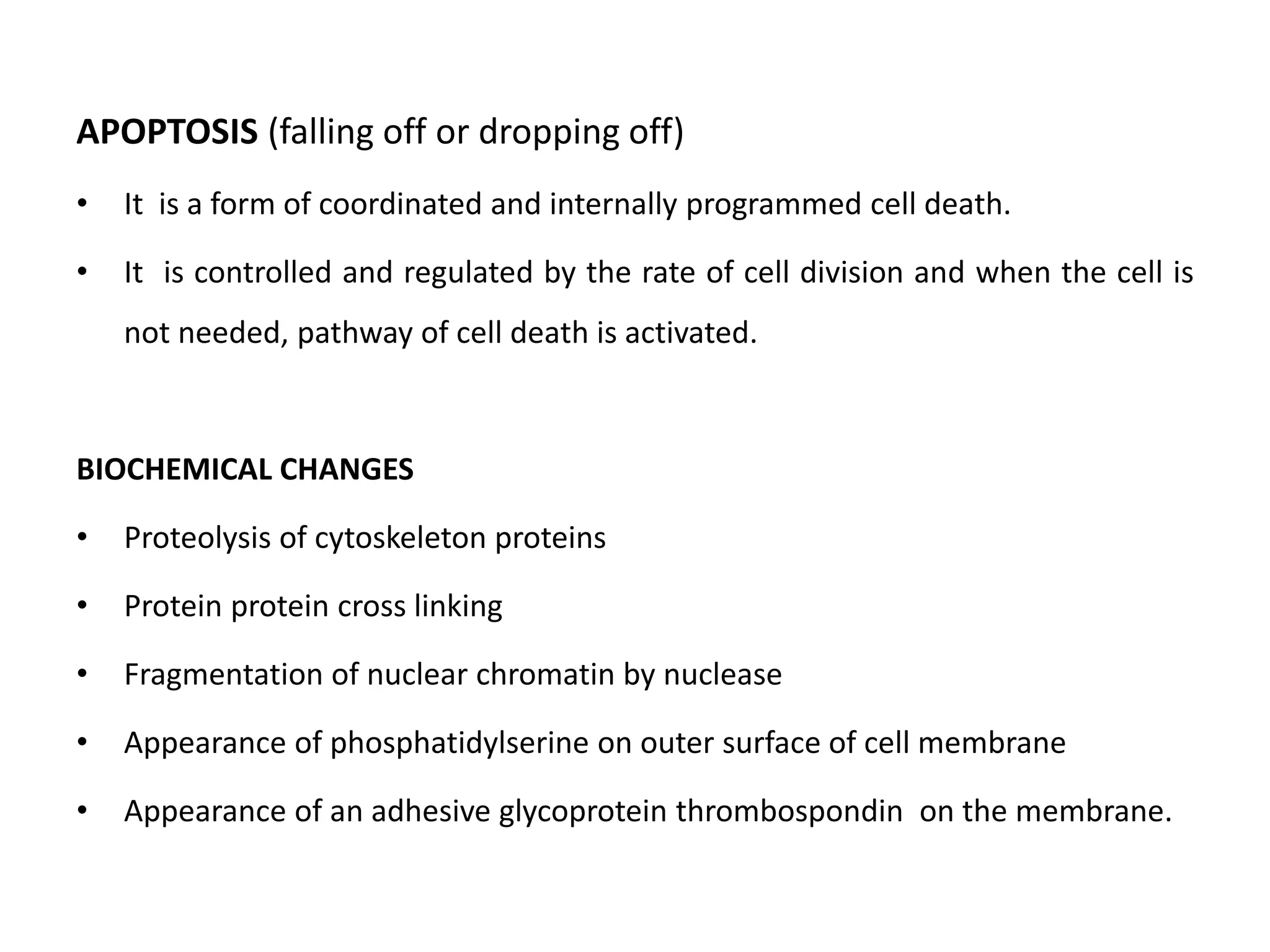
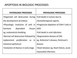
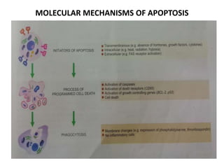
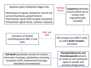
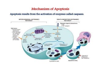
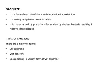
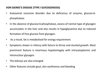
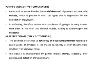
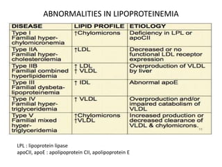
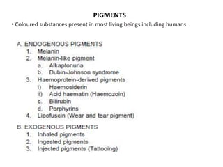
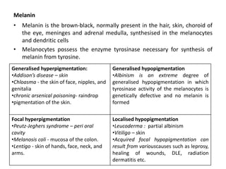
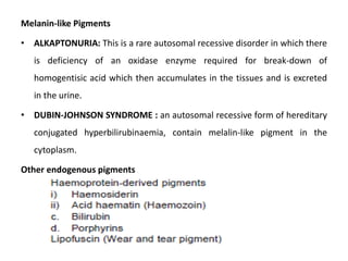
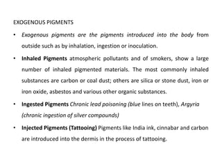
This document discusses various types of cell death and intracellular accumulations. It describes apoptosis as programmed cell death regulated by cell division. Biochemical changes in apoptosis include chromatin condensation and phosphatidylserine exposure. Apoptosis occurs physiologically in development and involution and pathologically in disease states. Gangrene is necrosis with bacterial infection and can be dry, wet, or gas gangrene. Intracellular accumulations include lipids, proteins, glycogen, and pigments. Glycogen storage diseases result from glycogen breakdown defects. Lipoprotein abnormalities can cause hyperlipidemias. Endogenous pigments include melanin while exogenous pigments come from external sources like pollution, diet, or tattoos.
Call Girls Ooty Just Call 9907093804 Top Class Call Girl Service Available
Apoptosis and Gangrene: A Guide to Programmed Cell Death and Tissue Necrosis
1. APOPTOSIS (falling off or dropping off)
• It is a form of coordinated and internally programmed cell death.
• It is controlled and regulated by the rate of cell division and when the cell is
not needed, pathway of cell death is activated.
BIOCHEMICAL CHANGES
• Proteolysis of cytoskeleton proteins
• Protein protein cross linking
• Fragmentation of nuclear chromatin by nuclease
• Appearance of phosphatidylserine on outer surface of cell membrane
• Appearance of an adhesive glycoprotein thrombospondin on the membrane.
2. APOPTOSIS IN BIOLOGIC PROCESSES
PHYSIOLOGIC PROCESSES PATHOLOGIC PROCESSES
•Organized cell destruction during
the development of embryo
•Physiologic involution of cells in
hormone dependant tissues
eg. endometrial shedding
•Normal cell destruction followed by
replacement proliferation as in
intestinal epithelium
•Involution of thymus in early stage.
•Cell death in tumors due to
chemotherapeutic agents.
•Progressive depletion of CD4+T cells in
AIDS
•Cell death in viral infections
•Degenerative disease of CNS
eg. Alzheimer’s disease, Parkinson’s
disease
•Heart diseases eg. Heart failure, acute
myocardial infarction.
3. TECHNIQUES TO IDENTIFY AND COUNT APOPTOTIC CELLS
• Staining of chromatin condensation (hematoxylin, acridine
orange)
• Flow cytometry to visualize rapid cell shrinkage.
• DNA changes detected by in situ techniques or by gel
electrophoresis.
• Annexin V as marker for apoptotic cell membrane having
phosphatidylserine on the cell exterior.
4. MORPHOLOGIC FEATURES
• Involvement of single cells or small cluster of cells in the background of
viable cells.
• The apoptotic cells are round to oval shrunken masses of intensely
eosinophilic cytoplasm.
• The chromatin condensed or fragmented.
• The cell membrane may show convulsions or projections on the cell
surface.
• Membrane bound near-spherical bodies called apoptotic bodies are
formed.
• Unlike necrosis, there is no acute inflammatory reaction around apoptosis.
• Phagocytosis of apoptotic bodies by macrophages takes place at varying
speed.
6. Apoptosis gets initiated by triggers like
•Withdrawal of signals needed for normal cell
survival (hormones, growth factors)
•Extracellular signals (FAS receptor activation)
•Intracellular signals (heat, radiation, hypoxia)
Etiologic
factors
Caspases(proteolytic
enzyme which act on
nucleus and
organelles) gets
activated
FAS receptor (on CD8+T cells),
so called death receptor,
activation
Activation of Growth
controlling genes (BCL-2 and
p53)
BAX and
BAD
Cell death (proteolytic actions on nucleus,
chromatin clumping, cytoskeleton clumping,
disruption of ER, mitochondrial damage,
disturbed cell membrane)
Phosphatidylserine and
thrombospondin present
on inside on cell membrane
appears outside and
promote phagocytosis
7.
8. GANGRENE
• It is a form of necrosis of tissue with superadded putrefaction.
• It is usually coagulative due to ischemia.
• It is characterized by primarily inflammation by virulent bacteria resulting in
massive tissue necrosis
TYPES OF GANGRENE
There are 2 main two forms:
• Dry gangrene
• Wet gangrene
• Gas gangrene ( a variant form of wet gangrene)
9. DRY GANGRENE
• It is usually initiated in one of toes(farthest from blood supply) containing
so little blood
• It spreads slowly upwards until it reaches a point where the blood supply is
adequate to keep the tissue viable.
• Causes of Dry gangrene includes Raynauds disease, trauma, ergot
poisoning.
MORPHOLOGY : The affected part is dry, shrunken and dark black.
•It is black due to liberation of Hb from hemolysed RBC which is acted upon by
hydrogen sulphide produced by the bacteria resulting in the formation of black
iron sulfide.
•The gangrenous part will be completely separated from the normal cell and
eventually fall off if it is not removed.
10. WET GANGRENE
• It occurs in naturally moist tissues and organs such as mouth, bowel, lung,
cervix, etc
• Diabetic foot is another example of wet gangrene due to high sugar
content in the necrosed tissue which favours the growth of bacteria.
• Bed sore is also an example.
• It usually develops rapidly due to blockage of venous, less commonly,
arterial blood flow from thrombosis or embolism.
• The affective part is stuffed with blood which favors the rapid growth of
putrefactive bacteria.
• The toxic products formed from bacteria causes septicemia and finally
death.
11. MORPHOLOGY :
•Grossly, affected part is soft, swollen, rotten and dark.
•The classic example is gangrene of bowel
•The part is stained due to same mechanism as in dry gangrene.
•Histologically, there is ulceration of mucosa and intense inflammatory
infiltration.
•Lumen of bowel contains mucus and blood.
GAS GANGRENE
•It is a special form of wet gangrene caused by gas forming clostridia (G+ve
anaerobic bacteria) which gain entry into the tissues through open
contaminated wounds, especially in muscles, or as a complication of operation
on colon which normally contains clostridia.
•Clostridia produce various toxins which produce necrosis and edema.
12. MORPHOLOGY
• Grossly, the affected area is swollen, edematous, painful and crepitant
due to accumulation of gas bubbles within the tissues.
• Thus the affected tissue becomes dark blue and foul smelling.
• Microscopically, the muscle fibre undergo coagulative necrosis with
liquefaction.
• Large number of G+ve bacilli can be identified.
• At periphery, a zone of leukocyte infiltration, edema and congestion are
found.
• Capillary and venous thrombi can be seen.
13. INTRACELLULAR ACCUMULATION
Intracellular accumulation of fats
– Fatty change (steatosis)
– Cholestrol deposits
– Stromal fatty infiltration
• Cholestrol deposits: In hypercholestremia, cholestrol and its esters
accumulates in macrophages, which turns to foam cells.
Eg: Fibrofatty plaques of atherosclerosis
Clusters of foam cells in tumor like masses called xanthomas and
xanthelasma.
14. Stromal fatty infiltration
• Deposition of mature adipose cells in the stromal connective tissue.
• It usually occurs in obese patients
• Affected organs are heart and pancreas.
• Heart can be the site for fatty change as well as stromal fatty infiltration.
• Usually, it does not produce any dysfunction.
INTRA CELLULAR ACCUMULATION OF PROTEINS
• In Proteinuria : Accumulation of plasma proteins(pink hyaline droplets)
due to excess reabsorption in PCT
• Russel’s bodies: hyaline changes in RER due to synthesized
immunoglobulins.
15. • In α1-antitrypsin deficiency, the cytoplasm of hepatocytes shows eosinophilic
globular deposits of a mutant protein.
• Mallory’s body or alcoholic hyalin in the hepatocytes is intracellular
accumulation of intermediate filaments of cytokeratin and appear as
amorphous pink masses.
INTRACELLULAR ACCUMULATION OF GLYCOGEN
• In diabetes mellitus, there is intracellular accumulation of glycogen because of
the impaired uptake of glucose.
• Glycogen deposits in diabetes mellitus are seen in epithelium of distal portion
of proximal convoluted tubule and descending loop of Henle, in the
hepatocytes, in beta cells of pancreatic islets, and in cardiac muscle cells
• In Glycogen storage disease also, excess glycogen will be accumulated.
16. GLYCOGEN STORAGE DISEASE (GLUCOGENOSIS)
• These are a group of inherited disorders in which there is defective
glucose metabolism resulting in excessive intracellular accumulation of
glycogen in various tissues
• Hepatic forms: von Gierke’s disease or type I glycogenosis) or due to lack
of hepatic enzymes necessary for breakdown of glycogen into glucose (e.g.
type VI glycogenosis).
• Myopathic forms :McArdle’s disease or type V glycogenosis
• Other forms: Pompe’s disease
17. VON GIERKE’S DISEASE (TYPE I GLYCOGENOSIS)
• Autosomal recessive disorder due to deficiency of enzyme, glucose-6-
phosphatase.
• In the absence of glucose-6-phosphatase, excess of normal type of glycogen
accumulates in the liver and also results in hypoglycaemia due to reduced
formation of free glucose from glycogen.
• As a result, fat is metabolised for energy requirement.
• Symptoms shown in infancy with failure to thrive and stunted growth. Most
prominent feature is enormous hepatomegaly with intracytoplasmic and
intranuclear glycogen.
• The kidneys are also enlarged.
• Other features include gout, skin xanthomas and bleeding
18. POMPE’S DISEASE (TYPE II GLYCOGENOSIS)
• Autosomal recessive disorder due to deficiency of a lysosomal enzyme, acid
maltase, which is present in most cell types and is responsible for the
degradation of glycogen.
• Its deficiency, therefore, results in accumulation of glycogen in many tissues,
most often in the heart and skeletal muscle, leading to cardiomegaly and
hypotonia
McARDLE’S DISEASE (TYPE V GLYCOGENOSIS)
• The condition occurs due to deficiency of muscle phosphorylase resulting in
accumulation of glycogen in the muscle (deficiency of liver phosphorylase
results in type VI glycogenosis).
• The disease is characterised by painful muscle cramps, especially after
exercise, and detection of myoglobinuria
21. Melanin
• Melanin is the brown-black, normally present in the hair, skin, choroid of
the eye, meninges and adrenal medulla, synthesised in the melanocytes
and dendritic cells
• Melanocytes possess the enzyme tyrosinase necessary for synthesis of
melanin from tyrosine.
Generalised hyperpigmentation:
•Addison’s disease – skin
•Chloasma - the skin of face, nipples, and
genitalia
•chronic arsenical poisoning- raindrop
•pigmentation of the skin.
Generalised hypopigmentation
•Albinism is an extreme degree of
generalised hypopigmentation in which
tyrosinase activity of the melanocytes is
genetically defective and no melanin is
formed
Focal hyperpigmentation
•Peutz-Jeghers syndrome – peri oral
cavity
•Melanosis coli - mucosa of the colon.
•Lentigo - skin of hands, face, neck, and
arms.
Localised hypopigmentation
•Leucoderma : partial albinism
•Vitiligo – skin
•Acquired focal hypopigmentation can
result from variouscauses such as leprosy,
healing of wounds, DLE, radiation
dermatitis etc.
22. Melanin-like Pigments
• ALKAPTONURIA: This is a rare autosomal recessive disorder in which there
is deficiency of an oxidase enzyme required for break-down of
homogentisic acid which then accumulates in the tissues and is excreted
in the urine.
• DUBIN-JOHNSON SYNDROME : an autosomal recessive form of hereditary
conjugated hyperbilirubinaemia, contain melalin-like pigment in the
cytoplasm.
Other endogenous pigments
23. EXOGENOUS PIGMENTS
• Exogenous pigments are the pigments introduced into the body from
outside such as by inhalation, ingestion or inoculation.
• Inhaled Pigments atmospheric pollutants and of smokers, show a large
number of inhaled pigmented materials. The most commonly inhaled
substances are carbon or coal dust; others are silica or stone dust, iron or
iron oxide, asbestos and various other organic substances.
• Ingested Pigments Chronic lead poisoning (blue lines on teeth), Argyria
(chronic ingestion of silver compounds)
• Injected Pigments (Tattooing) Pigments like India ink, cinnabar and carbon
are introduced into the dermis in the process of tattooing.