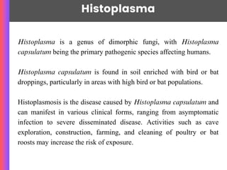
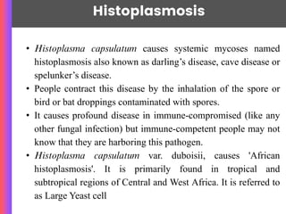
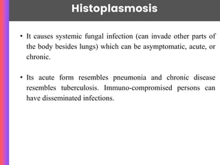
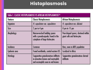
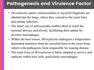
Histoplasma
~Nawang Sherpa
Histoplasma is a genus of dimorphic fungi, with Histoplasma
capsulatum being the primary pathogenic species affecting humans.
Histoplasma capsulatum is found in soil enriched with bird or bat
droppings, particularly in areas with high bird or bat populations.
Histoplasmosis is the disease caused by Histoplasma capsulatum and
can manifest in various clinical forms, ranging from asymptomatic
infection to severe disseminated disease. Activities such as cave
exploration, construction, farming, and cleaning of poultry or bat
roosts may increase the risk of exposure.
Histoplasma
Morphology
Histoplasma capsulatum, a mold with
septate hyphae bearing microconidia and
macroconidia.
These structures are typically observed in
cultures grown on specific fungal media,
such as Sabouraud agar.
Microconidia: Small,
unicellular conidia that are
round, elliptical, or pyriform
(pear-shaped).
Macroconidia: Large, multi-
septate conidia that are club- or
spindle-shaped.
TAXONOMY
Kingdom: Fungi
Phylum: Ascomycota
Class: Eurotiomycetes
Order: Onygenales
Family: Ajellomycetaceae
Genus: Histoplasma
Species: capsulatum
Histoplasmosis
~Nawang Sherpa
• Histoplasma capsulatum causes systemic mycoses named
histoplasmosis also known as darling’s disease, cave disease or
spelunker’s disease.
• People contract this disease by the inhalation of the spore or
bird or bat droppings contaminated with spores.
• It causes profound disease in immune-compromised (like any
other fungal infection) but immune-competent people may not
know that they are harboring this pathogen.
• Histoplasma capsulatum var. duboisii, causes 'African
histoplasmosis'. It is primarily found in tropical and
subtropical regions of Central and West Africa. It is referred to
as Large Yeast cell
Histoplasmosis
~Nawang Sherpa
• It causes systemic fungal infection (can invade other parts of
the body besides lungs) which can be asymptomatic, acute, or
chronic.
• Its acute form resembles pneumonia and chronic disease
resembles tuberculosis. Immuno-compromised persons can
have disseminated infections.
Histoplasmosis
Pathogenesis and Virulence Factor
• Histoplasma spores (microconidia) or mycelial fragments are
inhaled into the lungs, where they convert to the yeast form
and initiate infection.
• The small size of microconidia enables them to reach the
terminal airways and alveoli, facilitating their uptake by
alveolar macrophages.
• Within the host tissues, Histoplasma undergoes a temperature-
dependent transition from the mycelial form to the yeast form,
which is the pathogenic form responsible for causing disease.
• The yeast form of Histoplasma is better adapted to survive and
replicate within host cells, particularly macrophages.
Pathogenesis and Virulence factor
Histoplasma has several mechanisms to evade host
immune responses, including:
• Inhibition of phagolysosome fusion within
macrophages, allowing the fungus to survive and
replicate within phagosomes.
• Modulation of host immune signaling pathways to
suppress pro-inflammatory responses and promote
immune evasion.
• Resistance to oxidative stress and other host
antimicrobial mechanisms.
Pathogenesis and Virulence factor
Most infections of classical histoplasmosis are asymptomatic and
heal spontaneously, leaving behind an area of miliary calcification
( Case 1) .
Some infected individuals develop pulmonary disease which
resembles tuberculosis. Disseminated histoplasmosis develops
only in a minority of infected individuals.
Infection of the reticuloendothelial system manifests as
lymphadenopathy, hepatosplenomegaly, fever and anemia with a
high rate of fatality. Granulomatous and ulcerative lesions may
develop on the skin and mucous membranes.
Pathogenesis and Virulence factor
African histoplasmosis involves mainly the skin, subcutaneous
tissues and bones. The lungs are not commonly affected and
disseminated disease is infrequent.
Histoplasma capsulatum var. duboisii infection can also involve
the lymph nodes, leading to lymphadenitis. Enlarged, tender
lymph nodes may be palpable in affected individuals, particularly
in the cervical or inguinal regions.
Clinical manifestation (Histoplasmosis
Histoplasma capsulatum, can manifest in various clinical forms,
each with distinct presentations and outcomes:
Acute Pulmonary Histoplasmosis:
• Acute pulmonary histoplasmosis typically presents as a self-
limited flu-like illness with symptoms appearing 3 to 17 days
after exposure to the fungus. Symptoms may include fever,
chills, headache, non-productive cough, chest discomfort,
myalgias, and fatigue.
Clinical manifestation (Histoplasmosis
Chronic Pulmonary Histoplasmosis:
• Chronic pulmonary histoplasmosis primarily affects older
individuals with underlying lung disease, such as chronic
obstructive pulmonary disease (COPD) or emphysema.
Patients may present with chronic cough, dyspnea, fatigue,
weight loss, and occasional hemoptysis.
Clinical manifestation (Histoplasmosis
Disseminated Histoplasmosis:
• Disseminated histoplasmosis occurs when the infection
spreads from the lungs to other organs via the bloodstream or
lymphatics.
• It typically affects immunocompromised individuals, such as
those with HIV/AIDS or organ transplant recipients. Clinical
manifestations may include fever, weight loss,
hepatosplenomegaly, lymphadenopathy, anemia,
thrombocytopenia, and involvement of the bone marrow,
CNS, skin, and adrenal glands.
Clinical manifestation (Histoplasmosis
Ocular Histoplasmosis:
• Ocular histoplasmosis refers to ocular complications
associated with histoplasmosis, including chorioretinitis and
ocular neovascularization.
• Chorioretinitis: Characterized by the presence of punched-
out chorioretinal scars (histo spots) in the posterior pole of the
retina, often without associated inflammation.
• Ocular Neovascularization: Can lead to vision loss due to
the development of choroidal neovascular membranes and
subsequent macular scarring, a condition known as presumed
ocular histoplasmosis syndrome (POHS).
Clinical manifestation (African histop..)
Cutaneous and Subcutaneous Lesions:
• African histoplasmosis often presents with localized
cutaneous or subcutaneous lesions, which may appear as
nodules, ulcers, abscesses, or plaques.
• These lesions can vary in size, shape, and appearance and
may be single or multiple. They are typically painless but
may become tender or ulcerate over time.
Lymphadenitis:
• Histoplasma duboisii infection can also involve the lymph
nodes, leading to lymphadenitis. Enlarged, firm, and non-
tender lymph nodes may be palpable in affected individuals,
particularly in the cervical or inguinal regions.
Lab Diagnosis ( Histoplasmosis
Sample: Bone marrow, peripheral blood, and sputum (organisms
are less likely to be detected in the direct microscopic
examination of respiratory tract specimens).
• Microscopy and Staining: In the Wright or Giemsa stained
specimens, Histoplasma capsulatum is found
intracellularly within the cytoplasm of endothelial or
mononuclear cells.
⚬ H capsulatum yeasts: Small round or oval cells,
measuring 2-5 μm in diameter. They require
differentiation from the amastigotes of Leishmania
donovani which can also be found in mononuclear
cells.
Lab Diagnosis
• Culture*: Specimen must be cultured as soon as possible to
ensure optimal recovery of H. capsulatum and other
dimorphic fungi. It grows well in ordinary fungal culture
media but it may be overgrown by bacteria or rapidly
growing molds. It is a slow-growing mold at 25°C to 30°C
and commonly requires 2-4 weeks or more for colonies to
appear. The organism may, however, be recovered within 5
days or less if many yeast cells are present in the clinical
specimen.
• Colony characteristics: White, fluffy mold that turns brown to
buff with age. The organism may also produce wrinkle,
moist, heaped, yeast-like colonies that are soft and creamy
when grown at 37°C in certain media.
Lab Diagnosis
• Serodiagnosis
Antibodies formed during the course of systemic mycoses
increase in titre in progressive disease. Hence, serological tests
such as latex agglutination, complement fixation and
precipitation tests are useful in the diagnosis of histoplasmosis.
Titre is considered positive at reciprocal dilutions greater than 1
:8. A titre with dilutions greater than 1 :32 suggests active
histoplasmosis infection.
Antigens can be detected in urine and serum. These tests are
useful in immunocompromised individuals in whom antibody
formation is impaired
Lab Diagnosis
• Skin tests:
Delayed hypersensitivity develops following infection. It can be
demonstrated by skin testing with 'histoplasmin', which is
analogous to the tuberculin test for tuberculosis.
In histoplasmosis, skin tests are more specific than serological
methods.
Treatment
Antifungal therapy is the mainstay of treatment for
histoplasmosis, and the choice of antifungal agent and duration
of therapy may vary based on these factors.
Itraconazole, Fluconazole, Liposomal Amphotericin B,
Medically Important Histoplasma species .pptx

Medically Important Histoplasma species .pptx

  • 1.
  • 2.
    Histoplasma is agenus of dimorphic fungi, with Histoplasma capsulatum being the primary pathogenic species affecting humans. Histoplasma capsulatum is found in soil enriched with bird or bat droppings, particularly in areas with high bird or bat populations. Histoplasmosis is the disease caused by Histoplasma capsulatum and can manifest in various clinical forms, ranging from asymptomatic infection to severe disseminated disease. Activities such as cave exploration, construction, farming, and cleaning of poultry or bat roosts may increase the risk of exposure. Histoplasma
  • 3.
    Morphology Histoplasma capsulatum, amold with septate hyphae bearing microconidia and macroconidia. These structures are typically observed in cultures grown on specific fungal media, such as Sabouraud agar. Microconidia: Small, unicellular conidia that are round, elliptical, or pyriform (pear-shaped). Macroconidia: Large, multi- septate conidia that are club- or spindle-shaped.
  • 4.
    TAXONOMY Kingdom: Fungi Phylum: Ascomycota Class:Eurotiomycetes Order: Onygenales Family: Ajellomycetaceae Genus: Histoplasma Species: capsulatum
  • 5.
    Histoplasmosis ~Nawang Sherpa • Histoplasmacapsulatum causes systemic mycoses named histoplasmosis also known as darling’s disease, cave disease or spelunker’s disease. • People contract this disease by the inhalation of the spore or bird or bat droppings contaminated with spores. • It causes profound disease in immune-compromised (like any other fungal infection) but immune-competent people may not know that they are harboring this pathogen. • Histoplasma capsulatum var. duboisii, causes 'African histoplasmosis'. It is primarily found in tropical and subtropical regions of Central and West Africa. It is referred to as Large Yeast cell
  • 6.
    Histoplasmosis ~Nawang Sherpa • Itcauses systemic fungal infection (can invade other parts of the body besides lungs) which can be asymptomatic, acute, or chronic. • Its acute form resembles pneumonia and chronic disease resembles tuberculosis. Immuno-compromised persons can have disseminated infections.
  • 7.
  • 8.
    Pathogenesis and VirulenceFactor • Histoplasma spores (microconidia) or mycelial fragments are inhaled into the lungs, where they convert to the yeast form and initiate infection. • The small size of microconidia enables them to reach the terminal airways and alveoli, facilitating their uptake by alveolar macrophages. • Within the host tissues, Histoplasma undergoes a temperature- dependent transition from the mycelial form to the yeast form, which is the pathogenic form responsible for causing disease. • The yeast form of Histoplasma is better adapted to survive and replicate within host cells, particularly macrophages.
  • 9.
    Pathogenesis and Virulencefactor Histoplasma has several mechanisms to evade host immune responses, including: • Inhibition of phagolysosome fusion within macrophages, allowing the fungus to survive and replicate within phagosomes. • Modulation of host immune signaling pathways to suppress pro-inflammatory responses and promote immune evasion. • Resistance to oxidative stress and other host antimicrobial mechanisms.
  • 10.
    Pathogenesis and Virulencefactor Most infections of classical histoplasmosis are asymptomatic and heal spontaneously, leaving behind an area of miliary calcification ( Case 1) . Some infected individuals develop pulmonary disease which resembles tuberculosis. Disseminated histoplasmosis develops only in a minority of infected individuals. Infection of the reticuloendothelial system manifests as lymphadenopathy, hepatosplenomegaly, fever and anemia with a high rate of fatality. Granulomatous and ulcerative lesions may develop on the skin and mucous membranes.
  • 11.
    Pathogenesis and Virulencefactor African histoplasmosis involves mainly the skin, subcutaneous tissues and bones. The lungs are not commonly affected and disseminated disease is infrequent. Histoplasma capsulatum var. duboisii infection can also involve the lymph nodes, leading to lymphadenitis. Enlarged, tender lymph nodes may be palpable in affected individuals, particularly in the cervical or inguinal regions.
  • 12.
    Clinical manifestation (Histoplasmosis Histoplasmacapsulatum, can manifest in various clinical forms, each with distinct presentations and outcomes: Acute Pulmonary Histoplasmosis: • Acute pulmonary histoplasmosis typically presents as a self- limited flu-like illness with symptoms appearing 3 to 17 days after exposure to the fungus. Symptoms may include fever, chills, headache, non-productive cough, chest discomfort, myalgias, and fatigue.
  • 13.
    Clinical manifestation (Histoplasmosis ChronicPulmonary Histoplasmosis: • Chronic pulmonary histoplasmosis primarily affects older individuals with underlying lung disease, such as chronic obstructive pulmonary disease (COPD) or emphysema. Patients may present with chronic cough, dyspnea, fatigue, weight loss, and occasional hemoptysis.
  • 14.
    Clinical manifestation (Histoplasmosis DisseminatedHistoplasmosis: • Disseminated histoplasmosis occurs when the infection spreads from the lungs to other organs via the bloodstream or lymphatics. • It typically affects immunocompromised individuals, such as those with HIV/AIDS or organ transplant recipients. Clinical manifestations may include fever, weight loss, hepatosplenomegaly, lymphadenopathy, anemia, thrombocytopenia, and involvement of the bone marrow, CNS, skin, and adrenal glands.
  • 15.
    Clinical manifestation (Histoplasmosis OcularHistoplasmosis: • Ocular histoplasmosis refers to ocular complications associated with histoplasmosis, including chorioretinitis and ocular neovascularization. • Chorioretinitis: Characterized by the presence of punched- out chorioretinal scars (histo spots) in the posterior pole of the retina, often without associated inflammation. • Ocular Neovascularization: Can lead to vision loss due to the development of choroidal neovascular membranes and subsequent macular scarring, a condition known as presumed ocular histoplasmosis syndrome (POHS).
  • 16.
    Clinical manifestation (Africanhistop..) Cutaneous and Subcutaneous Lesions: • African histoplasmosis often presents with localized cutaneous or subcutaneous lesions, which may appear as nodules, ulcers, abscesses, or plaques. • These lesions can vary in size, shape, and appearance and may be single or multiple. They are typically painless but may become tender or ulcerate over time. Lymphadenitis: • Histoplasma duboisii infection can also involve the lymph nodes, leading to lymphadenitis. Enlarged, firm, and non- tender lymph nodes may be palpable in affected individuals, particularly in the cervical or inguinal regions.
  • 17.
    Lab Diagnosis (Histoplasmosis Sample: Bone marrow, peripheral blood, and sputum (organisms are less likely to be detected in the direct microscopic examination of respiratory tract specimens). • Microscopy and Staining: In the Wright or Giemsa stained specimens, Histoplasma capsulatum is found intracellularly within the cytoplasm of endothelial or mononuclear cells. ⚬ H capsulatum yeasts: Small round or oval cells, measuring 2-5 μm in diameter. They require differentiation from the amastigotes of Leishmania donovani which can also be found in mononuclear cells.
  • 18.
    Lab Diagnosis • Culture*:Specimen must be cultured as soon as possible to ensure optimal recovery of H. capsulatum and other dimorphic fungi. It grows well in ordinary fungal culture media but it may be overgrown by bacteria or rapidly growing molds. It is a slow-growing mold at 25°C to 30°C and commonly requires 2-4 weeks or more for colonies to appear. The organism may, however, be recovered within 5 days or less if many yeast cells are present in the clinical specimen. • Colony characteristics: White, fluffy mold that turns brown to buff with age. The organism may also produce wrinkle, moist, heaped, yeast-like colonies that are soft and creamy when grown at 37°C in certain media.
  • 19.
    Lab Diagnosis • Serodiagnosis Antibodiesformed during the course of systemic mycoses increase in titre in progressive disease. Hence, serological tests such as latex agglutination, complement fixation and precipitation tests are useful in the diagnosis of histoplasmosis. Titre is considered positive at reciprocal dilutions greater than 1 :8. A titre with dilutions greater than 1 :32 suggests active histoplasmosis infection. Antigens can be detected in urine and serum. These tests are useful in immunocompromised individuals in whom antibody formation is impaired
  • 20.
    Lab Diagnosis • Skintests: Delayed hypersensitivity develops following infection. It can be demonstrated by skin testing with 'histoplasmin', which is analogous to the tuberculin test for tuberculosis. In histoplasmosis, skin tests are more specific than serological methods.
  • 21.
    Treatment Antifungal therapy isthe mainstay of treatment for histoplasmosis, and the choice of antifungal agent and duration of therapy may vary based on these factors. Itraconazole, Fluconazole, Liposomal Amphotericin B,