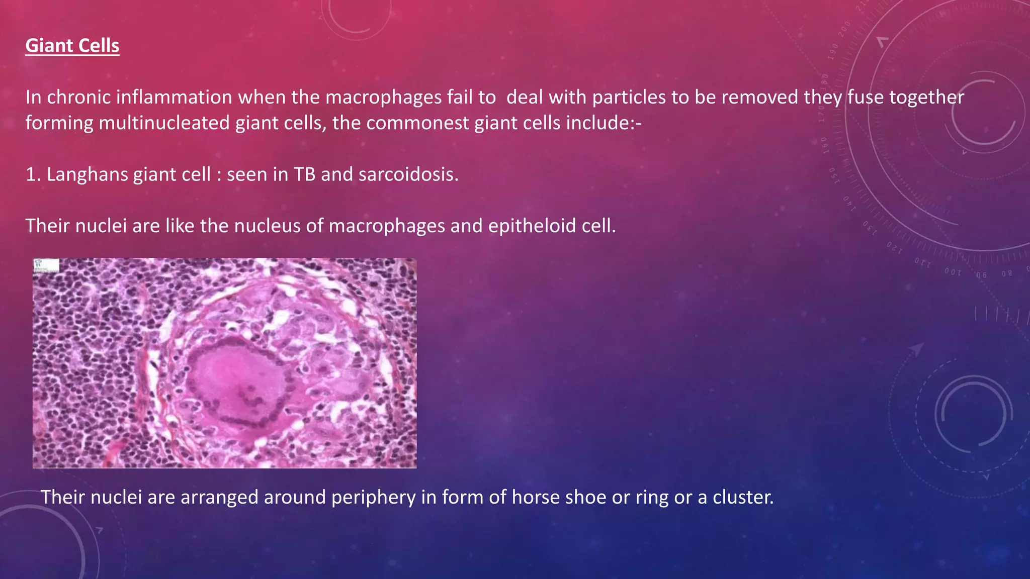
CHRONIC INFLAMMATION
DEPARTMENT OF PERIODONTICS
RAMA DENTAL COLLEGE HOSPITAL AND RESEARCH
CENTER
CONTENTS
Definition
Causes
General
Features
Types Cells Involved
Pathophysiology
Risks Factors
Symptoms Tests/Markers
Granulomatous
Inflammation
Inflammation &
Periodontology
Summary Conclusion
‘Inflammation’ is an observable
alteration in tissues associated
with changes in
vascular permeability and
dilation, often with the
infiltration of leukocytes into
affected tissues. (Carranza’
10th edition)
Chronic inflammation can be
considered to be inflammation
of prolonged duration in
which active inflammation,
tissue injury, and healing
proceed simultaneously.
(Robbins’s)
Chronic inflammation is defined
as prolonged process in which
tissue destruction
and inflammation occur at the
same time. (Harsh Mohan)
DEFINITION
CAUSES
Chronic inflammation
following acute
inflammation
• When the tissue destruction is
extensive , or the bacteria
present at the site of acute
inflammation.
• E.g. Osteomyelitis, pneumonia,
lung abscess.
Recurrent attacks of acute
inflammation
• When repeated episodes of
acute inflammation occur it
culminate in chronicity of the
process.
• E.g. in recurrent urinary tract
infection, repeated acute
infection.
Chronic inflammation
starting de novo
• When the infection with
organism of low pathogenicity
has chronic form of beginning.
• E.g. infection with
M.tuberculosis.
GENERAL FEATURES
1. Mononuclear cell infiltration :
* Chronic inflammatory lesions are infiltrated by mononuclear inflammatory cells like phagocytes and lymphoid cells.
* Phagocytes are represented by circulating monocytes, tissue macrophages, epithelioid cells and sometimes multinucleated
giant cells.
* Macrophages comprise of most important cells in chronic inflammation.
These may appear at the site of chronic inflammation from ;
Chemotactic factors for macrophages.
Local proliferation of macrophages.
Longer survival of macrophages at the site of inflammation.
The blood monocytes on reaching the extravascular space transforms into tissue macrophages. On activation they release acid
and neutral proteases, oxygen derived reactive metabolites and cytokines. These bring about tissue destruction,
neovascularization and fibrosis.
Other chronic inflammatory cells include lymphocytes, plasma cells, eosinophils and mast cells. In chronic inflammation,
lymphocytes and macrophages influence each other and release mediators of inflammation.
2.Tissue destruction or necrosis : It is very common and is brought about by activated macrophages.
3. Proliferative changes : As a result of necrosis, proliferation of small blood vessels and fibroblasts stimulated resulting in
formation of inflammatory granulation tissue. Eventually, healing by fibrosis and collagen laying takes place.
TYPES
1. Non-specific
2. Specific
Non-specific : When the irritant substance produces non specific inflammatory reaction with formation of granulation
tissue and healing by fibrosis.
E.g. Chronic osteomyelitis, chronic ulcer.
Specific : When the injurious agent causes a characteristic histological response.
E.g. tuberculosis, leprosy, and syphilis.
Descriptive Classification :
•Chronic non-specific inflammation : Characterized by non-specific inflammatory cell infiltration
eg. Chronic osteomyelitis, lung abcess.
•Chronic granulomatous inflammation : Characterized by formation of granulomas
eg. Tuberculosis, leprosy
CELLS INVOLVED
Macrophages Derived from blood monocytes.
Functions :-
1.Phagocytosis and destruction of debris & bacteria.
2.Processing and presentation of antigen to immune system
3.Control of other cells by cytokine release.
Secretes :-
• Acid and neutral proteases
• Complement components ( C1 to C5), coagulation factor V and VIII and tissue factor.
• Reactive oxygen species and Nitric oxide.
• AA metabolites
• Cytokines – IL1, TNF, Growth factors
Secretory products of macrophages
•ENZYMES
~ Neutral proteases- for example, collagenase, elastase, plasminogen activator, angiotensin converting
enzymne, enzymes denaturating proteoglycans and myelin.
~ Acid hydrolases- for example, phosphatases, sulphatases, proteases, ribonucleases.
~ Lysozyme
•PLASMA PROTEINS
~ Complement components (eg. C1, C2, C3, C4, C5; Properdin, Factors B, D)
~ Coagulation factors (eg. Factor V, VII, tissue factor)
•Oxygen metabolites (eg.Superoxides, hydrogen peroxide, hydroxyl radicals)
•Cytokines (IL-1, INF, IL-8)
•Growth Factors ( PDGF, EGF,FGF,TGF-beta)
Mononuclear Phagocyte System (MPS)
The name "mononuclear phagocyte system" was proposed in 1969 to describe the group of highly phagocytic mononuclear cells and their precursors
which are widely distributed in the body, related by morphology and function, and which originate from the bone marrow.
Kupffer cells (liver)
Sinus Histiocytes (spleen)
Microglia (CNS)
Alveolar Macrophages (lung)
•Mononuclear phagocytic cells are derived from precursor cells in the bone marrow. These precursors develop into monocytes and dendritic cells,
phagocytic cells that are released into the bloodstream. Some monocytes and dendritic cells remain in the general blood circulation, but most of them
enter body tissues.
•In tissues, monocytes develop into much larger phagocytic cells known as macrophages. The great majority of macrophages remain as stationary cells
within tissue, where they filter out and destroy foreign particles. Some of them break away, however, and wander through the circulation and within
the intercellular spaces.
•Each phagocytic cell can engulf and destroy microorganisms, cells, and even tiny fragments of foreign objects. Several mobile macrophages can
surround larger foreign objects and coalesce into a single phagocytic cell. Thus, by their phagocytosis of foreign substances, macrophages, monocytes,
and dendritic cells form an important first line of defense against harmful particles that have reached the body’s interior.
•Cells of the mononuclear phagocyte system also participate in immune reactions, in which a complex set of events is targeted at a specific foreign
substance. Through phagocytosis, macrophages reveal antigens (surface molecules) on foreign substances. Antigens stimulate immune reactions that
are directed by white blood cells known as lymphocytes .B lymphocytes (or B cells) synthesize and secrete antibodies with the help of T lymphocytes
(or T cells). The production of antibodies, in turn, greatly stimulates the phagocytic activity of the cells of the mononuclear phagocyte system.
Lymphocytes
Sometimes called ‘chronic inflammatory cells’
Functions:-
Mainly immunological.
•B lymphocytes differentiate to produce antibodies.
•T lymphocytes involved in control & some cytotoxic functions.
•Three main types of lymphocytes are distinguishable :
1.T cell
2.B cell
3.Natural killer (NK) cells
•T CELL
Recognize diverse antigens through low-affinity transmembrane complex , the T-cell antigen receptor (TCR).
Further 2 types :
CD4 - initiate and help immune responses by providing proliferation and differentiation signals
CD8 – predominantly cytotoxic , involved in controlling intracellular antigens (bacteria, fungi, viruses)
•B CELL
Help control extracellular antigens through high-affinity antigen receptor, the B-cell antigen receptor (BCR).
High-affinity enables B cell to bind and ingest antigen without antigen presentation.
Before antigen exposure B-cell express IgM as part of BCR, after antigen exposure , some B cell differentiate to form Plasma cells.
Plasma cell cause production and secretion of antibodies of IgM isotype and other in presence of T cell form memory B cell.
•NATURAL KILLER (NK) CELLS
Kill certain tumour and virally infected cells.
Giant Cells
In chronic inflammation when the macrophages fail to deal with particles to be removed they fuse together
forming multinucleated giant cells, the commonest giant cells include:-
1. Langhans giant cell : seen in TB and sarcoidosis.
Their nuclei are like the nucleus of macrophages and epitheloid cell.
Their nuclei are arranged around periphery in form of horse shoe or ring or a cluster.
2.Foreign body giant cells : may contain up to
100 nuclei which are scattered throughout the cytoplasm
(seen in leprosy).
3. Touton giant cells: These are multinucleated cells.
They have vacuolated cytoplasm due to lipid content e.g. xanthoma.
Three main ideas have been suggested for inflammatory giant cell formation.
• First, it was proposed that fusion may be an immune-mediated phenomenon, giant cell production being stimulated by lymphokines.
• The second suggestion is that fusion occurs between "young" macrophages and "older" cells, the latter having existed for some time in
the granulomatous environment acquiring chromosomal abnormalities and changes in the macrophage surface.
• The third proposal is that giant cells form as a result of simultaneous attempted phagocytosis,' during which two macrophages attempt
to ingest the same particle. The endosome margins of one macrophage, instead of fusing together around the particle, fuse with the
endosome margins of a second macrophage, resulting in fusion of the two cells.
Fig: Accumulation of Macrophages in chronic
inflammation.
Plasma cells:
•Differentiated antibody-producing B lymphocytes.
Eosinophils:
•Parasitic infestations, Allergic reactions particularly immune reactions mediated by IgE, some tumours.
•Their emigration is driven by adhesion molecules similar to neutrophils and specific chemokines (e.g. Eotaxin)
derived from leukocytes or epithelial cells.
•Eosinophil-specific granules contain major basic protein (MBP), a highly charged cationic protein toxic to
parasites (but also cause epithelial cell lysis).
Mast cells:
•Widely distributed in body.
•Participate in both acute and chronic inflammation.
•Mast cells are “armed” with IgE, for when triggered it releases histamines and AA metabolites (that cause early
vascular changes in acute inflammation).
•Also affect some cytokines such as TNF to participate in chronic inflammation.
OTHER CELLS
PATHOPHYSIOLOGY
•Most of the features of acute inflammation continue as the inflammation becomes chronic, including the
expansion of blood vessels (vasodilation), increase in blood flow, capillary permeability and migration of
neutrophils into the infected tissue through the capillary wall (diapedesis).
• However, the composition of the white blood cells changes soon and the macrophages and lymphocytes
begin to replace short-lived neutrophils.
•Thus the hallmarks of chronic inflammation are the infiltration of the primary inflammatory cells such as
macrophages, lymphocytes, and plasma cells in the tissue site, producing inflammatory cytokines, growth
factors, enzymes and hence contributing to the progression of tissue damage and secondary repair including
fibrosis and granuloma formation, etc.
•In response to foreign or self-antigens, the tissue immune cells such as macrophages and dendritic cells
release cytokines such as IL-1 and TNF-α. These cytokines induce the injury-site-endothelial cells to release
Selectins and Integrins which stimulate chemotaxis and diapedesis of the circulating leukocytes.
•In addition to the recruitment of leukocytes, the tissue macrophages, and dendritic cells also play a role in
the clearing of the antigen by phagocytosis, the release of cytokines and serving as antigen-presenting-cells to
lymphocytes.
•Once the circulating leukocytes enter the local injury site, they are activated by various cytokines and
chemokines secreted by the macrophages and dendritic cells.
•On activation, the leukocytes further release cytokines and mediators of inflammation.
•Neutrophils are the initial cells and most predominant in the acute phase of inflammation.
•Neutrophils contain granules rich with lysozyme, matrix metalloproteinases, myeloperoxidase
which are released on the foreign or self-antigen leading to its destruction.
•Neutrophils also destroy the antigen by phagocytosis, the release of reactive oxygen species
and cytokines such as IL-1, IL-6, and TNF-α.
•Lymphocytes including T-lymphocytes and B-lymphocytes are the next line of defense, and they
play a crucial role in mediating inflammation by several complex mechanisms including secreting
of cytokines, costimulation of lymphocytes, and production of antibodies and immune
complexes.
•Circulating platelets can also play a role in inflammation by platelet aggregation, thrombus
formation and degranulation releasing chemokines and inflammatory mediators.
RISK FACTORS ASSOCIATED WITH CHRONIC INFLAMMATION
Several risk factors promote a low-level inflammatory response.
These include:
1. Age : Increasing age is positively correlated with elevated levels of several inflammatory molecules. The age-associated
increase in inflammatory molecules may be due to mitochondrial dysfunction or free radical accumulation over time and other
age-related factors like an increase in visceral body fat.
2. Obesity: Many studies have reported that fat tissue is an endocrine organ, secreting multiple adipokines and other
inflammatory mediators. Some reports show that the body mass index of an individual is proportional to the amount of pro-
inflammatory cytokines secreted. Metabolic syndrome typifies this well.
3. Diet : Diet rich in saturated fat, trans-fats, or refined sugar is associated with higher production of pro-inflammatory
molecules, especially in individuals with diabetes or overweight individuals.
4. Smoking : Cigarette smoking is associated with lowering the production of anti-inflammatory molecules and inducing
inflammation.
5. Low Sex Hormones : Studies show that sex hormones like testosterone and estrogen can suppress the production and
secretion of several pro-inflammatory markers and it has been observed that maintaining sex hormone levels reduces the risk
of several inflammatory diseases.
6. Stress and Sleep Disorders : Both physical and emotional stress is associated with inflammatory cytokine release. Stress
can also cause sleep disorders. Since individuals with irregular sleep schedules are more likely to have chronic inflammation
than consistent sleepers, sleep disorders are also considered as one of the independent risk factors for chronic inflammation.
Some of the common signs and symptoms that develop during chronic
inflammation are listed below.
•Body pain, arthralgia, myalgia
•Chronic fatigue and insomnia
•Depression, anxiety and mood disorders
•Gastrointestinal complications like constipation, diarrhea, and acid reflux
•Weight gain or weight loss
•Frequent infections
SYMPTOMS OF CHRONIC INFLAMMATION
TESTS/MARKERS FOR CHRONIC INFLAMMATION
Unfortunately, there are no highly effective laboratory measures to assess patients for chronic
inflammation and diagnoses are only undertaken when the inflammation occurs in association with
another medical condition.
1.Serum protein electrophoresis (SPE) can show concomitant hypoalbuminemia and polyclonal
increase in all gamma globulins (polyclonal gammopathy).
2.The two blood tests that are inexpensive and good markers of systemic inflammation include high-
sensitivity C-reactive protein (hsCRP) and fibrinogen. High levels of hs-CRP indicate inflammation, but
it is not a specific marker for chronic inflammation since it is also elevated in acute inflammation
resulting from a recent injury or sickness. The normal serum levels for hsCRP is less than 0.55 mg/L in
men and less than 1.0 mg/L in women. The normal levels of fibrinogen are 200 to 300 mg/dl. SAA
(Serum Amyloid A) can also mark inflammation but is not a standardized test.
3.Detecting pro-inflammatory cytokines like tumor necrosis factor-alpha (TNF-alpha), interleukin-1
beta (IL-1beta), interleukin-6 (IL-6), and interleukin-8 (IL-8) is an expensive method but may identify
specific factors causing chronic inflammation. Again, the assays are not standardized like hs-CRP,
fibrinogen, and SPE.
GRANULOMATOUS INFLAMMATION
The word ‘granuloma’ is derived from ‘granule’ meaning circumscribed lesion , and ‘oma’ which is suffix commonly
used for true tumors but here it indicates inflammatory mass or collection of macrophages.
Granuloma is defined as circumscribed, tiny lesion, about 1mm in diameter, composed predominantly of modified
macrophages called epitheloid cells, and are rimmed at the periphery of lymphoid cell.
Granuloma formation is usually regarded as a means of defending the host from persistent irritants of either
exogenous or endogenous origin. The causative agent is walled off and sequestered by cells of macrophage
lineage allowing it to be contained, if not destroyed altogether.
Granulomatous inflammation is characterized by aggregates of activated macrophages that assume a squamous
cell-like (epithelioid) appearance.
Granuloma:
Epithelioid cells fuse to form giant cells containing 20 or more nuclei.
The nuclei arranged either peripherally (Langhans-type giant cell ) or haphazardly (foreign body-type giant cell).
These giant cells can be found either at the periphery or the center of the granuloma.
Fig : Mechanism of evolution of a granuloma
On basis of histopathology:-
1.Immune granulomas –
•Caused by insoluble particles that are capable of inducing a cell- mediated immune response. •Macrophages
are transformed into Epitheloid cells and multinucleated giant cells
Examples: Bacteria, Tuberculosis (high incidence due to drug resistant strains) ; Leprosy ; Parasites ;
Schistosomiasis ;Fungi – Histoplasmosis, Blastomycosis.
2.Foreign Body Granulomas –
•Does not belong to either an inflammatory or immune response. •Epitheloid cells and giant cells are
apposed to the surface and encompass the foreign body.
•The foreign body is usually found in the center of the granuloma.
Examples: Metal/Dust Berylliosis Silicosis Foreign body Splinter Suture; Sarcoidosis -Bad systemic disease,
probably autoimmune disease .
Types of Granulomatous Inflammation:
Composition of Granuloma
1) Epitheloid cells:
-Epithelial cell like appearance.
-Modified macrophages which are somewhat elongated.
-Pale staining abundant cytoplasm.
-Cell membrane closely apposed.
-Weakly phagocytic.
2) Multinucleated Gaint Cells:
-formed by fusion of adjacent epithelioid cells.
-may have 20 or more nuclei.
-these nuclei may be arranged at the periphery like the horseshoe or as a ring, or may be clustered at the two poles ( Langhan’s gaint cells),
- or they may be present centrally ( foreign body gaint cells)
- the former are Commonly Seen in tuberclosis while the later our commonly in foreign body tissue reaction.
-Like epithelioid cells , these giant cells our weakly Phagocytic.
-But, produce secretory products while help in removing the invading agents.
3) Lymphoid cells:
-As a cell mediated immune reaction to antigen, The host response by lymphocytes is integral to Composition of a granuloma.
-Plasma cells indicative of accelerated humoral immune response are present in some type of granuloma.
4) Necrosis:
-Feature of some granulomatous condition eg. Central caseation necrosis in TB. So, called because of it's dry cheese like appearance.
5) Fibrosis:
-Feature of healing by proliferating fibroblasts at the periphery of granuloma.
Pathogenesis of Granulation
Pathogen enter the host
Bacteria are phagocytosed by
macrophages
Macrophages start losing their
motility, they accumulate at the site
of injury and transform themselves
into nodular collections (their
lysosomal enzymes are unable to
denature the chemical structure of
the irritant)
A localized inflammatory response
recruits more mononuclear cells
The granuloma consisting of a kernel
of infected macrophages surrounded
by foamy macrophages and a ring of
lymphocytes and a fibrous cuff forms.
Fate of Chronic Inflammation
INFLAMMATION AND PERIODONTOLOGY
•The inflammatory process significantly affects the periodontium in following way:
➢Bacteria colonize the tooth surface around the gingival margin and interproximal
areas.
➢Plaque biofilm releases a variety of biologically active products as endotoxins,
cytokines and protein toxins.
➢These molecules penetrate the gingival epithelium and initiate a host response.
INFLAMMATORY MEDIATORS PROMINENT IN GINGIVAL TISSUE
•Produced by:-
-activated resident leukocytes.
-infiltrating leukocytes.
-complement cascade.
-kinin system.
•These mediators are found in elevated values in gingival sulcus and GCF.
•These values decrease after the successful periodontal therapy.
•IL -1 is a major mediator in periodontitis.
~ IL-1 ß- macrophages and PMN’s.
~ IL-1 - α keratinocytes of junctional and pocket epithelium.
•Suppressed by bacterial metabolites such as:-
~ butyric acid
~propionic acid
~IL-1 antagonist produced by mast cells.
•IL- 1 upregulates complement and Fc receptor on neutrophils and monocytes , along with adhesion molecule on fibroblast and leukocyte.
•IL- 2 to 5 are involved in lymphocyte clonal expansion and the differentiation of B cell into antibody producing plasma cells.
•The major sources of prostaglandins and leukotrienes in inflamed periodontal tissue are activated macrophages and PMN. Can also be produced by fibroblast.
•Prostaglandins mainly PGE2 make up the primary pathway of alveolar bone destruction during periodontitis.
INFLAMMATORY REACTION DURING PERIODONTITIS
•Beneath the pocket epithelium, connective tissue shows neutrophils, macrophages, plasma cells, and
lymphocytes infiltrating the area.
•Infiltrate extends from epithelium to crest of alveolar bone.
•Increased number of plasma cell- leads to irreversible changes in attachment level.
•Disease progression- rapid- with activation of processes with catabolic outcome.
SUMMARY
•Inflammation is a part of biological complex response of vascular tissue to harmful stimuli like pathogens, damaged
cells or irritants.
•It is a protective attempt to remove injurious stimuli and to initiate the healing process.
•It can be classified into acute and chronic inflammation . Acute inflammation is an initial response of the body to
harmful stimuli, and is achieved by increased movement of plasma and leukocytes from the blood into the injured
tissue.
•A cascade of biochemical events propagates and matures the inflammatory response involving the local vascular
system, and the various cells within the vascular tissues.
•Prolonged inflammation known as chronic inflammation leads to progressive shift in the types of cells present at the
site of the inflammation and is characterised by simultaneous destruction and healing of tissue from inflammatory
process.
•Granulomatous inflammation is a type of chronic inflammation characterised by granuloma formation.
•Both gingival and periodontal tissue can undergo inflammation leading to gingivitis and periodontitis.
Inflammation is an important process occurring in the
body required for maintaining tissue health under any
harmful stimuli by producing products against the stimuli.
Inflammatory process is closely related to the process of
repair, it serves to destroy, dilute or wall off the injurious
agents, but sets into a motion of series of events that , as
far as possible, heal and reconstitute the damaged tissue.
Fundamentally it is a protective response. Without
inflammation , infection would go unchecked, wounds
would never heal and injured organ might remain
permanent festering sores.
Granulomatous inflammation represents a distinctive
tissue reaction to an irritant in which the central cell is the
mononuclear phagocyte cell, but which can be modified
by other phenomena, especially hypersensitivity.
CONCLUSION
Chronic inflammation

Chronic inflammation

  • 1.
    CHRONIC INFLAMMATION DEPARTMENT OFPERIODONTICS RAMA DENTAL COLLEGE HOSPITAL AND RESEARCH CENTER
  • 2.
    CONTENTS Definition Causes General Features Types Cells Involved Pathophysiology RisksFactors Symptoms Tests/Markers Granulomatous Inflammation Inflammation & Periodontology Summary Conclusion
  • 3.
    ‘Inflammation’ is anobservable alteration in tissues associated with changes in vascular permeability and dilation, often with the infiltration of leukocytes into affected tissues. (Carranza’ 10th edition) Chronic inflammation can be considered to be inflammation of prolonged duration in which active inflammation, tissue injury, and healing proceed simultaneously. (Robbins’s) Chronic inflammation is defined as prolonged process in which tissue destruction and inflammation occur at the same time. (Harsh Mohan) DEFINITION
  • 4.
    CAUSES Chronic inflammation following acute inflammation •When the tissue destruction is extensive , or the bacteria present at the site of acute inflammation. • E.g. Osteomyelitis, pneumonia, lung abscess. Recurrent attacks of acute inflammation • When repeated episodes of acute inflammation occur it culminate in chronicity of the process. • E.g. in recurrent urinary tract infection, repeated acute infection. Chronic inflammation starting de novo • When the infection with organism of low pathogenicity has chronic form of beginning. • E.g. infection with M.tuberculosis.
  • 5.
    GENERAL FEATURES 1. Mononuclearcell infiltration : * Chronic inflammatory lesions are infiltrated by mononuclear inflammatory cells like phagocytes and lymphoid cells. * Phagocytes are represented by circulating monocytes, tissue macrophages, epithelioid cells and sometimes multinucleated giant cells. * Macrophages comprise of most important cells in chronic inflammation. These may appear at the site of chronic inflammation from ; Chemotactic factors for macrophages. Local proliferation of macrophages. Longer survival of macrophages at the site of inflammation. The blood monocytes on reaching the extravascular space transforms into tissue macrophages. On activation they release acid and neutral proteases, oxygen derived reactive metabolites and cytokines. These bring about tissue destruction, neovascularization and fibrosis. Other chronic inflammatory cells include lymphocytes, plasma cells, eosinophils and mast cells. In chronic inflammation, lymphocytes and macrophages influence each other and release mediators of inflammation. 2.Tissue destruction or necrosis : It is very common and is brought about by activated macrophages. 3. Proliferative changes : As a result of necrosis, proliferation of small blood vessels and fibroblasts stimulated resulting in formation of inflammatory granulation tissue. Eventually, healing by fibrosis and collagen laying takes place.
  • 6.
    TYPES 1. Non-specific 2. Specific Non-specific: When the irritant substance produces non specific inflammatory reaction with formation of granulation tissue and healing by fibrosis. E.g. Chronic osteomyelitis, chronic ulcer. Specific : When the injurious agent causes a characteristic histological response. E.g. tuberculosis, leprosy, and syphilis. Descriptive Classification : •Chronic non-specific inflammation : Characterized by non-specific inflammatory cell infiltration eg. Chronic osteomyelitis, lung abcess. •Chronic granulomatous inflammation : Characterized by formation of granulomas eg. Tuberculosis, leprosy
  • 7.
  • 8.
    Functions :- 1.Phagocytosis anddestruction of debris & bacteria. 2.Processing and presentation of antigen to immune system 3.Control of other cells by cytokine release. Secretes :- • Acid and neutral proteases • Complement components ( C1 to C5), coagulation factor V and VIII and tissue factor. • Reactive oxygen species and Nitric oxide. • AA metabolites • Cytokines – IL1, TNF, Growth factors
  • 9.
    Secretory products ofmacrophages •ENZYMES ~ Neutral proteases- for example, collagenase, elastase, plasminogen activator, angiotensin converting enzymne, enzymes denaturating proteoglycans and myelin. ~ Acid hydrolases- for example, phosphatases, sulphatases, proteases, ribonucleases. ~ Lysozyme •PLASMA PROTEINS ~ Complement components (eg. C1, C2, C3, C4, C5; Properdin, Factors B, D) ~ Coagulation factors (eg. Factor V, VII, tissue factor) •Oxygen metabolites (eg.Superoxides, hydrogen peroxide, hydroxyl radicals) •Cytokines (IL-1, INF, IL-8) •Growth Factors ( PDGF, EGF,FGF,TGF-beta)
  • 11.
    Mononuclear Phagocyte System(MPS) The name "mononuclear phagocyte system" was proposed in 1969 to describe the group of highly phagocytic mononuclear cells and their precursors which are widely distributed in the body, related by morphology and function, and which originate from the bone marrow. Kupffer cells (liver) Sinus Histiocytes (spleen) Microglia (CNS) Alveolar Macrophages (lung) •Mononuclear phagocytic cells are derived from precursor cells in the bone marrow. These precursors develop into monocytes and dendritic cells, phagocytic cells that are released into the bloodstream. Some monocytes and dendritic cells remain in the general blood circulation, but most of them enter body tissues. •In tissues, monocytes develop into much larger phagocytic cells known as macrophages. The great majority of macrophages remain as stationary cells within tissue, where they filter out and destroy foreign particles. Some of them break away, however, and wander through the circulation and within the intercellular spaces. •Each phagocytic cell can engulf and destroy microorganisms, cells, and even tiny fragments of foreign objects. Several mobile macrophages can surround larger foreign objects and coalesce into a single phagocytic cell. Thus, by their phagocytosis of foreign substances, macrophages, monocytes, and dendritic cells form an important first line of defense against harmful particles that have reached the body’s interior. •Cells of the mononuclear phagocyte system also participate in immune reactions, in which a complex set of events is targeted at a specific foreign substance. Through phagocytosis, macrophages reveal antigens (surface molecules) on foreign substances. Antigens stimulate immune reactions that are directed by white blood cells known as lymphocytes .B lymphocytes (or B cells) synthesize and secrete antibodies with the help of T lymphocytes (or T cells). The production of antibodies, in turn, greatly stimulates the phagocytic activity of the cells of the mononuclear phagocyte system.
  • 12.
    Lymphocytes Sometimes called ‘chronicinflammatory cells’ Functions:- Mainly immunological. •B lymphocytes differentiate to produce antibodies. •T lymphocytes involved in control & some cytotoxic functions. •Three main types of lymphocytes are distinguishable : 1.T cell 2.B cell 3.Natural killer (NK) cells •T CELL Recognize diverse antigens through low-affinity transmembrane complex , the T-cell antigen receptor (TCR). Further 2 types : CD4 - initiate and help immune responses by providing proliferation and differentiation signals CD8 – predominantly cytotoxic , involved in controlling intracellular antigens (bacteria, fungi, viruses) •B CELL Help control extracellular antigens through high-affinity antigen receptor, the B-cell antigen receptor (BCR). High-affinity enables B cell to bind and ingest antigen without antigen presentation. Before antigen exposure B-cell express IgM as part of BCR, after antigen exposure , some B cell differentiate to form Plasma cells. Plasma cell cause production and secretion of antibodies of IgM isotype and other in presence of T cell form memory B cell.
  • 13.
    •NATURAL KILLER (NK)CELLS Kill certain tumour and virally infected cells.
  • 14.
    Giant Cells In chronicinflammation when the macrophages fail to deal with particles to be removed they fuse together forming multinucleated giant cells, the commonest giant cells include:- 1. Langhans giant cell : seen in TB and sarcoidosis. Their nuclei are like the nucleus of macrophages and epitheloid cell. Their nuclei are arranged around periphery in form of horse shoe or ring or a cluster.
  • 15.
    2.Foreign body giantcells : may contain up to 100 nuclei which are scattered throughout the cytoplasm (seen in leprosy). 3. Touton giant cells: These are multinucleated cells. They have vacuolated cytoplasm due to lipid content e.g. xanthoma.
  • 16.
    Three main ideashave been suggested for inflammatory giant cell formation. • First, it was proposed that fusion may be an immune-mediated phenomenon, giant cell production being stimulated by lymphokines. • The second suggestion is that fusion occurs between "young" macrophages and "older" cells, the latter having existed for some time in the granulomatous environment acquiring chromosomal abnormalities and changes in the macrophage surface. • The third proposal is that giant cells form as a result of simultaneous attempted phagocytosis,' during which two macrophages attempt to ingest the same particle. The endosome margins of one macrophage, instead of fusing together around the particle, fuse with the endosome margins of a second macrophage, resulting in fusion of the two cells. Fig: Accumulation of Macrophages in chronic inflammation.
  • 17.
    Plasma cells: •Differentiated antibody-producingB lymphocytes. Eosinophils: •Parasitic infestations, Allergic reactions particularly immune reactions mediated by IgE, some tumours. •Their emigration is driven by adhesion molecules similar to neutrophils and specific chemokines (e.g. Eotaxin) derived from leukocytes or epithelial cells. •Eosinophil-specific granules contain major basic protein (MBP), a highly charged cationic protein toxic to parasites (but also cause epithelial cell lysis). Mast cells: •Widely distributed in body. •Participate in both acute and chronic inflammation. •Mast cells are “armed” with IgE, for when triggered it releases histamines and AA metabolites (that cause early vascular changes in acute inflammation). •Also affect some cytokines such as TNF to participate in chronic inflammation. OTHER CELLS
  • 18.
    PATHOPHYSIOLOGY •Most of thefeatures of acute inflammation continue as the inflammation becomes chronic, including the expansion of blood vessels (vasodilation), increase in blood flow, capillary permeability and migration of neutrophils into the infected tissue through the capillary wall (diapedesis). • However, the composition of the white blood cells changes soon and the macrophages and lymphocytes begin to replace short-lived neutrophils. •Thus the hallmarks of chronic inflammation are the infiltration of the primary inflammatory cells such as macrophages, lymphocytes, and plasma cells in the tissue site, producing inflammatory cytokines, growth factors, enzymes and hence contributing to the progression of tissue damage and secondary repair including fibrosis and granuloma formation, etc. •In response to foreign or self-antigens, the tissue immune cells such as macrophages and dendritic cells release cytokines such as IL-1 and TNF-α. These cytokines induce the injury-site-endothelial cells to release Selectins and Integrins which stimulate chemotaxis and diapedesis of the circulating leukocytes. •In addition to the recruitment of leukocytes, the tissue macrophages, and dendritic cells also play a role in the clearing of the antigen by phagocytosis, the release of cytokines and serving as antigen-presenting-cells to lymphocytes. •Once the circulating leukocytes enter the local injury site, they are activated by various cytokines and chemokines secreted by the macrophages and dendritic cells.
  • 19.
    •On activation, theleukocytes further release cytokines and mediators of inflammation. •Neutrophils are the initial cells and most predominant in the acute phase of inflammation. •Neutrophils contain granules rich with lysozyme, matrix metalloproteinases, myeloperoxidase which are released on the foreign or self-antigen leading to its destruction. •Neutrophils also destroy the antigen by phagocytosis, the release of reactive oxygen species and cytokines such as IL-1, IL-6, and TNF-α. •Lymphocytes including T-lymphocytes and B-lymphocytes are the next line of defense, and they play a crucial role in mediating inflammation by several complex mechanisms including secreting of cytokines, costimulation of lymphocytes, and production of antibodies and immune complexes. •Circulating platelets can also play a role in inflammation by platelet aggregation, thrombus formation and degranulation releasing chemokines and inflammatory mediators.
  • 20.
    RISK FACTORS ASSOCIATEDWITH CHRONIC INFLAMMATION Several risk factors promote a low-level inflammatory response. These include: 1. Age : Increasing age is positively correlated with elevated levels of several inflammatory molecules. The age-associated increase in inflammatory molecules may be due to mitochondrial dysfunction or free radical accumulation over time and other age-related factors like an increase in visceral body fat. 2. Obesity: Many studies have reported that fat tissue is an endocrine organ, secreting multiple adipokines and other inflammatory mediators. Some reports show that the body mass index of an individual is proportional to the amount of pro- inflammatory cytokines secreted. Metabolic syndrome typifies this well. 3. Diet : Diet rich in saturated fat, trans-fats, or refined sugar is associated with higher production of pro-inflammatory molecules, especially in individuals with diabetes or overweight individuals. 4. Smoking : Cigarette smoking is associated with lowering the production of anti-inflammatory molecules and inducing inflammation. 5. Low Sex Hormones : Studies show that sex hormones like testosterone and estrogen can suppress the production and secretion of several pro-inflammatory markers and it has been observed that maintaining sex hormone levels reduces the risk of several inflammatory diseases. 6. Stress and Sleep Disorders : Both physical and emotional stress is associated with inflammatory cytokine release. Stress can also cause sleep disorders. Since individuals with irregular sleep schedules are more likely to have chronic inflammation than consistent sleepers, sleep disorders are also considered as one of the independent risk factors for chronic inflammation.
  • 21.
    Some of thecommon signs and symptoms that develop during chronic inflammation are listed below. •Body pain, arthralgia, myalgia •Chronic fatigue and insomnia •Depression, anxiety and mood disorders •Gastrointestinal complications like constipation, diarrhea, and acid reflux •Weight gain or weight loss •Frequent infections SYMPTOMS OF CHRONIC INFLAMMATION
  • 22.
    TESTS/MARKERS FOR CHRONICINFLAMMATION Unfortunately, there are no highly effective laboratory measures to assess patients for chronic inflammation and diagnoses are only undertaken when the inflammation occurs in association with another medical condition. 1.Serum protein electrophoresis (SPE) can show concomitant hypoalbuminemia and polyclonal increase in all gamma globulins (polyclonal gammopathy). 2.The two blood tests that are inexpensive and good markers of systemic inflammation include high- sensitivity C-reactive protein (hsCRP) and fibrinogen. High levels of hs-CRP indicate inflammation, but it is not a specific marker for chronic inflammation since it is also elevated in acute inflammation resulting from a recent injury or sickness. The normal serum levels for hsCRP is less than 0.55 mg/L in men and less than 1.0 mg/L in women. The normal levels of fibrinogen are 200 to 300 mg/dl. SAA (Serum Amyloid A) can also mark inflammation but is not a standardized test. 3.Detecting pro-inflammatory cytokines like tumor necrosis factor-alpha (TNF-alpha), interleukin-1 beta (IL-1beta), interleukin-6 (IL-6), and interleukin-8 (IL-8) is an expensive method but may identify specific factors causing chronic inflammation. Again, the assays are not standardized like hs-CRP, fibrinogen, and SPE.
  • 23.
    GRANULOMATOUS INFLAMMATION The word‘granuloma’ is derived from ‘granule’ meaning circumscribed lesion , and ‘oma’ which is suffix commonly used for true tumors but here it indicates inflammatory mass or collection of macrophages. Granuloma is defined as circumscribed, tiny lesion, about 1mm in diameter, composed predominantly of modified macrophages called epitheloid cells, and are rimmed at the periphery of lymphoid cell. Granuloma formation is usually regarded as a means of defending the host from persistent irritants of either exogenous or endogenous origin. The causative agent is walled off and sequestered by cells of macrophage lineage allowing it to be contained, if not destroyed altogether. Granulomatous inflammation is characterized by aggregates of activated macrophages that assume a squamous cell-like (epithelioid) appearance. Granuloma: Epithelioid cells fuse to form giant cells containing 20 or more nuclei. The nuclei arranged either peripherally (Langhans-type giant cell ) or haphazardly (foreign body-type giant cell). These giant cells can be found either at the periphery or the center of the granuloma.
  • 24.
    Fig : Mechanismof evolution of a granuloma
  • 25.
    On basis ofhistopathology:- 1.Immune granulomas – •Caused by insoluble particles that are capable of inducing a cell- mediated immune response. •Macrophages are transformed into Epitheloid cells and multinucleated giant cells Examples: Bacteria, Tuberculosis (high incidence due to drug resistant strains) ; Leprosy ; Parasites ; Schistosomiasis ;Fungi – Histoplasmosis, Blastomycosis. 2.Foreign Body Granulomas – •Does not belong to either an inflammatory or immune response. •Epitheloid cells and giant cells are apposed to the surface and encompass the foreign body. •The foreign body is usually found in the center of the granuloma. Examples: Metal/Dust Berylliosis Silicosis Foreign body Splinter Suture; Sarcoidosis -Bad systemic disease, probably autoimmune disease . Types of Granulomatous Inflammation:
  • 26.
  • 27.
    1) Epitheloid cells: -Epithelialcell like appearance. -Modified macrophages which are somewhat elongated. -Pale staining abundant cytoplasm. -Cell membrane closely apposed. -Weakly phagocytic. 2) Multinucleated Gaint Cells: -formed by fusion of adjacent epithelioid cells. -may have 20 or more nuclei. -these nuclei may be arranged at the periphery like the horseshoe or as a ring, or may be clustered at the two poles ( Langhan’s gaint cells), - or they may be present centrally ( foreign body gaint cells) - the former are Commonly Seen in tuberclosis while the later our commonly in foreign body tissue reaction. -Like epithelioid cells , these giant cells our weakly Phagocytic. -But, produce secretory products while help in removing the invading agents. 3) Lymphoid cells: -As a cell mediated immune reaction to antigen, The host response by lymphocytes is integral to Composition of a granuloma. -Plasma cells indicative of accelerated humoral immune response are present in some type of granuloma. 4) Necrosis: -Feature of some granulomatous condition eg. Central caseation necrosis in TB. So, called because of it's dry cheese like appearance. 5) Fibrosis: -Feature of healing by proliferating fibroblasts at the periphery of granuloma.
  • 28.
    Pathogenesis of Granulation Pathogenenter the host Bacteria are phagocytosed by macrophages Macrophages start losing their motility, they accumulate at the site of injury and transform themselves into nodular collections (their lysosomal enzymes are unable to denature the chemical structure of the irritant) A localized inflammatory response recruits more mononuclear cells The granuloma consisting of a kernel of infected macrophages surrounded by foamy macrophages and a ring of lymphocytes and a fibrous cuff forms. Fate of Chronic Inflammation
  • 29.
    INFLAMMATION AND PERIODONTOLOGY •Theinflammatory process significantly affects the periodontium in following way: ➢Bacteria colonize the tooth surface around the gingival margin and interproximal areas. ➢Plaque biofilm releases a variety of biologically active products as endotoxins, cytokines and protein toxins. ➢These molecules penetrate the gingival epithelium and initiate a host response.
  • 30.
    INFLAMMATORY MEDIATORS PROMINENTIN GINGIVAL TISSUE •Produced by:- -activated resident leukocytes. -infiltrating leukocytes. -complement cascade. -kinin system. •These mediators are found in elevated values in gingival sulcus and GCF. •These values decrease after the successful periodontal therapy. •IL -1 is a major mediator in periodontitis. ~ IL-1 ß- macrophages and PMN’s. ~ IL-1 - α keratinocytes of junctional and pocket epithelium. •Suppressed by bacterial metabolites such as:- ~ butyric acid ~propionic acid ~IL-1 antagonist produced by mast cells. •IL- 1 upregulates complement and Fc receptor on neutrophils and monocytes , along with adhesion molecule on fibroblast and leukocyte. •IL- 2 to 5 are involved in lymphocyte clonal expansion and the differentiation of B cell into antibody producing plasma cells. •The major sources of prostaglandins and leukotrienes in inflamed periodontal tissue are activated macrophages and PMN. Can also be produced by fibroblast. •Prostaglandins mainly PGE2 make up the primary pathway of alveolar bone destruction during periodontitis.
  • 31.
    INFLAMMATORY REACTION DURINGPERIODONTITIS •Beneath the pocket epithelium, connective tissue shows neutrophils, macrophages, plasma cells, and lymphocytes infiltrating the area. •Infiltrate extends from epithelium to crest of alveolar bone. •Increased number of plasma cell- leads to irreversible changes in attachment level. •Disease progression- rapid- with activation of processes with catabolic outcome.
  • 32.
    SUMMARY •Inflammation is apart of biological complex response of vascular tissue to harmful stimuli like pathogens, damaged cells or irritants. •It is a protective attempt to remove injurious stimuli and to initiate the healing process. •It can be classified into acute and chronic inflammation . Acute inflammation is an initial response of the body to harmful stimuli, and is achieved by increased movement of plasma and leukocytes from the blood into the injured tissue. •A cascade of biochemical events propagates and matures the inflammatory response involving the local vascular system, and the various cells within the vascular tissues. •Prolonged inflammation known as chronic inflammation leads to progressive shift in the types of cells present at the site of the inflammation and is characterised by simultaneous destruction and healing of tissue from inflammatory process. •Granulomatous inflammation is a type of chronic inflammation characterised by granuloma formation. •Both gingival and periodontal tissue can undergo inflammation leading to gingivitis and periodontitis.
  • 33.
    Inflammation is animportant process occurring in the body required for maintaining tissue health under any harmful stimuli by producing products against the stimuli. Inflammatory process is closely related to the process of repair, it serves to destroy, dilute or wall off the injurious agents, but sets into a motion of series of events that , as far as possible, heal and reconstitute the damaged tissue. Fundamentally it is a protective response. Without inflammation , infection would go unchecked, wounds would never heal and injured organ might remain permanent festering sores. Granulomatous inflammation represents a distinctive tissue reaction to an irritant in which the central cell is the mononuclear phagocyte cell, but which can be modified by other phenomena, especially hypersensitivity. CONCLUSION