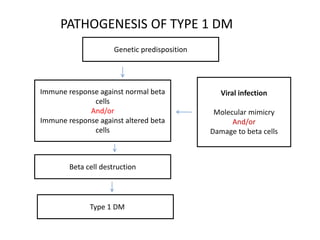
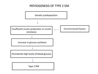
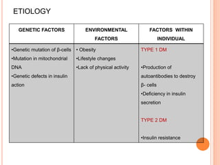
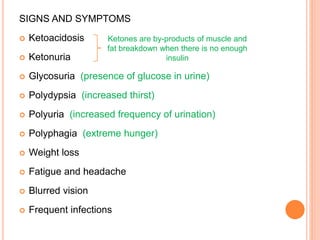
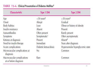
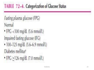
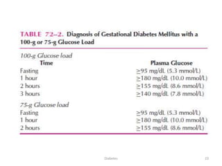
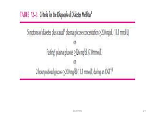
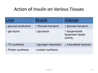
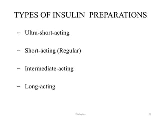
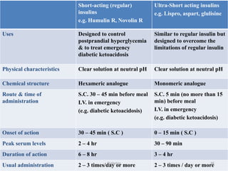
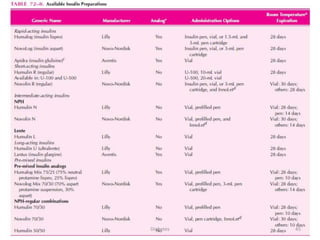
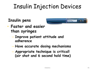
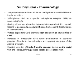
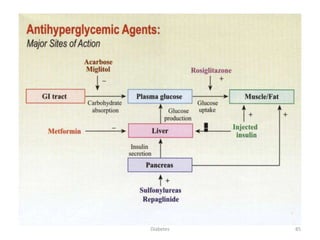
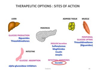
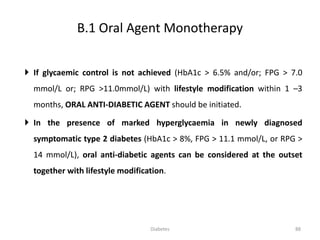
Diabetes- Pharmacotherapy
1Diabetes
Dibetes Mellitus
• Diabetes mellitus (DM) is a group of metabolic disorders characterized
by hyperglycemia; is associated with abnormalities in carbohydrate,
fat and protein metabolism; and results in chronic complications
including microvascular, macrovascular, and neuropathic disorders.
• DM is the leading cause of blindness in adults aged 20 to 74 years,
and the leading contributor to development of end-stage renal
disease.
• Finally, a cardiovascular event is responsible for 75% of deaths in
individuals with type 2 DM.
2Diabetes
CLASSIFICATION OF DIABETES
• TYPE 1 DIABETES
• TYPE 2 DIABETES
• GESTATIONAL DIABETES MELLITUS
• Maturity onset diabetes of youth (MODY)
3Diabetes
TYPE 1 DIABETES
• This form of diabetes results from autoimmune destruction of the β
cells of the pancreas.
• Markers of immune destruction of the β cell are present at the time of
diagnosis in 90% of individuals and include islet cell antibodies,
antibodies to glutamic acid decarboxylase, and antibodies to insulin.
• While this form of diabetes usually occurs in children and adolescents,
it can occur at any age.
• Younger individuals typically have a rapid rate of β-cell destruction and
present with ketoacidosis, while adults often maintain sufficient
insulin secretion to prevent ketoacidosis for many years, which is often
referred to as latent autoimmune diabetes in adults (LADA).
4Diabetes
TYPE 2 DIABETES
• This form of diabetes is characterized by insulin resistance and at least
initially, a relative lack of insulin secretion.
• Most individuals with type 2 diabetes exhibit abdominal obesity which
itself causes insulin resistance.
• In addition, hypertension, dyslipidemia (high triglyceride levels and
low HDL-cholesterol levels), and elevated inhibitor plasminogen
activator-1 (PAI-1) levels are often present in these individuals.
• This clustering of abnormalities is referred to as the “insulin resistance
syndrome” or the “metabolic syndrome.”
5Diabetes
GESTATIONAL DIABETES MELLITUS
• Gestational diabetes mellitus (GDM) is defined as glucose intolerance
which is first recognized during pregnancy.
• Gestational diabetes complicates about 7% of all pregnancies.
• Clinical detection is important, as therapy will reduce perinatal
morbidity and mortality.
6Diabetes
Maturity onset diabetes of youth (MODY)
• Maturity onset diabetes of youth (MODY) is characterized by impaired
insulin secretion with minimal or no insulin resistance.
• Patients typically exhibit mild hyperglycemia at an early age.
• The disease is inherited in an autosomal dominant pattern with at least
three different loci identified to date.
• Genetic inability to convert proinsulin to insulin results in mild
hyperglycemia and is inherited in an autosomal dominant pattern.
• Similarly, the production of mutant insulin molecules has been
identified in a few families and results in mild glucose intolerance.
7Diabetes
PATHOGENESIS
8Diabetes
PATHOGENESIS OF TYPE 1 DM
Viral infection
Molecular mimicry
And/or
Damage to beta cells
Immune response against normal beta
cells
And/or
Immune response against altered beta
cells
Genetic predisposition
Beta cell destruction
TType 1 DM
Environmental factorsInsufficient insulin production or insulin
resistance
Genetic predisposition
Persistently high levels of blood glucose
TType 2 DM
Increase in glucose synthesis
PATHOGENESIS OF TYPE 2 DM
TYPE 1 DIABETES MELLITUS
• Type 1 DM is characterized by an absolute deficiency of insulin.
• Most often this is the result of an immune-mediated destruction of
pancreatic β cells, but rare unknown or idiopathic processes may contribute.
• What is evident are four main features:
• (1) A long preclinical period marked by the presence of immune markers
when β-cell destruction is thought to occur;
• (2) Hyperglycemia when 80% to 90% of β cells are destroyed;
• (3) Transient remission (the so-called, “honeymoon” phase); and
• (4) Established disease with associated risks for complications and death.
11Diabetes
TYPE 1 DIABETES MELLITUS
• Unknown is whether there is one or more inciting factors (e.g., cow’s
milk, or viral, dietary, or other environmental exposure) that initiate
the autoimmune process.
• The autoimmune process is mediated by macrophages and T
lymphocytes with circulating auto antibodies to various β-cell
antigens.
• The most commonly detected antibody associated with type 1 DM is
the islet cell antibody.
12Diabetes
TYPE 2 DIABETES MELLITUS
Normal Insulin Action
• In the fasting state 75% of total body glucose disposal takes place in
non–insulin dependent tissues: the brain and splanchnic tissues (liver
and gastrointestinal tissues).
• In fact, brain glucose uptake occurs at the same rate during fed and
fasting periods and is not altered in type 2 diabetes.
• The remaining 25% of glucose metabolism takes place in muscle,
which is dependent on insulin.
13Diabetes
Normal Insulin Action
• In the fasting state approximately 85% of glucose production is
derived from the liver, and the remaining amount is produced by the
kidney.
• In the fed state, carbohydrate ingestion increases the plasma glucose
concentration and stimulates insulin release from the pancreatic β
cells.
• The resultant hyperinsulinemia
• (1) Suppresses hepatic glucose production and
• (2) Stimulates glucose uptake by peripheral tissues.
Diabetes 14
Normal Insulin Action
• Small increments in the plasma insulin concentration exert a potent anti
lipolytic effect, leading to a marked reduction in the plasma free fatty acid
(FFA) level.
• The decline in plasma FFA concentration results in increased glucose uptake in
muscle and reduces hepatic glucose production.
• Thus a decrease in the plasma FFA concentration lowers plasma glucose by
both decreasing its production and enhancing the uptake in muscle.
Type 2 diabetic individuals are characterized by:
• (1) Defects in insulin secretion; and
• (2) Insulin resistance involving muscle, liver, and the adipocyte. Insulin
resistance is present even in lean type 2 diabetic individuals.
Diabetes 15
CELLULAR MECHANISMS OF INSULIN
RESISTANCE
Obesity and Insulin Resistance
• Visceral adipose tissue represents 20% of fat in men and 6% of fat in
women.
• This fat tissue has been shown to have a higher rate of lipolysis than
subcutaneous fat, resulting in an increase in free fatty acid production.
• These fatty acids are released into the portal circulation and drain into
the liver, where they stimulate the production of very-low-density
lipoproteins and decrease insulin sensitivity in peripheral tissues.
16Diabetes
ETIOLOGY
GENETIC FACTORS ENVIRONMENTAL
FACTORS
FACTORS WITHIN
INDIVIDUAL
•Genetic mutation of β-cells
•Mutation in mitochondrial
DNA
•Genetic defects in insulin
action
• Obesity
•Lifestyle changes
•Lack of physical activity
TYPE 1 DM
•Production of
autoantibodies to destroy
β- cells
•Deficiency in insulin
secretion
TYPE 2 DM
•Insulin resistance
SIGNS AND SYMPTOMS
 Ketoacidosis
 Ketonuria
 Glycosuria (presence of glucose in urine)
 Polydypsia (increased thirst)
 Polyuria (increased frequency of urination)
 Polyphagia (extreme hunger)
 Weight loss
 Fatigue and headache
 Blurred vision
 Frequent infections
Ketones are by-products of muscle and
fat breakdown when there is no enough
insulin
The Metabolic Syndrome
19Diabetes
CLINICAL PRESENTATIONS
AND DIAGNOSIS
20Diabetes
21Diabetes
22Diabetes
23Diabetes
24Diabetes
• The major components of the treatment of
diabetes are:
Management of DM
• Diet and ExerciseA
• Oral hypoglycaemic
therapyB
• Insulin TherapyC
25Diabetes
DIET
• Medical nutrition therapy is recommended for all persons with DM.
• For individuals with type 1 DM, the focus is on regulating insulin
administration with a balanced diet to achieve and maintain a
healthy body weight.
• Patients with type 2 DM often require caloric restriction to
promote weight loss.
26Diabetes
Diet
Multiple Insulin Injection
I
N
S
U
L
I
N
I
N
J
E
C
T
I
O
N
25 years male IBW 60 kgm
Carbohydrate (65%) 390
Protein (10%) 60
Fat (25%) 150
Carbohydrate 100 gm
Protein (10%) 15 gm
Fat (25%) 17 gm
Diet Carbohy. Protein Fat _
Arabian bread 30 gm --- ---
Cheese 5 gm 10 gm 10 gm
Honey 50 gm 2 gm 3 gm
Glass of milk 10 gm 5 gm 5 gm_
Total 95 gm 17 gm 18 gm
Carbohydrate (65%) 520
Protein (10%) 80
Fat (25%) 200
Carbohydrate 130 gm
Protein (10%) 20 gm
Fat (25%) 22 gm
Diet Carbohy. Protein Fat _
Rice 80 gm --- 6 gm
chicken 5 gm 15 gm 12 gm
Salad 30 gm 4 gm 4 gm
Orange 10 gm --- ---___
Total 125 gm 19 gm 22 gm
Carbohydrate (65%) 260
Protein (10%) 40
Fat (25%) 100
Carbohydrate 65 gm
Protein (10%) 10 gm
Fat (25%) 11 gm
Diet Carbohy. Protein Fat _
Tuna sandwich 45 gm 12 gm 10 gm
Apple 15 gm --- ---
Tea --- --- --- _
Total 95 gm 17 gm 18 gm
60 Kg X 30 kcal = 1800 kcal
Breakfast 600 kcal Lunch 800 kcal Dinner 400 kcal
The total calories intake depends on patients age and activity but have to
related to the desirable body weight.
Total daily calories = IBW X Estimated daily energy
Add 300 kcal/day during pregnancy.
Add 500 kcal/day during lactation.
Fibers, sweeteners, vitamins, and minerals.
27Diabetes
Exercise
• In general, most patients with DM can
benefit from increased activity.
• Aerobic exercise improves insulin
resistance and glycemic control in the
majority of individuals, and reduces
cardiovascular risk factors, contributes to
weight loss or maintenance, and
improves wellbeing.
• Start exercise slowly in previously
sedentary patients.
28Diabetes
Insulin
• Insulin is an anabolic and anti catabolic hormone.
• It plays major roles in protein, carbohydrate, and fat metabolism.
• Endogenously produced insulin is cleaved from the larger proinsulin
peptide in the β cell to the active peptide of insulin and C-peptide,
which can be used as a marker for endogenous insulin production.
• All commercially available insulin preparations contain only the active
insulin peptide.
29Diabetes
Structure of Insulin
30Diabetes
Sources of Insulin
• Beta islet cells of pancreas
– 30 to 40 units per day
Bovine – differs three amino acids from human
Porcine - differs one amino acid from human
Three forms of insulin
EMP – Enzyme modified porcine
PRB – Proinsulin recombinant in bacteria
PYR – Precursor insulin yeast recombinant
31Diabetes
Endogenous insulin is secreted from
cells in the pancreas
Islet of Langerhans
Alpha cell: 20%, glucagon
Beta cell: 75%, insulin
Delta cell: 5%, somatostatin (Inhibits
the release of GH and TSH)
32Diabetes
33
-60 0 60 120 180 240
20.0
18.3
16.6
15.0
13.3
6.1
4.4
140
130
120
110
100
90
120
90
60
30
0
Glucose
(mmol/l)
Insulin
(mU/l)
Glucagon
(ng/l)
Meal
Time (min)
Type 2 diabetes
Normal subjects
Source : Müller et al. N Engl J Med. 1970
INSULIN AND GLUCAGON DYNAMICS IN RESPONSE TO MEALS IN
NORMAL SUBJECTS AND TYPE 2 DIABETES
Diabetes
Action of Insulin on Various Tissues
Liver Muscle Adipose
↓ glucose production ↑ Glucose transport ↑ glucose transport
↑ glycolysis ↑ glycolysis ↑ lipogenesis&
lipoprotein lipase
activity
↑ TG synthesis ↑ glycogen deposition ↓ intracellular lipolysis
↑ Protein synthesis ↑ protein synthesis
34Diabetes
TYPES OF INSULIN PREPARATIONS
– Ultra-short-acting
– Short-acting (Regular)
– Intermediate-acting
– Long-acting
35Diabetes
Short-acting (regular)
insulins
e.g. Humulin R, Novolin R
Ultra-Short acting insulins
e.g. Lispro, aspart, glulisine
Uses Designed to control
postprandial hyperglycemia
& to treat emergency
diabetic ketoacidosis
Similar to regular insulin but
designed to overcome the
limitations of regular insulin
Physical characteristics Clear solution at neutral pH Clear solution at neutral pH
Chemical structure Hexameric analogue Monomeric analogue
Route & time of
administration
S.C. 30 – 45 min before meal
I.V. in emergency
(e.g. diabetic ketoacidosis)
S.C. 5 min (no more than 15
min) before meal
I.V. in emergency
(e.g. diabetic ketoacidosis)
Onset of action 30 – 45 min ( S.C ) 0 – 15 min ( S.C )
Peak serum levels 2 – 4 hr 30 – 90 min
Duration of action 6 – 8 hr 3 – 4 hr
Usual administration 2 – 3 times/day or more 2 – 3 times / day or more36Diabetes
3. Intermediate - acting insulin
e.g. isophane (NPH)
Turbid suspension
Injected S.C.(Only)
Onset of action 1 - 2 hr
Peak serum level 5 - 7 hr
Duration of action 13 - 18 hr
Insulin mixtures
75/25 70/30 50/50 ( NPH / Regular )
37Diabetes
3. Intermediate - acting insulin (contd.)
Lente insulin
Turbid suspension
Mixture of 30% semilente insulin
70% ultralente insulin
Injected S.C. (only)
Onset of action 1 - 3 hr
Peak serum level 4 - 8 hr
Duration of action 13 - 20 hr
38Diabetes
3. Intermediate - acting insulins (contd.)
Lente and NPH insulins
Are roughly equivalent in biological effects.
They are usually given once or twice a day.
N.B: They are not used during emergencies
(e.g. diabetic ketoacidosis).
39Diabetes
4. Long acting insulin
e.g.Insulin glargine
Onset of action 2 hr
Absorbed less rapidly than NPH & Lente insulins.
Duration of action upto 24 hr
Designed to overcome the deficiencies of intermediate acting insulins
Advantages over intermediate-acting insulins:
• Constant circulating insulin over 24hr with no pronounced peak.
• More safe than NPH & Lente insulins due to reduced risk of
hypoglycemia(esp.nocturnal hypoglycemia).
• Clear solution that does not require resuspention before administration.
40Diabetes
First calculate total daily dose of insulin
Body weight in kgs / 2
For example in an 80-kg diabetic requiring 40 units per day, start with:
• 08:00 a.m. --- 10 units regular insulin s/c ½ hr before breakfast.
• 02:00 p.m. --- 10 units regular insulin s/c ½ hr before lunch.
• 08:00 p.m. --- 10 units regular insulin s/c ½ hr before dinner.
• 11:00 p.m. --- 10 units NPH/ lantus insulin s/c
41Diabetes
Dose adjustment
• For adjustment of dosage, check fasting blood sugar the next day
and adjust the dose of night time NPH Insulin accordingly
• i.e. keep on increasing the dose of NPH by approximately 2
units daily until you achieve a normal fasting blood glucose
level of 80-110 mg/dl.
• Once the fasting blood glucose has been controlled, check 6-
Point blood sugar as follows:
– Fasting.
– 2 hours after breakfast.
– Before lunch (and noon insulin)
– 2 hours after lunch.
– Before dinner (AND EVENING INSULIN)
– 2 hours after dinner
42Diabetes
COMPLICATIONS OF INSULIN THERAPY
• Severe Hypoglycemia (< 50 mg/dl ) – Life threatening
Overdose of insulin
Excessive (unusual) physical exercise
A meal is missed
• Weight gain
• Local or systemic allergic reactions (rare)
• Lipodystrophy at injection sites
• Insulin resistance
• Hypokalemia
43Diabetes
Severe insulin reaction
(Hypoglycemic Shock)
Diabetic coma
(Diabetic Ketoacidosis)
Onset Rapid Slow- Over several days
Insulin Excess Too little
Acidosis &
dehydration
No Ketoacidosis
Signs and symptoms
B.P. Normal or elevated Subnormal or in shock
Respiration Normal or shallow Deep & air hunger
Skin Pale & Sweating Hot & dry
CNS Tremors, mental
confusion, sometimes
convulsions
General depression
Blood sugar Lower than 70
mg/100cc
Elevated above 200
mg/100cc
Ketones Normal Elevated 44Diabetes
45Diabetes
Insulin Injection Devices
Insulin pens
• Faster and easier
than syringes
– Improve patient attitude and
adherence
– Have accurate dosing mechanisms
– Appropriate technique is critical!
(air shot and 6 second hold time)
46Diabetes
Oral hypoglycemic drugs
OHA
47Diabetes
Sulfonylureas - Pharmacology
• The primary mechanism of action of sulfonylureas is enhancement of
insulin secretion.
• Sulfonylureas bind to a specific sulfonylurea receptor (SUR) on
pancreatic β cells.
• Binding closes an adenosine triphosphate–dependent K+ channel,
leading to decreased potassium influx and subsequent depolarization
of the membrane.
• Voltage-dependent Ca+2 channels open and allow an inward flux of
Ca+2.
• Increases in intracellular Ca+2 cause translocation of secretory
granules of insulin to the cell surface and resultant exocytosis of the
granule of insulin.
• Elevated secretion of insulin from the pancreas travels via the portal
vein and subsequently suppresses hepatic glucose production.
48Diabetes
Sulfonylureas - Pharmacology
49Diabetes
50
Sulfonylureas (Oral Hypoglycemic drugs)
Tolbutamide Acetohexamide
Tolazamide
Chlorpropamide Glipizide
Glyburide
(Glibenclamide)
Glimepiride
Short
acting
First generation
Intermediate
acting
Long
acting
Long
acting
Short
acting
Second generation
Diabetes
51Diabetes
52
SIDE EFFECTS OF SULPHONYLUREAS
1) Nausea, vomiting, abdominal pain, diarrhea
2) Hypoglycaemia
3) Dilutional hyponatraemia & water intoxication (Chlorpropamide)
4) Disulfiram-like reaction with alcohol (Chlorpropamide)
5) Weight gain
6) Blood dyscrasias
(not common; less than 1% of patients)
- Agranulocytosis
- Haemolytic anaemia
- Thrombocytopenia
7) Cholestatic obstructive jaundice (uncommon)
8) Dermatitis (Mild)
9) Muscle weakness, headache, vertigo (not common)
10) Increased cardio-vascular mortality with longterm use ??
Diabetes
CONTRAINDICATIONS OF SULPHONYLUREAS
1) Type 1 DM ( insulin dependent)
2) Parenchymal disease of the liver or kidney
3) Pregnancy, lactation
4) Major stress
53Diabetes
DRUGS THAT AUGMENT THE HYPOGLYCEMIC
ACTION OF SULPHONYLUREAS
 WARFARIN
 SULFONAMIDES
 SALICYLATES
 PHENYLBUTAZONE
 PROPRANOLOL
 ALCOHOL
 CHLORAMPHENICOL
 FLUCONAZOLE
54Diabetes
DRUGS THAT ANTAGONIZE THE HYPOGLYCEMIC
ACTION OF SULPHONYLUREAS
 DIURETICS (THIAZIDE, FUROSEMIDE)
 DIAZOXIDE
 CORTICOSTEROIDS
 ORAL CONTRACEPTIVES
 PHENYTOIN, PHENOBARB., RIFAMPIN
 ALCOHOL ( chronic pts )
55Diabetes
Pharmacology
 Metformin is the only biguanide available.
 Metformin has been used clinically for 45 years.
 Metformin enhances insulin sensitivity of both hepatic and
peripheral (muscle) tissues.
 This allows for an increased uptake of glucose into these insulin-
sensitive tissues.
 Metformin has no direct effect on the β cells, though insulin levels
are reduced, reflecting increases in insulin sensitivity.
56Diabetes
Actions of Metformin
57Diabetes
METFORMIN
Initiate:
 After meals
 250 to 500mg twice or thrice a day
 Increase gradually if required in 1 or 2 weeks
 Mild loose stools in 10% initially, which reduces gradually
 Persistent loose stools in 5%
 Sustained released forms: more effective- vehicle excreted in stool
Advantages:
 Perpetuates weight loss
 Can be combined with insulin to reduce insulin requirements
58Diabetes
Diabetes
Bio-avalability (% of dose) 50% to 60%
C max (g/ml) 1.0 to 1.5
t max (in hours) 1.9 to 3.0
Plasma ½ life (t ½) 2.0 to 5.4
Renal clearance (ml/min) 400 to 600
Total clearance (ml/min) 1,300
Metformin - Pharmacokinetics
59
METFORMIN
Side effects:
 Occurs in 20-25 % of patients.
 Include.. Diarrhea, abdominal discomfort, nausea, metallic taste
and decreased absorption of vitamin B12.
Contraindications
 Patients with renal or hepatic impairment.
 Past history of lactic acidosis.
 Malabsorption or GI disturbances
 Low BMI---?less than 21kg/m2…….marked weight loss
 Organ Failure: Creatinine: >1.4mg/dl
Liver failure: Acute/Chronic
Cardiac Failure
Hypotension/Sepsis
Active Vitamin B12 Deficiency
GI intolerance 60Diabetes
α-GlucosidaseInhibitors
Pharmacology
Currently, there are two α-glucosidase inhibitors available in the United
States (acarbose and miglitol).
α-Glucosidase inhibitors competitively inhibit enzymes (maltase,
isomaltase, sucrase, and glucoamylase) in the small intestine, delaying
the breakdown of sucrose and complex carbohydrates.
They do not cause any malabsorption of these nutrients. The net effect
from this action is to reduce the postprandial blood glucose rise.
61Diabetes
α-GlucosidaseInhibitors
62Diabetes
Efficacy
Postprandial glucose concentrations are reduced (40 to 50 mg/dL), while
fasting glucose levels are relatively unchanged (∼10% reduction).
Efficacy on glycemic control is modest (average reductions in HbA1c of
0.3% to 1%), affecting primarily postprandial glycemic excursions.
Thus patients near target HbA1c levels with near normal fasting plasma
glucose levels, but high postprandial levels, may be candidates for
therapy.
α-Glucosidase Inhibitors
63Diabetes
Dosing and Administration.
Dosing for both miglitol and acarbose are similar. Initiate with a very low
dose (25 mg with one meal a day); increase very gradually (over several
months) to a maximum of 50 mg three times a day for patients ≤60 kg or
100 mg three times a day for patients >60 kg .
Both α-glucosidase inhibitors should be taken with the first bite of the
meal so that drug may be present to inhibit enzyme activity. Only
patients consuming a diet high in complex carbohydrates will have
significant reductions in glucose levels.
α-Glucosidase inhibitors are contraindicated in patients with short-bowel
syndrome or inflammatory bowel disease, and neither should be
administered in patients with serum creatinine >2 mg/dL, as this
population has not been studied.
α-Glucosidase Inhibitors
64Diabetes
α-GlucosidaseInhibitors
65Diabetes
Adverse Effects
The gastrointestinal side effects, such as flatulence, bloating,
abdominal discomfort, and diarrhea, are very common and greatly
limit the use of α-glucosidase inhibitors.
Mechanistically, these side effects are caused by distal intestinal
degradation of undigested carbohydrate by the microflora, which
results in gas (CO2 and methane) production.
α-Glucosidase inhibitors should be initiated at a low dose and
titrated slowly to reduce gastrointestinal intolerance.
α-GlucosidaseInhibitors
66Diabetes
ALPHA-GLUCOSIDASE INHIBITORS
• Postprandial hyperglycemia
• Inhibits glycoside hydrolases
– glucosidase
• Taken at beginning of a meal
• Gastrointestinal side effects
– Common
• Higher efficacy than Voglibose
• Postprandial hyperglycemia
• Inhibit glycoside hydrolases
• Taken at beginning of a meal
• Miglitol is systemically absorbed
– Excreted by the kidneys
• Voglibose is newest:
– less side effects
– economical
Acarbose Miglitol & Voglibose
Acarbose
Miglitol Voglibose
67Diabetes
Pharmacology
Though the binding site is adjacent to the binding site of
sulfonylureas, nateglinide and repaglinide stimulate insulin
secretion from the β cells of the pancreas, similarly to sulfonylureas.
Repaglinide, a benzoic acid derivative, and nateglinide, a
phenylalanine amino acid derivative, both require the presence of
glucose to stimulate insulin secretion.
As glucose levels diminish to normal, stimulated insulin secretion
diminishes.
Short-Acting Insulin Secretagogues
68Diabetes
Efficacy
In monotherapy, both significantly reduce postprandial glucose
excursions and reduce HbA1c levels.
Repaglinide, dosed 4 mg three times a day
Nateglinide, dosed 120 mg three times a day
The lower efficacy of these agents vs. sulphonylureas should be
considered when patients are >1% above their HbA1c goal.
These agents can be used to provide increased insulin secretion during
meals, when it is needed, in patients close to glycemic goals.
Short-Acting Insulin Secretagogues
69Diabetes
Short-Acting Insulin Secretagogues
70Diabetes
Adverse Effects
Hypoglycemia is the main side effect noted with both agents.
Hypoglycemic risk appears to be less vs. sulfonylurea.
In part, this is due to the glucose-sensitive release of insulin. If the
glucose concentration is normal, less glucose-stimulated release of
insulin will occur.
Short-Acting Insulin Secretagogues
71Diabetes
Current Medications for Treatment of Diabetes
Sulfonylureas
Meglitinides
Metformin
Thiazolidinediones
α-Glucosidase inhibitors
Insulin
Hypoglycemia, weight gain
GI intolerance
Edema, weight gain
GI intolerance
Hypoglycemia, weight gain
Drug Class Side Effects
72Diabetes
Role of Active GLP-1 and GIP in Glucose
Homeostasis
• Glucagon-Like-Peptide (GLP-1) and
• Glucose - Dependent-Insulotropic-Polypeptide (GIP)
-are secreted in response to food intake (glucose-dependent
mechanism)
• GLP-1 and GIP - (↑) stimulate insulin biosynthesis & secretion
• GLP-1 inhibits - (↓) glucagon secretion
• GLP-1 and GIP - (↓)lower blood sugar level
73Diabetes
74
ROLE OF INCRETINS IN GLUCOSE HOMEOSTASIS
Ingestion of food
Beta cells
Alpha cells
Release of gut
hormones :
Incretins
Pancreas
Glucose-dependent
 Insulin from beta cells
(GLP-1 and GIP)
Glucose
uptake by
muscles
Glucose
production
by liver
Blood
glucose
Glucose dependent
 Glucagon from
alpha cells
(GLP-1)
Active
GLP-1 & GIP
DPP-4
enzyme
Inactive
GIP
Inactive
GLP-1
Diabetes
75
NEW DRUG TARGET
• Incretins :
– Gut hormones that enhance insulin secretion in reponse to food
– Glucose - dependant insulin secretion
• Medication affecting the incretin system :
– GLP1 analogues : Exenatide
– DPP4 Inhibitors : Sitagliptin
Diabetes
Incretin mimetics
Exenatide
• The first incretin-related therapy available for patients with type 2
diabetes.
• Naturally occurring peptide from the saliva of the Gila Monster.
• Has an approximate 50% amino acid homology with GLP-1.
• Binds to GLP-1 receptors and behaves as GLP-1.
76Diabetes
77Diabetes
Dipeptidyl peptidase-4 (DPP-4) inhibitor
78Diabetes
• During a meal the incretins glucagon-like peptide 1 (GLP-1) and glucose-
dependent gastric inhibitory polypeptide (GIP) are released from the small
intestine into the vasculature.
• The hormones regulate insulin secretion in a glucose-dependent manner. GLP-
1 has many roles in the human body; it stimulates insulin biosynthesis,
inhibits glucagon secretion, slows gastric emptying, reduces appetite and
stimulates regeneration of islet β-cells.
• GIP and GLP-1 have extremely short plasma half-lives due to a very rapid
inactivation.
• The enzyme responsible for the metabolism is DPP-4. Inhibition of DPP-4 leads
to potentiation of endogenous GIP and GLP-1 and hence improves treatment
of type 2 diabetes.
Dipeptidyl peptidase-4 (DPP-4) inhibitor
79Diabetes
Dipeptidyl peptidase-4 (DPP-4) inhibitor
Drugs belonging to this class are :
Sitagliptin (FDA approved 2006, marketed by Merck & Co)
Vildagliptin(EU approved 2007, marketed in the EU by Novartis)
Saxagliptin (FDA approved in 2009)
linagliptin (FDA approved in 2011, marketed by Eli Lilly Co)
Dutogliptin (being developed by Phenomix Corporation),
Phase IIIgemigliptin (being developed by LG Life Sciences, Korea)
alogliptin ( FDA approved 2013, marketed by Takeda Pharmaceutical Company)
80Diabetes
Adverse effects
• Nasopharyngitis
• Headache
• Nausea
• Hypersensitivity
• Hypersensitivity reactions and
• Pancreatitis
81Diabetes
Thiazolidinediones
Pharmacology
• Thiazolidinediones are also referred to as TZDs or glitazones. Pioglitazone
and rosiglitazone are the two recently banned thiazolidinediones for the
treatment of type 2 DM.
• Thiazolidinediones work by binding to the peroxisome proliferator
activator receptor-γ (PPAR-γ), which are primarily located on fat cells and
vascular cells.
82Diabetes
Thiazolidinediones
Efficacy
• Pioglitazone and rosiglitazone, given for about 6 months, reduce HbA1c
values∼1.5% and reduce FPG levels by approximately 60 to 70 mg/dL at maximal
doses.
• Glycemic-lowering onset is slow, and maximal glycemic-lowering effects may not
be seen until 3 to 4 months of therapy.
• It is important to inform patients of this fact and that they should not stop therapy
even if minimal glucose lowering is initially encountered.
• The efficacy of both drugs is dependent on sufficient insulinemia. If there is
insufficient endogenous insulin production (β-cell function) or exogenous insulin
delivery via injections, neither will lower glucose concentrations efficiently.
83Diabetes
Thiazolidinediones
• Adverse Effects
• Troglitazone, the first thiazolidinedione approved, caused idiosyncratic
hepatotoxicity and had 28 deaths from liver failure, which prompted
removal from the U.S. market in March 2000.
• Approximately 1.9% of patients placed on troglitazone had alanine
aminotransferase (ALT) levels more than three times the upper limit of
normal.
• The incidence, using these criteria for elevated liver enzymes, with
pioglitazone (0.25%) and rosiglitazone (0.2%) has been low.
• Pioglitazone – Bladder cancer
• Rosiglitazone – Increased risk of cardiovascular events
84Diabetes
85Diabetes
86
GLUCOSE ABSORPTION
GLUCOSE PRODUCTION
Biguanides
Thiazolidinediones
MUSCLE
PERIPHERAL
GLUCOSE UPTAKE
Thiazolidinediones
(Biguanides)
PANCREAS
INSULIN Secretion
Sulfonylureas
Meglitinides
Insulin
Amylin
INTESTINAL HORMONES
Incretin
ADIPOSE TISSUELIVER
alpha-glucosidase inhibitors
INTESTINE
THERAPEUTIC OPTIONS : SITES OF ACTION
Diabetes
ADA/EASD Consensus Algorithm for
Management of Diabetes
At diagnosis:
Lifestyle
+
Metformin
Lifestyle+Metformin
+
Pioglitazone
(No hypoglycemia, edema,
CHF, bone loss)
Lifestyle+Metformin
+
Sulfonylurea
Lifestyle+Metformin
+
Intensive insulin
Lifestyle+Metformin
+
Basal Insulin
Lifestyle+Metformin
+
GLP1
(No hypoglycemia, wt loss,
Nausea/vomiting)
Lifestyle+Metformin
+
Pioglitazone
+
Sulfonylurea
Lifestyle+Metformin
+
Basal Insulin
Tier 2: less well-validated
therapies
Tier 1: Well-validated core therapies
Step 1 Step 2 Step 3
Amylin agonists, Glinides
DPP-4 inhibitors may be
appropriate in selected
patients
*Useful when
hypoglycemia is to be
avoided
87Diabetes
 If glycaemic control is not achieved (HbA1c > 6.5% and/or; FPG > 7.0
mmol/L or; RPG >11.0mmol/L) with lifestyle modification within 1 –3
months, ORAL ANTI-DIABETIC AGENT should be initiated.
 In the presence of marked hyperglycaemia in newly diagnosed
symptomatic type 2 diabetes (HbA1c > 8%, FPG > 11.1 mmol/L, or RPG >
14 mmol/L), oral anti-diabetic agents can be considered at the outset
together with lifestyle modification.
B.1 Oral Agent Monotherapy
88Diabetes
As first line therapy:
 Obese type 2 patients, consider use of metformin, acarbose or TZD.
 Non-obese type 2 patients, consider the use of insulin secretagogues
 Metformin is the drug of choice in overweight/obese patients.
 TZDs and acarbose are acceptable alternatives in those who are
intolerant to metformin.
 If monotherapy fails, a combination of TZDs, acarbose and metformin is
recommended. If targets are still not achieved, insulin secretagogues may
be added
B.1 Oral Agent Monotherapy (cont.)
89Diabetes
Combination oral agents is indicated in:
• Newly diagnosed symptomatic patients with HbA1c >10
• Patients who are not reaching targets after 3 months on monotherapy
B.2 Combination Oral Agents
90Diabetes
 If targets have not been reached after optimal dose of combination therapy
for 3 months, consider adding intermediate-acting/long-acting insulin
(BIDS).
 Combination of insulin+ oral anti-diabetic agents (BIDS) has been
shown to improve glycaemic control in those not achieving target despite
maximal combination oral anti-diabetic agents.
 Combining insulin and the following oral anti-diabetic agents has been
shown to be effective in people with type 2 diabetes:
◦ Biguanide (metformin)
◦ Insulin secretagogues (sulphonylureas)
◦ Insulin sensitizers (TZDs)(the combination of a TZD plus insulin is not
an approved indication)
◦ α-glucosidase inhibitor (acarbose)
 Insulin dose can be increased until target FPG is achieved.
B.3 Combination Oral Agents and Insulin
91Diabetes
Diabetes
Management
Algorithm
92Diabetes
93Diabetes

Diabetes pharmacotherapy(1)

  • 1.
  • 2.
    Dibetes Mellitus • Diabetesmellitus (DM) is a group of metabolic disorders characterized by hyperglycemia; is associated with abnormalities in carbohydrate, fat and protein metabolism; and results in chronic complications including microvascular, macrovascular, and neuropathic disorders. • DM is the leading cause of blindness in adults aged 20 to 74 years, and the leading contributor to development of end-stage renal disease. • Finally, a cardiovascular event is responsible for 75% of deaths in individuals with type 2 DM. 2Diabetes
  • 3.
    CLASSIFICATION OF DIABETES •TYPE 1 DIABETES • TYPE 2 DIABETES • GESTATIONAL DIABETES MELLITUS • Maturity onset diabetes of youth (MODY) 3Diabetes
  • 4.
    TYPE 1 DIABETES •This form of diabetes results from autoimmune destruction of the β cells of the pancreas. • Markers of immune destruction of the β cell are present at the time of diagnosis in 90% of individuals and include islet cell antibodies, antibodies to glutamic acid decarboxylase, and antibodies to insulin. • While this form of diabetes usually occurs in children and adolescents, it can occur at any age. • Younger individuals typically have a rapid rate of β-cell destruction and present with ketoacidosis, while adults often maintain sufficient insulin secretion to prevent ketoacidosis for many years, which is often referred to as latent autoimmune diabetes in adults (LADA). 4Diabetes
  • 5.
    TYPE 2 DIABETES •This form of diabetes is characterized by insulin resistance and at least initially, a relative lack of insulin secretion. • Most individuals with type 2 diabetes exhibit abdominal obesity which itself causes insulin resistance. • In addition, hypertension, dyslipidemia (high triglyceride levels and low HDL-cholesterol levels), and elevated inhibitor plasminogen activator-1 (PAI-1) levels are often present in these individuals. • This clustering of abnormalities is referred to as the “insulin resistance syndrome” or the “metabolic syndrome.” 5Diabetes
  • 6.
    GESTATIONAL DIABETES MELLITUS •Gestational diabetes mellitus (GDM) is defined as glucose intolerance which is first recognized during pregnancy. • Gestational diabetes complicates about 7% of all pregnancies. • Clinical detection is important, as therapy will reduce perinatal morbidity and mortality. 6Diabetes
  • 7.
    Maturity onset diabetesof youth (MODY) • Maturity onset diabetes of youth (MODY) is characterized by impaired insulin secretion with minimal or no insulin resistance. • Patients typically exhibit mild hyperglycemia at an early age. • The disease is inherited in an autosomal dominant pattern with at least three different loci identified to date. • Genetic inability to convert proinsulin to insulin results in mild hyperglycemia and is inherited in an autosomal dominant pattern. • Similarly, the production of mutant insulin molecules has been identified in a few families and results in mild glucose intolerance. 7Diabetes
  • 8.
  • 9.
    PATHOGENESIS OF TYPE1 DM Viral infection Molecular mimicry And/or Damage to beta cells Immune response against normal beta cells And/or Immune response against altered beta cells Genetic predisposition Beta cell destruction TType 1 DM
  • 10.
    Environmental factorsInsufficient insulinproduction or insulin resistance Genetic predisposition Persistently high levels of blood glucose TType 2 DM Increase in glucose synthesis PATHOGENESIS OF TYPE 2 DM
  • 11.
    TYPE 1 DIABETESMELLITUS • Type 1 DM is characterized by an absolute deficiency of insulin. • Most often this is the result of an immune-mediated destruction of pancreatic β cells, but rare unknown or idiopathic processes may contribute. • What is evident are four main features: • (1) A long preclinical period marked by the presence of immune markers when β-cell destruction is thought to occur; • (2) Hyperglycemia when 80% to 90% of β cells are destroyed; • (3) Transient remission (the so-called, “honeymoon” phase); and • (4) Established disease with associated risks for complications and death. 11Diabetes
  • 12.
    TYPE 1 DIABETESMELLITUS • Unknown is whether there is one or more inciting factors (e.g., cow’s milk, or viral, dietary, or other environmental exposure) that initiate the autoimmune process. • The autoimmune process is mediated by macrophages and T lymphocytes with circulating auto antibodies to various β-cell antigens. • The most commonly detected antibody associated with type 1 DM is the islet cell antibody. 12Diabetes
  • 13.
    TYPE 2 DIABETESMELLITUS Normal Insulin Action • In the fasting state 75% of total body glucose disposal takes place in non–insulin dependent tissues: the brain and splanchnic tissues (liver and gastrointestinal tissues). • In fact, brain glucose uptake occurs at the same rate during fed and fasting periods and is not altered in type 2 diabetes. • The remaining 25% of glucose metabolism takes place in muscle, which is dependent on insulin. 13Diabetes
  • 14.
    Normal Insulin Action •In the fasting state approximately 85% of glucose production is derived from the liver, and the remaining amount is produced by the kidney. • In the fed state, carbohydrate ingestion increases the plasma glucose concentration and stimulates insulin release from the pancreatic β cells. • The resultant hyperinsulinemia • (1) Suppresses hepatic glucose production and • (2) Stimulates glucose uptake by peripheral tissues. Diabetes 14
  • 15.
    Normal Insulin Action •Small increments in the plasma insulin concentration exert a potent anti lipolytic effect, leading to a marked reduction in the plasma free fatty acid (FFA) level. • The decline in plasma FFA concentration results in increased glucose uptake in muscle and reduces hepatic glucose production. • Thus a decrease in the plasma FFA concentration lowers plasma glucose by both decreasing its production and enhancing the uptake in muscle. Type 2 diabetic individuals are characterized by: • (1) Defects in insulin secretion; and • (2) Insulin resistance involving muscle, liver, and the adipocyte. Insulin resistance is present even in lean type 2 diabetic individuals. Diabetes 15
  • 16.
    CELLULAR MECHANISMS OFINSULIN RESISTANCE Obesity and Insulin Resistance • Visceral adipose tissue represents 20% of fat in men and 6% of fat in women. • This fat tissue has been shown to have a higher rate of lipolysis than subcutaneous fat, resulting in an increase in free fatty acid production. • These fatty acids are released into the portal circulation and drain into the liver, where they stimulate the production of very-low-density lipoproteins and decrease insulin sensitivity in peripheral tissues. 16Diabetes
  • 17.
    ETIOLOGY GENETIC FACTORS ENVIRONMENTAL FACTORS FACTORSWITHIN INDIVIDUAL •Genetic mutation of β-cells •Mutation in mitochondrial DNA •Genetic defects in insulin action • Obesity •Lifestyle changes •Lack of physical activity TYPE 1 DM •Production of autoantibodies to destroy β- cells •Deficiency in insulin secretion TYPE 2 DM •Insulin resistance
  • 18.
    SIGNS AND SYMPTOMS Ketoacidosis  Ketonuria  Glycosuria (presence of glucose in urine)  Polydypsia (increased thirst)  Polyuria (increased frequency of urination)  Polyphagia (extreme hunger)  Weight loss  Fatigue and headache  Blurred vision  Frequent infections Ketones are by-products of muscle and fat breakdown when there is no enough insulin
  • 19.
  • 20.
  • 21.
  • 22.
  • 23.
  • 24.
  • 25.
    • The majorcomponents of the treatment of diabetes are: Management of DM • Diet and ExerciseA • Oral hypoglycaemic therapyB • Insulin TherapyC 25Diabetes
  • 26.
    DIET • Medical nutritiontherapy is recommended for all persons with DM. • For individuals with type 1 DM, the focus is on regulating insulin administration with a balanced diet to achieve and maintain a healthy body weight. • Patients with type 2 DM often require caloric restriction to promote weight loss. 26Diabetes
  • 27.
    Diet Multiple Insulin Injection I N S U L I N I N J E C T I O N 25years male IBW 60 kgm Carbohydrate (65%) 390 Protein (10%) 60 Fat (25%) 150 Carbohydrate 100 gm Protein (10%) 15 gm Fat (25%) 17 gm Diet Carbohy. Protein Fat _ Arabian bread 30 gm --- --- Cheese 5 gm 10 gm 10 gm Honey 50 gm 2 gm 3 gm Glass of milk 10 gm 5 gm 5 gm_ Total 95 gm 17 gm 18 gm Carbohydrate (65%) 520 Protein (10%) 80 Fat (25%) 200 Carbohydrate 130 gm Protein (10%) 20 gm Fat (25%) 22 gm Diet Carbohy. Protein Fat _ Rice 80 gm --- 6 gm chicken 5 gm 15 gm 12 gm Salad 30 gm 4 gm 4 gm Orange 10 gm --- ---___ Total 125 gm 19 gm 22 gm Carbohydrate (65%) 260 Protein (10%) 40 Fat (25%) 100 Carbohydrate 65 gm Protein (10%) 10 gm Fat (25%) 11 gm Diet Carbohy. Protein Fat _ Tuna sandwich 45 gm 12 gm 10 gm Apple 15 gm --- --- Tea --- --- --- _ Total 95 gm 17 gm 18 gm 60 Kg X 30 kcal = 1800 kcal Breakfast 600 kcal Lunch 800 kcal Dinner 400 kcal The total calories intake depends on patients age and activity but have to related to the desirable body weight. Total daily calories = IBW X Estimated daily energy Add 300 kcal/day during pregnancy. Add 500 kcal/day during lactation. Fibers, sweeteners, vitamins, and minerals. 27Diabetes
  • 28.
    Exercise • In general,most patients with DM can benefit from increased activity. • Aerobic exercise improves insulin resistance and glycemic control in the majority of individuals, and reduces cardiovascular risk factors, contributes to weight loss or maintenance, and improves wellbeing. • Start exercise slowly in previously sedentary patients. 28Diabetes
  • 29.
    Insulin • Insulin isan anabolic and anti catabolic hormone. • It plays major roles in protein, carbohydrate, and fat metabolism. • Endogenously produced insulin is cleaved from the larger proinsulin peptide in the β cell to the active peptide of insulin and C-peptide, which can be used as a marker for endogenous insulin production. • All commercially available insulin preparations contain only the active insulin peptide. 29Diabetes
  • 30.
  • 31.
    Sources of Insulin •Beta islet cells of pancreas – 30 to 40 units per day Bovine – differs three amino acids from human Porcine - differs one amino acid from human Three forms of insulin EMP – Enzyme modified porcine PRB – Proinsulin recombinant in bacteria PYR – Precursor insulin yeast recombinant 31Diabetes
  • 32.
    Endogenous insulin issecreted from cells in the pancreas Islet of Langerhans Alpha cell: 20%, glucagon Beta cell: 75%, insulin Delta cell: 5%, somatostatin (Inhibits the release of GH and TSH) 32Diabetes
  • 33.
    33 -60 0 60120 180 240 20.0 18.3 16.6 15.0 13.3 6.1 4.4 140 130 120 110 100 90 120 90 60 30 0 Glucose (mmol/l) Insulin (mU/l) Glucagon (ng/l) Meal Time (min) Type 2 diabetes Normal subjects Source : Müller et al. N Engl J Med. 1970 INSULIN AND GLUCAGON DYNAMICS IN RESPONSE TO MEALS IN NORMAL SUBJECTS AND TYPE 2 DIABETES Diabetes
  • 34.
    Action of Insulinon Various Tissues Liver Muscle Adipose ↓ glucose production ↑ Glucose transport ↑ glucose transport ↑ glycolysis ↑ glycolysis ↑ lipogenesis& lipoprotein lipase activity ↑ TG synthesis ↑ glycogen deposition ↓ intracellular lipolysis ↑ Protein synthesis ↑ protein synthesis 34Diabetes
  • 35.
    TYPES OF INSULINPREPARATIONS – Ultra-short-acting – Short-acting (Regular) – Intermediate-acting – Long-acting 35Diabetes
  • 36.
    Short-acting (regular) insulins e.g. HumulinR, Novolin R Ultra-Short acting insulins e.g. Lispro, aspart, glulisine Uses Designed to control postprandial hyperglycemia & to treat emergency diabetic ketoacidosis Similar to regular insulin but designed to overcome the limitations of regular insulin Physical characteristics Clear solution at neutral pH Clear solution at neutral pH Chemical structure Hexameric analogue Monomeric analogue Route & time of administration S.C. 30 – 45 min before meal I.V. in emergency (e.g. diabetic ketoacidosis) S.C. 5 min (no more than 15 min) before meal I.V. in emergency (e.g. diabetic ketoacidosis) Onset of action 30 – 45 min ( S.C ) 0 – 15 min ( S.C ) Peak serum levels 2 – 4 hr 30 – 90 min Duration of action 6 – 8 hr 3 – 4 hr Usual administration 2 – 3 times/day or more 2 – 3 times / day or more36Diabetes
  • 37.
    3. Intermediate -acting insulin e.g. isophane (NPH) Turbid suspension Injected S.C.(Only) Onset of action 1 - 2 hr Peak serum level 5 - 7 hr Duration of action 13 - 18 hr Insulin mixtures 75/25 70/30 50/50 ( NPH / Regular ) 37Diabetes
  • 38.
    3. Intermediate -acting insulin (contd.) Lente insulin Turbid suspension Mixture of 30% semilente insulin 70% ultralente insulin Injected S.C. (only) Onset of action 1 - 3 hr Peak serum level 4 - 8 hr Duration of action 13 - 20 hr 38Diabetes
  • 39.
    3. Intermediate -acting insulins (contd.) Lente and NPH insulins Are roughly equivalent in biological effects. They are usually given once or twice a day. N.B: They are not used during emergencies (e.g. diabetic ketoacidosis). 39Diabetes
  • 40.
    4. Long actinginsulin e.g.Insulin glargine Onset of action 2 hr Absorbed less rapidly than NPH & Lente insulins. Duration of action upto 24 hr Designed to overcome the deficiencies of intermediate acting insulins Advantages over intermediate-acting insulins: • Constant circulating insulin over 24hr with no pronounced peak. • More safe than NPH & Lente insulins due to reduced risk of hypoglycemia(esp.nocturnal hypoglycemia). • Clear solution that does not require resuspention before administration. 40Diabetes
  • 41.
    First calculate totaldaily dose of insulin Body weight in kgs / 2 For example in an 80-kg diabetic requiring 40 units per day, start with: • 08:00 a.m. --- 10 units regular insulin s/c ½ hr before breakfast. • 02:00 p.m. --- 10 units regular insulin s/c ½ hr before lunch. • 08:00 p.m. --- 10 units regular insulin s/c ½ hr before dinner. • 11:00 p.m. --- 10 units NPH/ lantus insulin s/c 41Diabetes
  • 42.
    Dose adjustment • Foradjustment of dosage, check fasting blood sugar the next day and adjust the dose of night time NPH Insulin accordingly • i.e. keep on increasing the dose of NPH by approximately 2 units daily until you achieve a normal fasting blood glucose level of 80-110 mg/dl. • Once the fasting blood glucose has been controlled, check 6- Point blood sugar as follows: – Fasting. – 2 hours after breakfast. – Before lunch (and noon insulin) – 2 hours after lunch. – Before dinner (AND EVENING INSULIN) – 2 hours after dinner 42Diabetes
  • 43.
    COMPLICATIONS OF INSULINTHERAPY • Severe Hypoglycemia (< 50 mg/dl ) – Life threatening Overdose of insulin Excessive (unusual) physical exercise A meal is missed • Weight gain • Local or systemic allergic reactions (rare) • Lipodystrophy at injection sites • Insulin resistance • Hypokalemia 43Diabetes
  • 44.
    Severe insulin reaction (HypoglycemicShock) Diabetic coma (Diabetic Ketoacidosis) Onset Rapid Slow- Over several days Insulin Excess Too little Acidosis & dehydration No Ketoacidosis Signs and symptoms B.P. Normal or elevated Subnormal or in shock Respiration Normal or shallow Deep & air hunger Skin Pale & Sweating Hot & dry CNS Tremors, mental confusion, sometimes convulsions General depression Blood sugar Lower than 70 mg/100cc Elevated above 200 mg/100cc Ketones Normal Elevated 44Diabetes
  • 45.
  • 46.
    Insulin Injection Devices Insulinpens • Faster and easier than syringes – Improve patient attitude and adherence – Have accurate dosing mechanisms – Appropriate technique is critical! (air shot and 6 second hold time) 46Diabetes
  • 47.
  • 48.
    Sulfonylureas - Pharmacology •The primary mechanism of action of sulfonylureas is enhancement of insulin secretion. • Sulfonylureas bind to a specific sulfonylurea receptor (SUR) on pancreatic β cells. • Binding closes an adenosine triphosphate–dependent K+ channel, leading to decreased potassium influx and subsequent depolarization of the membrane. • Voltage-dependent Ca+2 channels open and allow an inward flux of Ca+2. • Increases in intracellular Ca+2 cause translocation of secretory granules of insulin to the cell surface and resultant exocytosis of the granule of insulin. • Elevated secretion of insulin from the pancreas travels via the portal vein and subsequently suppresses hepatic glucose production. 48Diabetes
  • 49.
  • 50.
    50 Sulfonylureas (Oral Hypoglycemicdrugs) Tolbutamide Acetohexamide Tolazamide Chlorpropamide Glipizide Glyburide (Glibenclamide) Glimepiride Short acting First generation Intermediate acting Long acting Long acting Short acting Second generation Diabetes
  • 51.
  • 52.
    52 SIDE EFFECTS OFSULPHONYLUREAS 1) Nausea, vomiting, abdominal pain, diarrhea 2) Hypoglycaemia 3) Dilutional hyponatraemia & water intoxication (Chlorpropamide) 4) Disulfiram-like reaction with alcohol (Chlorpropamide) 5) Weight gain 6) Blood dyscrasias (not common; less than 1% of patients) - Agranulocytosis - Haemolytic anaemia - Thrombocytopenia 7) Cholestatic obstructive jaundice (uncommon) 8) Dermatitis (Mild) 9) Muscle weakness, headache, vertigo (not common) 10) Increased cardio-vascular mortality with longterm use ?? Diabetes
  • 53.
    CONTRAINDICATIONS OF SULPHONYLUREAS 1)Type 1 DM ( insulin dependent) 2) Parenchymal disease of the liver or kidney 3) Pregnancy, lactation 4) Major stress 53Diabetes
  • 54.
    DRUGS THAT AUGMENTTHE HYPOGLYCEMIC ACTION OF SULPHONYLUREAS  WARFARIN  SULFONAMIDES  SALICYLATES  PHENYLBUTAZONE  PROPRANOLOL  ALCOHOL  CHLORAMPHENICOL  FLUCONAZOLE 54Diabetes
  • 55.
    DRUGS THAT ANTAGONIZETHE HYPOGLYCEMIC ACTION OF SULPHONYLUREAS  DIURETICS (THIAZIDE, FUROSEMIDE)  DIAZOXIDE  CORTICOSTEROIDS  ORAL CONTRACEPTIVES  PHENYTOIN, PHENOBARB., RIFAMPIN  ALCOHOL ( chronic pts ) 55Diabetes
  • 56.
    Pharmacology  Metformin isthe only biguanide available.  Metformin has been used clinically for 45 years.  Metformin enhances insulin sensitivity of both hepatic and peripheral (muscle) tissues.  This allows for an increased uptake of glucose into these insulin- sensitive tissues.  Metformin has no direct effect on the β cells, though insulin levels are reduced, reflecting increases in insulin sensitivity. 56Diabetes
  • 57.
  • 58.
    METFORMIN Initiate:  After meals 250 to 500mg twice or thrice a day  Increase gradually if required in 1 or 2 weeks  Mild loose stools in 10% initially, which reduces gradually  Persistent loose stools in 5%  Sustained released forms: more effective- vehicle excreted in stool Advantages:  Perpetuates weight loss  Can be combined with insulin to reduce insulin requirements 58Diabetes
  • 59.
    Diabetes Bio-avalability (% ofdose) 50% to 60% C max (g/ml) 1.0 to 1.5 t max (in hours) 1.9 to 3.0 Plasma ½ life (t ½) 2.0 to 5.4 Renal clearance (ml/min) 400 to 600 Total clearance (ml/min) 1,300 Metformin - Pharmacokinetics 59
  • 60.
    METFORMIN Side effects:  Occursin 20-25 % of patients.  Include.. Diarrhea, abdominal discomfort, nausea, metallic taste and decreased absorption of vitamin B12. Contraindications  Patients with renal or hepatic impairment.  Past history of lactic acidosis.  Malabsorption or GI disturbances  Low BMI---?less than 21kg/m2…….marked weight loss  Organ Failure: Creatinine: >1.4mg/dl Liver failure: Acute/Chronic Cardiac Failure Hypotension/Sepsis Active Vitamin B12 Deficiency GI intolerance 60Diabetes
  • 61.
    α-GlucosidaseInhibitors Pharmacology Currently, there aretwo α-glucosidase inhibitors available in the United States (acarbose and miglitol). α-Glucosidase inhibitors competitively inhibit enzymes (maltase, isomaltase, sucrase, and glucoamylase) in the small intestine, delaying the breakdown of sucrose and complex carbohydrates. They do not cause any malabsorption of these nutrients. The net effect from this action is to reduce the postprandial blood glucose rise. 61Diabetes
  • 62.
  • 63.
    Efficacy Postprandial glucose concentrationsare reduced (40 to 50 mg/dL), while fasting glucose levels are relatively unchanged (∼10% reduction). Efficacy on glycemic control is modest (average reductions in HbA1c of 0.3% to 1%), affecting primarily postprandial glycemic excursions. Thus patients near target HbA1c levels with near normal fasting plasma glucose levels, but high postprandial levels, may be candidates for therapy. α-Glucosidase Inhibitors 63Diabetes
  • 64.
    Dosing and Administration. Dosingfor both miglitol and acarbose are similar. Initiate with a very low dose (25 mg with one meal a day); increase very gradually (over several months) to a maximum of 50 mg three times a day for patients ≤60 kg or 100 mg three times a day for patients >60 kg . Both α-glucosidase inhibitors should be taken with the first bite of the meal so that drug may be present to inhibit enzyme activity. Only patients consuming a diet high in complex carbohydrates will have significant reductions in glucose levels. α-Glucosidase inhibitors are contraindicated in patients with short-bowel syndrome or inflammatory bowel disease, and neither should be administered in patients with serum creatinine >2 mg/dL, as this population has not been studied. α-Glucosidase Inhibitors 64Diabetes
  • 65.
  • 66.
    Adverse Effects The gastrointestinalside effects, such as flatulence, bloating, abdominal discomfort, and diarrhea, are very common and greatly limit the use of α-glucosidase inhibitors. Mechanistically, these side effects are caused by distal intestinal degradation of undigested carbohydrate by the microflora, which results in gas (CO2 and methane) production. α-Glucosidase inhibitors should be initiated at a low dose and titrated slowly to reduce gastrointestinal intolerance. α-GlucosidaseInhibitors 66Diabetes
  • 67.
    ALPHA-GLUCOSIDASE INHIBITORS • Postprandialhyperglycemia • Inhibits glycoside hydrolases – glucosidase • Taken at beginning of a meal • Gastrointestinal side effects – Common • Higher efficacy than Voglibose • Postprandial hyperglycemia • Inhibit glycoside hydrolases • Taken at beginning of a meal • Miglitol is systemically absorbed – Excreted by the kidneys • Voglibose is newest: – less side effects – economical Acarbose Miglitol & Voglibose Acarbose Miglitol Voglibose 67Diabetes
  • 68.
    Pharmacology Though the bindingsite is adjacent to the binding site of sulfonylureas, nateglinide and repaglinide stimulate insulin secretion from the β cells of the pancreas, similarly to sulfonylureas. Repaglinide, a benzoic acid derivative, and nateglinide, a phenylalanine amino acid derivative, both require the presence of glucose to stimulate insulin secretion. As glucose levels diminish to normal, stimulated insulin secretion diminishes. Short-Acting Insulin Secretagogues 68Diabetes
  • 69.
    Efficacy In monotherapy, bothsignificantly reduce postprandial glucose excursions and reduce HbA1c levels. Repaglinide, dosed 4 mg three times a day Nateglinide, dosed 120 mg three times a day The lower efficacy of these agents vs. sulphonylureas should be considered when patients are >1% above their HbA1c goal. These agents can be used to provide increased insulin secretion during meals, when it is needed, in patients close to glycemic goals. Short-Acting Insulin Secretagogues 69Diabetes
  • 70.
  • 71.
    Adverse Effects Hypoglycemia isthe main side effect noted with both agents. Hypoglycemic risk appears to be less vs. sulfonylurea. In part, this is due to the glucose-sensitive release of insulin. If the glucose concentration is normal, less glucose-stimulated release of insulin will occur. Short-Acting Insulin Secretagogues 71Diabetes
  • 72.
    Current Medications forTreatment of Diabetes Sulfonylureas Meglitinides Metformin Thiazolidinediones α-Glucosidase inhibitors Insulin Hypoglycemia, weight gain Hypoglycemia, weight gain GI intolerance Edema, weight gain GI intolerance Hypoglycemia, weight gain Drug Class Side Effects 72Diabetes
  • 73.
    Role of ActiveGLP-1 and GIP in Glucose Homeostasis • Glucagon-Like-Peptide (GLP-1) and • Glucose - Dependent-Insulotropic-Polypeptide (GIP) -are secreted in response to food intake (glucose-dependent mechanism) • GLP-1 and GIP - (↑) stimulate insulin biosynthesis & secretion • GLP-1 inhibits - (↓) glucagon secretion • GLP-1 and GIP - (↓)lower blood sugar level 73Diabetes
  • 74.
    74 ROLE OF INCRETINSIN GLUCOSE HOMEOSTASIS Ingestion of food Beta cells Alpha cells Release of gut hormones : Incretins Pancreas Glucose-dependent  Insulin from beta cells (GLP-1 and GIP) Glucose uptake by muscles Glucose production by liver Blood glucose Glucose dependent  Glucagon from alpha cells (GLP-1) Active GLP-1 & GIP DPP-4 enzyme Inactive GIP Inactive GLP-1 Diabetes
  • 75.
    75 NEW DRUG TARGET •Incretins : – Gut hormones that enhance insulin secretion in reponse to food – Glucose - dependant insulin secretion • Medication affecting the incretin system : – GLP1 analogues : Exenatide – DPP4 Inhibitors : Sitagliptin Diabetes
  • 76.
    Incretin mimetics Exenatide • Thefirst incretin-related therapy available for patients with type 2 diabetes. • Naturally occurring peptide from the saliva of the Gila Monster. • Has an approximate 50% amino acid homology with GLP-1. • Binds to GLP-1 receptors and behaves as GLP-1. 76Diabetes
  • 77.
  • 78.
    Dipeptidyl peptidase-4 (DPP-4)inhibitor 78Diabetes
  • 79.
    • During ameal the incretins glucagon-like peptide 1 (GLP-1) and glucose- dependent gastric inhibitory polypeptide (GIP) are released from the small intestine into the vasculature. • The hormones regulate insulin secretion in a glucose-dependent manner. GLP- 1 has many roles in the human body; it stimulates insulin biosynthesis, inhibits glucagon secretion, slows gastric emptying, reduces appetite and stimulates regeneration of islet β-cells. • GIP and GLP-1 have extremely short plasma half-lives due to a very rapid inactivation. • The enzyme responsible for the metabolism is DPP-4. Inhibition of DPP-4 leads to potentiation of endogenous GIP and GLP-1 and hence improves treatment of type 2 diabetes. Dipeptidyl peptidase-4 (DPP-4) inhibitor 79Diabetes
  • 80.
    Dipeptidyl peptidase-4 (DPP-4)inhibitor Drugs belonging to this class are : Sitagliptin (FDA approved 2006, marketed by Merck & Co) Vildagliptin(EU approved 2007, marketed in the EU by Novartis) Saxagliptin (FDA approved in 2009) linagliptin (FDA approved in 2011, marketed by Eli Lilly Co) Dutogliptin (being developed by Phenomix Corporation), Phase IIIgemigliptin (being developed by LG Life Sciences, Korea) alogliptin ( FDA approved 2013, marketed by Takeda Pharmaceutical Company) 80Diabetes
  • 81.
    Adverse effects • Nasopharyngitis •Headache • Nausea • Hypersensitivity • Hypersensitivity reactions and • Pancreatitis 81Diabetes
  • 82.
    Thiazolidinediones Pharmacology • Thiazolidinediones arealso referred to as TZDs or glitazones. Pioglitazone and rosiglitazone are the two recently banned thiazolidinediones for the treatment of type 2 DM. • Thiazolidinediones work by binding to the peroxisome proliferator activator receptor-γ (PPAR-γ), which are primarily located on fat cells and vascular cells. 82Diabetes
  • 83.
    Thiazolidinediones Efficacy • Pioglitazone androsiglitazone, given for about 6 months, reduce HbA1c values∼1.5% and reduce FPG levels by approximately 60 to 70 mg/dL at maximal doses. • Glycemic-lowering onset is slow, and maximal glycemic-lowering effects may not be seen until 3 to 4 months of therapy. • It is important to inform patients of this fact and that they should not stop therapy even if minimal glucose lowering is initially encountered. • The efficacy of both drugs is dependent on sufficient insulinemia. If there is insufficient endogenous insulin production (β-cell function) or exogenous insulin delivery via injections, neither will lower glucose concentrations efficiently. 83Diabetes
  • 84.
    Thiazolidinediones • Adverse Effects •Troglitazone, the first thiazolidinedione approved, caused idiosyncratic hepatotoxicity and had 28 deaths from liver failure, which prompted removal from the U.S. market in March 2000. • Approximately 1.9% of patients placed on troglitazone had alanine aminotransferase (ALT) levels more than three times the upper limit of normal. • The incidence, using these criteria for elevated liver enzymes, with pioglitazone (0.25%) and rosiglitazone (0.2%) has been low. • Pioglitazone – Bladder cancer • Rosiglitazone – Increased risk of cardiovascular events 84Diabetes
  • 85.
  • 86.
    86 GLUCOSE ABSORPTION GLUCOSE PRODUCTION Biguanides Thiazolidinediones MUSCLE PERIPHERAL GLUCOSEUPTAKE Thiazolidinediones (Biguanides) PANCREAS INSULIN Secretion Sulfonylureas Meglitinides Insulin Amylin INTESTINAL HORMONES Incretin ADIPOSE TISSUELIVER alpha-glucosidase inhibitors INTESTINE THERAPEUTIC OPTIONS : SITES OF ACTION Diabetes
  • 87.
    ADA/EASD Consensus Algorithmfor Management of Diabetes At diagnosis: Lifestyle + Metformin Lifestyle+Metformin + Pioglitazone (No hypoglycemia, edema, CHF, bone loss) Lifestyle+Metformin + Sulfonylurea Lifestyle+Metformin + Intensive insulin Lifestyle+Metformin + Basal Insulin Lifestyle+Metformin + GLP1 (No hypoglycemia, wt loss, Nausea/vomiting) Lifestyle+Metformin + Pioglitazone + Sulfonylurea Lifestyle+Metformin + Basal Insulin Tier 2: less well-validated therapies Tier 1: Well-validated core therapies Step 1 Step 2 Step 3 Amylin agonists, Glinides DPP-4 inhibitors may be appropriate in selected patients *Useful when hypoglycemia is to be avoided 87Diabetes
  • 88.
     If glycaemiccontrol is not achieved (HbA1c > 6.5% and/or; FPG > 7.0 mmol/L or; RPG >11.0mmol/L) with lifestyle modification within 1 –3 months, ORAL ANTI-DIABETIC AGENT should be initiated.  In the presence of marked hyperglycaemia in newly diagnosed symptomatic type 2 diabetes (HbA1c > 8%, FPG > 11.1 mmol/L, or RPG > 14 mmol/L), oral anti-diabetic agents can be considered at the outset together with lifestyle modification. B.1 Oral Agent Monotherapy 88Diabetes
  • 89.
    As first linetherapy:  Obese type 2 patients, consider use of metformin, acarbose or TZD.  Non-obese type 2 patients, consider the use of insulin secretagogues  Metformin is the drug of choice in overweight/obese patients.  TZDs and acarbose are acceptable alternatives in those who are intolerant to metformin.  If monotherapy fails, a combination of TZDs, acarbose and metformin is recommended. If targets are still not achieved, insulin secretagogues may be added B.1 Oral Agent Monotherapy (cont.) 89Diabetes
  • 90.
    Combination oral agentsis indicated in: • Newly diagnosed symptomatic patients with HbA1c >10 • Patients who are not reaching targets after 3 months on monotherapy B.2 Combination Oral Agents 90Diabetes
  • 91.
     If targetshave not been reached after optimal dose of combination therapy for 3 months, consider adding intermediate-acting/long-acting insulin (BIDS).  Combination of insulin+ oral anti-diabetic agents (BIDS) has been shown to improve glycaemic control in those not achieving target despite maximal combination oral anti-diabetic agents.  Combining insulin and the following oral anti-diabetic agents has been shown to be effective in people with type 2 diabetes: ◦ Biguanide (metformin) ◦ Insulin secretagogues (sulphonylureas) ◦ Insulin sensitizers (TZDs)(the combination of a TZD plus insulin is not an approved indication) ◦ α-glucosidase inhibitor (acarbose)  Insulin dose can be increased until target FPG is achieved. B.3 Combination Oral Agents and Insulin 91Diabetes
  • 92.
  • 93.