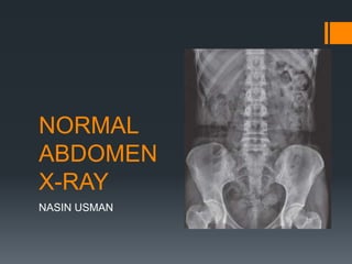
SlideShare a Scribd company logo
NORMAL
ABDOMEN
X-RAY
NASIN USMAN
PLANES AND REGIONS
 EXTEND: Inferior surface of diaphragm (superior) to the pelvic inlet
(inferior) and contained by muscles of abdominal walls.
 PLANES: Divided into nine regions by two transverse and two
parasagittal planes
I. Transpyloric plane: midway between the suprasternal notch
and the symphysis pubis (level of L1 vertebra and tips of Rt and
Lt 9th CC)
II. Transtubercular plane: level of tubercles of iliac crest and
upper border of L5
III. 2 X Parasagittal planes: run at Right angles to the transverse
planes vertically passing through a point midway between ASIS
and symphysis pubis on each side in the mid clavicular line.
 REGIONS:
Five basic densities on x-
rays
 Gas: Black
 Fat: Dark grey
 Soft tissue: Light grey
 Bone / calcification: White
 Metal: Intense white
Abdominal Organs
 Liver
 right upper quadrant
 extends to the hemidiaphragm and past the midline
 Chilaiditi’s syndrome
 Spleen
 left upper quadrant
 extends to the hemidiaphragm
 Its lower pole may be outlined by fat
 Measurement of its length from the dome of the diaphragm
to the tip. This is usually less than 14 cm
 Relationship of the spleen to the ninth, tenth and eleventh
ribs
 Normal gallbladder or biliary system are not visible. Gas
may be seen in the extrahepatic ducts in the elderly where
the ampullary tone is low, after sphincterotomy, or after
surgical anastomosis of bile ducts to small bowel
 Pancreas is not visible unless calcified. If calcification is
distributed throughout the gland it is seen as a transverse
structure at L 1 level, with a larger head on the right side and
a body and tail extending to the left and upwards.
 Psoas muscle
 symmetrical triangles either side of the lumbar spine
 Arise from the transverse processes of lumbar vertebrae
and combine with iliacus muscles to insert to lesser
trochanter of femur
 narrowest near the diaphragm, widest at the pelvis
 Stomach
 left of midline, beneath hemidiaphragm
 Gastric fundus fixed in location: within 2.5cm of left
hemidiaphragm.
 sometimes just a small volume of gas in the fundus
 do not mistake a rim of gas for pneumoperitoneum
 Kidneys
 sit on the psoas muscles at level of T12 to L3
 often just see the rounded lower pole
 Perirenal fat often makes part or all of the renal outlines
visible
 Renal size is variable, with a normal range of 10 – 15 cm on
a radiograph or approximately three-and-a-half vertebral
bodies in height
 The left kidney is usually larger, but a difference in size of
more than 2 cm is abnormal
 The kidneys are relatively larger in the child (approximately
four vertebral bodies in height)
 Adrenal glands visible only if calcified.
 Small bowel
 less than 3 cm wide
 tends to be central
 only seen if it contains gas
 3 or more air fluid levels - abnormal
 mucosal folds (valvulae conniventes) traverse the
bowel lumen
 Large bowel
 less than 6 cm wide, caecum and sigmoid up to 9
cm
 peripheral
 ascending and descending colon in fixed positions
laterally
 transverse colon and sigmoid variable position on a
mesentery
 Haustral folds do not go all the way across the
lumen
 Any air fluid levels – abnormal (?)
 Numerous gas – fluid levels may be normal and
18% of normal films have fluid levels in the
caecum
 contains faeces - mottled appearance
THE 3/6/9
RULE
VALVULAE CONNIVENTES HAUSTRAL FOLDS
 Faecoliths or fluid levels of the appendix may be visible on plain films of
the abdomen in the right iliac fossa in approximately 10% of individuals.
 Haustra:
I. The sacculation of the colon by the taeniae coli gives rise
to septa called haustra
II. The haustra are fixed anatomical structures in the
proximal colon, but in the distal colon require active con-
traction for their formation
III. Haustra may be absent distal to the midtransverse colon.
 Normal portal veins are not visible
• Gas in the portal vein and its radicles may occur in cases of
ischaemic bowel
• Portal vein gas may also be seen in well patients after
insertion of feeding tubes into the jejunum because of
physical mucosal damage caused by tunnelling of the tube.
 Lung bases
 pulmonary vessels in the bases projected over
upper abdomen
 Also look for free intra abdominal air below
the diaphragm, costophrenic angles, or for a
raised or flattened diaphragm.
Bladder: has variable appearance depending on how full it is. It has
the same density as other soft tissue structures, due to its water content.
Bones and Joints
 Spine
 lower thoracic and lumbar spine should be of similar height
 intervertebral disc spaces should be similar
 spinous processes should be visible
 Lower ribs
 Sacrum and pelvis
 Sacroiliac Joints And Hip Joints are often visualised on
abdominal radiographs. Make sure that you look at the bones
to check for other causes of abdominal pain. Evidence of
discitis, bony metastases etc.
 Bones can be used as landmarks for invisible soft tissue
structures. E.g. the transverse processes of the lumbar
vertebrae(L2 to L5) act as landmarks for the course of the
ureters. The vesico-ureteric junctions are located at the level
of the ischial spines.
Vessels
 Aorta is visible only if calcified It is then seen as linear
calcification vertically in the midline and to the left
 The shadow of the inferior vena cava can be identified as it
pierces the right hemidiaphragm and enters the heart. On a
lateral chest radiograph it identifies a hemidiaphragm as
being the right-sided one
 Factors affecting position and surface marking of organs:
a) Body build
b) Phase of respiration
c) Posture
d) Age: loss of tone of abdominal musculature
e) Pathology of organs
f) Contents of hollow viscera
g) Presence of abnormal mass
h) Normal variants within the population
Normal Variant
 Riedel’s Lobe
I. is a tongue-like, inferior
projection of the right lobe of
the liver beyond the level of
the most inferior costal
cartilage on cross-sectional
images.
II. It is not considered a true
accessory lobe of the liver
but an anatomical variant of
the right lobe of the liver.
Referral criteria
 A preliminary evaluation of bowel gas in an emergent setting: 50%
sensitivity for acute bowel obstruction
 Evaluation of radiopaque tubes and lines
 Evaluation for radiopaque foreign bodies
 Evaluation for post procedural intraperitoneal/retroperitoneal free
gas
 Monitoring the amount of bowel gas in postoperative ileus
 Monitoring the passage of contrast through the bowel
 Monitoring renal calculi: 80 – 90% sens if radiolucent stone
Procedure
 The patient should be gowned with minimum clothing.
 Radiopaque materials (zippers, belts, etc.) should be removed.
 If relevant, enteric tube suction should be avoided before the
study. Ideally, the patient's bladder should be emptied as well.
 Abdominal radiographs may be obtained in the radiology
department or may be performed portably. Portable abdominal
radiographs may be necessary due to patient immobility but are
of much poorer quality.
 Gonadal shielding may be provided for men
 Views should generally include either the diaphragm or inferior
pubic ramus
projections
 Basic: Antero-posterior - supine
 Alternative: Postero-anterior - prone
 Supplementary: * Antero-posterior – erect
* Antero-posterior or Postero-anterior
- left lateral decubitus
* Lateral – dorsal decubitus
* Anterior/Posterior obliques
AP Supine
 POSITION of patient:
I. Supine with pelvis adjusted so that ASIS are equidistant from
the tabletop. Arms placed alongside the trunk or above the
head. Median sagittal plane right angle to table.
II. CR casette positioned so that region below symphysis pubis
included.
III. Centre of image receptor located 1 cm below line joining iliac
crests.
IV. Ideally respiration arrested on full expiration.
 Picture Criteria:
I. Bowel pattern should be demonstrable with minimal
unsharpness
II. Diaphragm to symphysis pubis
III. Lateral abdominal wall and peritoneal fat layer
IV. Sharply demonstrated outline of psoas muscles, lower
border of liver, kidney.
V. Ribs and spinous processes of lumbar vertebrae
VI. Whole of urinary tract
VII. The abdomen should be free from rotation with symmetry
of the: ribs (superior), iliac crests (middle), obturator
foramen (inferior)
Free intraperitoneal gas may outline the
umbilical ligaments and falciform ligament
making them visible, thus making a
diagnosis of pneumoperitoneum possible
on a supine radiograph.
PA PRONE
 When kidneys are not of primary interest
 Reduces gonad dose
 POSITION of the patient:
I. Prone with median sagittal plane at right angles to table
II. Arms up beside head and both legs extended.
III. CR, equipment setting and picture criteria same as supine
projection.
PA ERECT
 Valuable projection in assessing air fluid levels,
and free air in the abdominal cavity.
 Perforation of a hollow abdominal viscus:
most sensitive to detect the presence of free
gas in the abdomen IS ERECT CHEST X-RAY
AND NOT ABDOMEN ERECT.
 POSITION of the patient:
I. Patient stands with back against the receptor or vertical
Bucky
II. Legs separated well apart to maintain comfortable position
III. Pelvis is adjusted so that the ASIS are equidistant
IV. Horizontal central ray directed perpendicular to midpoint
at the level of iliac crests.
 Picture criteria same as that of supine with both domes of
diaphragm visible to ensure any free air in the peritoneal
cavity.
Air fluid
levels
Lateral
 For identification and localization of foreign bodies.
 POSITION of the patient:
I. Patient turned onto the side of examination with hands
resting near the head
II. Hips and knees flexed for stability
III. Median sagittal plane parallel to table
IV. Vertebral column positioned over midline of the table
V. Immobilization band applied across pelvis
VI. Cassette centered at the level of iliac crest
VII. Vertical central ray directed to the center of
the cassette.
 Picture criteria: The prevertebral space along
with the abdominal aorta.
Lateral Decubitus
 Performed as an alternative to the PA erect view to assess
for free gas in the abdominal cavity if the patient is unable to
sit or stand.
 POSITION of the patient:
I. Patient in lateral recumbent position
II. Elbows and arms flexed and hands resting near head
III. Cassette positioned in vertical bucky against the posterior
aspect of the trunk.
IV. Central ray is directed perpendicular to the midpoint at the
level of iliac crest with x-ray tube horizontally
 Picture Criteria: elevated lateral abdominal wall included
on the image to detect any free intraperitoneal gas.
Dorsal Decubitus
 Used when it is unsafe to perform both a PA erect or a lateral
decubitus view
 This projection requires no patient movement.
 Xray beam: 5 cm above the iliac crests at the midcoronal plane
of the patient
 Picture Criteria:
I. The anterior abdominal wall and the diaphragms are
included on the image to detect any free intraperitoneal gas.
II. There should be no blurring of the bowel gas due to
respiratory motion.
III. Due to the high exposure of this examination and the need to
demonstrate soft tissue, the use of an aluminium filter over
the anterior portion of the patient is advantageous to even
out density and filter out higher energy x-rays
Pediatric Abdominal X-ray
Pockets of gas scattered
in several areas such as
Small bowel
Colon
Rectum
No excessive dilated
bowel
No air fluid levels
Imaging
 Film or IR size: 14 x 17 inches
 Moving or stationary grid
 65 – 80 kVr range
 mAs 30
Contraindications
 Pregnancy is a relative contraindication
I. Ten day rule : Whenever possible, one should confine the
radiological examination of the lower abdomen and pelvis
to the 10-day interval following the onset of menstruation.
Now this is applied only to examinations falling under high
dose.
II. 28 day rule: In case if the women
confirms she is certain she is not
pregnant and the LMP is within
28 days, it is regarded as safe.
Things to look for
 Name, Date
 Position of film and view
 Adequate area covered or not
 Bowel preparation
 Pre- Peritoneal fat lines
 Visualized organs are normal in size
 Visualized bones and joints are normal
 Visualized shadows
 Any Radio opacity
 Any artifacts
 Any calcification
Abd xray

More Related Content

What's hot

Presentation2.pptx, radiological imaging of gastric lesions.
Presentation2.pptx, radiological imaging of gastric lesions.Presentation2.pptx, radiological imaging of gastric lesions.
Presentation2.pptx, radiological imaging of gastric lesions.Abdellah Nazeer
 
Abdominal x ray
Abdominal x rayAbdominal x ray
Abdominal x ray
Mathew Joseph
 
Intravenous urography (IVU)
Intravenous urography (IVU)Intravenous urography (IVU)
Intravenous urography (IVU)
Dr Abdalla M. Gamal
 
Imaging of abdominal trauma
Imaging of abdominal traumaImaging of abdominal trauma
Imaging of abdominal trauma
Dev Lakhera
 
radiology.Plain abd.(dr.kawa)
radiology.Plain abd.(dr.kawa)radiology.Plain abd.(dr.kawa)
radiology.Plain abd.(dr.kawa)student
 
Antegrade and retrograde pyelography
Antegrade and retrograde pyelographyAntegrade and retrograde pyelography
Antegrade and retrograde pyelography
Ramayya Pramila
 
Imaging of the abdomen & the gastrointestinal
Imaging of the abdomen & the gastrointestinalImaging of the abdomen & the gastrointestinal
Imaging of the abdomen & the gastrointestinalHidayat Shariff
 
Radiology of digestive system
Radiology of digestive systemRadiology of digestive system
Radiology of digestive systemghalan
 
Kidney ultrasound
Kidney  ultrasoundKidney  ultrasound
Kidney ultrasound
Safi. Khan
 
Presentation1.pptx, radiological imaging of esophageal lesions.
Presentation1.pptx, radiological imaging of esophageal lesions.Presentation1.pptx, radiological imaging of esophageal lesions.
Presentation1.pptx, radiological imaging of esophageal lesions.Abdellah Nazeer
 
Presentation1, abdominal ultrasound anatomy.
Presentation1, abdominal ultrasound anatomy.Presentation1, abdominal ultrasound anatomy.
Presentation1, abdominal ultrasound anatomy.
Abdellah Nazeer
 
barium studies in gi pathologies
barium studies in gi pathologiesbarium studies in gi pathologies
barium studies in gi pathologies
Ahmad Jawad
 
Mrcp Radiology
Mrcp RadiologyMrcp Radiology
Mrcp Radiology
kunalj000
 
Presentation1.pptx, ultrasound examination of the liver and gall bladder.
Presentation1.pptx, ultrasound examination of the liver and gall bladder.Presentation1.pptx, ultrasound examination of the liver and gall bladder.
Presentation1.pptx, ultrasound examination of the liver and gall bladder.Abdellah Nazeer
 
Anterograde/Retrograde urethrography (RGU/MCU)
Anterograde/Retrograde urethrography (RGU/MCU)Anterograde/Retrograde urethrography (RGU/MCU)
Anterograde/Retrograde urethrography (RGU/MCU)
Shubham Singhal
 
Normal chest xray
Normal chest xrayNormal chest xray
Normal chest xray
Sai Kumar Sai
 
Abdominal xray - imaging and interpretation
Abdominal xray - imaging and interpretation Abdominal xray - imaging and interpretation
Abdominal xray - imaging and interpretation
ArushiGupta119
 
Invertogram ANORECTAL MALFORMATION ( ARM ) PRANAYA
Invertogram ANORECTAL MALFORMATION ( ARM ) PRANAYAInvertogram ANORECTAL MALFORMATION ( ARM ) PRANAYA
Invertogram ANORECTAL MALFORMATION ( ARM ) PRANAYA
PRANAYA PANIGRAHI
 

What's hot (20)

Presentation2.pptx, radiological imaging of gastric lesions.
Presentation2.pptx, radiological imaging of gastric lesions.Presentation2.pptx, radiological imaging of gastric lesions.
Presentation2.pptx, radiological imaging of gastric lesions.
 
Abdominal x ray
Abdominal x rayAbdominal x ray
Abdominal x ray
 
Intravenous urography (IVU)
Intravenous urography (IVU)Intravenous urography (IVU)
Intravenous urography (IVU)
 
Imaging of abdominal trauma
Imaging of abdominal traumaImaging of abdominal trauma
Imaging of abdominal trauma
 
radiology.Plain abd.(dr.kawa)
radiology.Plain abd.(dr.kawa)radiology.Plain abd.(dr.kawa)
radiology.Plain abd.(dr.kawa)
 
Antegrade and retrograde pyelography
Antegrade and retrograde pyelographyAntegrade and retrograde pyelography
Antegrade and retrograde pyelography
 
Imaging of the abdomen & the gastrointestinal
Imaging of the abdomen & the gastrointestinalImaging of the abdomen & the gastrointestinal
Imaging of the abdomen & the gastrointestinal
 
Radiology of digestive system
Radiology of digestive systemRadiology of digestive system
Radiology of digestive system
 
Kidney ultrasound
Kidney  ultrasoundKidney  ultrasound
Kidney ultrasound
 
Presentation1.pptx, radiological imaging of esophageal lesions.
Presentation1.pptx, radiological imaging of esophageal lesions.Presentation1.pptx, radiological imaging of esophageal lesions.
Presentation1.pptx, radiological imaging of esophageal lesions.
 
Presentation1, abdominal ultrasound anatomy.
Presentation1, abdominal ultrasound anatomy.Presentation1, abdominal ultrasound anatomy.
Presentation1, abdominal ultrasound anatomy.
 
barium studies in gi pathologies
barium studies in gi pathologiesbarium studies in gi pathologies
barium studies in gi pathologies
 
Mrcp Radiology
Mrcp RadiologyMrcp Radiology
Mrcp Radiology
 
Abdomen radiography
Abdomen radiographyAbdomen radiography
Abdomen radiography
 
Liver ultrasound
Liver ultrasoundLiver ultrasound
Liver ultrasound
 
Presentation1.pptx, ultrasound examination of the liver and gall bladder.
Presentation1.pptx, ultrasound examination of the liver and gall bladder.Presentation1.pptx, ultrasound examination of the liver and gall bladder.
Presentation1.pptx, ultrasound examination of the liver and gall bladder.
 
Anterograde/Retrograde urethrography (RGU/MCU)
Anterograde/Retrograde urethrography (RGU/MCU)Anterograde/Retrograde urethrography (RGU/MCU)
Anterograde/Retrograde urethrography (RGU/MCU)
 
Normal chest xray
Normal chest xrayNormal chest xray
Normal chest xray
 
Abdominal xray - imaging and interpretation
Abdominal xray - imaging and interpretation Abdominal xray - imaging and interpretation
Abdominal xray - imaging and interpretation
 
Invertogram ANORECTAL MALFORMATION ( ARM ) PRANAYA
Invertogram ANORECTAL MALFORMATION ( ARM ) PRANAYAInvertogram ANORECTAL MALFORMATION ( ARM ) PRANAYA
Invertogram ANORECTAL MALFORMATION ( ARM ) PRANAYA
 

Similar to Abd xray

Normal abd xray rjj.pptx
Normal abd xray rjj.pptxNormal abd xray rjj.pptx
Normal abd xray rjj.pptx
rohanjohnjacob
 
Radiological anatomy of_abdomen[1]
Radiological anatomy of_abdomen[1]Radiological anatomy of_abdomen[1]
Radiological anatomy of_abdomen[1]
suriyaprakash nagarajan
 
cxr.ppt
cxr.pptcxr.ppt
Oesophagus ppt for ss
Oesophagus ppt for ssOesophagus ppt for ss
Oesophagus ppt for ss
sateesh kumar atmakuri
 
Mj final seminar 20 01-17
Mj final seminar 20 01-17Mj final seminar 20 01-17
Mj final seminar 20 01-17
Dr. madan jakhar
 
role of radiology in pediatric.pptx
role of radiology in pediatric.pptxrole of radiology in pediatric.pptx
role of radiology in pediatric.pptx
dypradio
 
Learn Chest X-Ray With Its Normal Positioning & Radio-Anatomy
Learn Chest X-Ray With Its Normal Positioning & Radio-AnatomyLearn Chest X-Ray With Its Normal Positioning & Radio-Anatomy
Learn Chest X-Ray With Its Normal Positioning & Radio-Anatomy
Dr.Santosh Atreya
 
Thorax and abdomen & pelvis
Thorax and abdomen & pelvisThorax and abdomen & pelvis
Thorax and abdomen & pelvisMpdodz
 
chest-x-ray.pptx
chest-x-ray.pptxchest-x-ray.pptx
chest-x-ray.pptx
VasanthakohilaMuthuk
 
Radiographic interpretation
Radiographic interpretationRadiographic interpretation
Radiographic interpretation
Prince Avi
 
Liver ultrasound, step by step, part 1
Liver ultrasound, step by step, part 1Liver ultrasound, step by step, part 1
Liver ultrasound, step by step, part 1
Durre Sabih
 
Chest x ray
Chest x rayChest x ray
Chest x ray
Dr,saket Jain
 
Radiography OF Abdomen -NISCHAL_NMC.pptx
Radiography OF Abdomen -NISCHAL_NMC.pptxRadiography OF Abdomen -NISCHAL_NMC.pptx
Radiography OF Abdomen -NISCHAL_NMC.pptx
nisalsilakar
 
Anatomy of the stomach
Anatomy of the stomachAnatomy of the stomach
Anatomy of the stomach
drsukriti1
 
Thorax-XRAY and CT
Thorax-XRAY and CTThorax-XRAY and CT
Thorax-XRAY and CT
dypradio
 
Anatomy of esophgus
Anatomy of esophgusAnatomy of esophgus
Anatomy of esophgus
Anish Choudhary
 
Wayang kulit, no 1 a fundamentals
Wayang kulit, no 1 a fundamentalsWayang kulit, no 1 a fundamentals
Wayang kulit, no 1 a fundamentals
ycche19
 
Anatomy of gastroesophagial junction with specail reference to hiatus hernia...
Anatomy of gastroesophagial junction  with specail reference to hiatus hernia...Anatomy of gastroesophagial junction  with specail reference to hiatus hernia...
Anatomy of gastroesophagial junction with specail reference to hiatus hernia...
Rana Singh
 
Interpretation of chest xray ppt
Interpretation of chest xray pptInterpretation of chest xray ppt
Interpretation of chest xray ppt
Rithwik Karumuri
 
Approach to cxr.pptx
Approach to cxr.pptxApproach to cxr.pptx
Approach to cxr.pptx
MohammadMamunuzzaman2
 

Similar to Abd xray (20)

Normal abd xray rjj.pptx
Normal abd xray rjj.pptxNormal abd xray rjj.pptx
Normal abd xray rjj.pptx
 
Radiological anatomy of_abdomen[1]
Radiological anatomy of_abdomen[1]Radiological anatomy of_abdomen[1]
Radiological anatomy of_abdomen[1]
 
cxr.ppt
cxr.pptcxr.ppt
cxr.ppt
 
Oesophagus ppt for ss
Oesophagus ppt for ssOesophagus ppt for ss
Oesophagus ppt for ss
 
Mj final seminar 20 01-17
Mj final seminar 20 01-17Mj final seminar 20 01-17
Mj final seminar 20 01-17
 
role of radiology in pediatric.pptx
role of radiology in pediatric.pptxrole of radiology in pediatric.pptx
role of radiology in pediatric.pptx
 
Learn Chest X-Ray With Its Normal Positioning & Radio-Anatomy
Learn Chest X-Ray With Its Normal Positioning & Radio-AnatomyLearn Chest X-Ray With Its Normal Positioning & Radio-Anatomy
Learn Chest X-Ray With Its Normal Positioning & Radio-Anatomy
 
Thorax and abdomen & pelvis
Thorax and abdomen & pelvisThorax and abdomen & pelvis
Thorax and abdomen & pelvis
 
chest-x-ray.pptx
chest-x-ray.pptxchest-x-ray.pptx
chest-x-ray.pptx
 
Radiographic interpretation
Radiographic interpretationRadiographic interpretation
Radiographic interpretation
 
Liver ultrasound, step by step, part 1
Liver ultrasound, step by step, part 1Liver ultrasound, step by step, part 1
Liver ultrasound, step by step, part 1
 
Chest x ray
Chest x rayChest x ray
Chest x ray
 
Radiography OF Abdomen -NISCHAL_NMC.pptx
Radiography OF Abdomen -NISCHAL_NMC.pptxRadiography OF Abdomen -NISCHAL_NMC.pptx
Radiography OF Abdomen -NISCHAL_NMC.pptx
 
Anatomy of the stomach
Anatomy of the stomachAnatomy of the stomach
Anatomy of the stomach
 
Thorax-XRAY and CT
Thorax-XRAY and CTThorax-XRAY and CT
Thorax-XRAY and CT
 
Anatomy of esophgus
Anatomy of esophgusAnatomy of esophgus
Anatomy of esophgus
 
Wayang kulit, no 1 a fundamentals
Wayang kulit, no 1 a fundamentalsWayang kulit, no 1 a fundamentals
Wayang kulit, no 1 a fundamentals
 
Anatomy of gastroesophagial junction with specail reference to hiatus hernia...
Anatomy of gastroesophagial junction  with specail reference to hiatus hernia...Anatomy of gastroesophagial junction  with specail reference to hiatus hernia...
Anatomy of gastroesophagial junction with specail reference to hiatus hernia...
 
Interpretation of chest xray ppt
Interpretation of chest xray pptInterpretation of chest xray ppt
Interpretation of chest xray ppt
 
Approach to cxr.pptx
Approach to cxr.pptxApproach to cxr.pptx
Approach to cxr.pptx
 

Recently uploaded

Evaluation of antidepressant activity of clitoris ternatea in animals
Evaluation of antidepressant activity of clitoris ternatea in animalsEvaluation of antidepressant activity of clitoris ternatea in animals
Evaluation of antidepressant activity of clitoris ternatea in animals
Shweta
 
Tom Selleck Health: A Comprehensive Look at the Iconic Actor’s Wellness Journey
Tom Selleck Health: A Comprehensive Look at the Iconic Actor’s Wellness JourneyTom Selleck Health: A Comprehensive Look at the Iconic Actor’s Wellness Journey
Tom Selleck Health: A Comprehensive Look at the Iconic Actor’s Wellness Journey
greendigital
 
The POPPY STUDY (Preconception to post-partum cardiovascular function in prim...
The POPPY STUDY (Preconception to post-partum cardiovascular function in prim...The POPPY STUDY (Preconception to post-partum cardiovascular function in prim...
The POPPY STUDY (Preconception to post-partum cardiovascular function in prim...
Catherine Liao
 
MANAGEMENT OF ATRIOVENTRICULAR CONDUCTION BLOCK.pdf
MANAGEMENT OF ATRIOVENTRICULAR CONDUCTION BLOCK.pdfMANAGEMENT OF ATRIOVENTRICULAR CONDUCTION BLOCK.pdf
MANAGEMENT OF ATRIOVENTRICULAR CONDUCTION BLOCK.pdf
Jim Jacob Roy
 
Ozempic: Preoperative Management of Patients on GLP-1 Receptor Agonists
Ozempic: Preoperative Management of Patients on GLP-1 Receptor Agonists  Ozempic: Preoperative Management of Patients on GLP-1 Receptor Agonists
Ozempic: Preoperative Management of Patients on GLP-1 Receptor Agonists
Saeid Safari
 
ARTHROLOGY PPT NCISM SYLLABUS AYURVEDA STUDENTS
ARTHROLOGY PPT NCISM SYLLABUS AYURVEDA STUDENTSARTHROLOGY PPT NCISM SYLLABUS AYURVEDA STUDENTS
ARTHROLOGY PPT NCISM SYLLABUS AYURVEDA STUDENTS
Dr. Vinay Pareek
 
POST OPERATIVE OLIGURIA and its management
POST OPERATIVE OLIGURIA and its managementPOST OPERATIVE OLIGURIA and its management
POST OPERATIVE OLIGURIA and its management
touseefaziz1
 
HOT NEW PRODUCT! BIG SALES FAST SHIPPING NOW FROM CHINA!! EU KU DB BK substit...
HOT NEW PRODUCT! BIG SALES FAST SHIPPING NOW FROM CHINA!! EU KU DB BK substit...HOT NEW PRODUCT! BIG SALES FAST SHIPPING NOW FROM CHINA!! EU KU DB BK substit...
HOT NEW PRODUCT! BIG SALES FAST SHIPPING NOW FROM CHINA!! EU KU DB BK substit...
GL Anaacs
 
THOA 2.ppt Human Organ Transplantation Act
THOA 2.ppt Human Organ Transplantation ActTHOA 2.ppt Human Organ Transplantation Act
THOA 2.ppt Human Organ Transplantation Act
DrSathishMS1
 
Superficial & Deep Fascia of the NECK.pptx
Superficial & Deep Fascia of the NECK.pptxSuperficial & Deep Fascia of the NECK.pptx
Superficial & Deep Fascia of the NECK.pptx
Dr. Rabia Inam Gandapore
 
Ophthalmology Clinical Tests for OSCE exam
Ophthalmology Clinical Tests for OSCE examOphthalmology Clinical Tests for OSCE exam
Ophthalmology Clinical Tests for OSCE exam
KafrELShiekh University
 
Antiulcer drugs Advance Pharmacology .pptx
Antiulcer drugs Advance Pharmacology .pptxAntiulcer drugs Advance Pharmacology .pptx
Antiulcer drugs Advance Pharmacology .pptx
Rohit chaurpagar
 
For Better Surat #ℂall #Girl Service ❤85270-49040❤ Surat #ℂall #Girls
For Better Surat #ℂall #Girl Service ❤85270-49040❤ Surat #ℂall #GirlsFor Better Surat #ℂall #Girl Service ❤85270-49040❤ Surat #ℂall #Girls
For Better Surat #ℂall #Girl Service ❤85270-49040❤ Surat #ℂall #Girls
Savita Shen $i11
 
Factory Supply Best Quality Pmk Oil CAS 28578–16–7 PMK Powder in Stock
Factory Supply Best Quality Pmk Oil CAS 28578–16–7 PMK Powder in StockFactory Supply Best Quality Pmk Oil CAS 28578–16–7 PMK Powder in Stock
Factory Supply Best Quality Pmk Oil CAS 28578–16–7 PMK Powder in Stock
rebeccabio
 
The Normal Electrocardiogram - Part I of II
The Normal Electrocardiogram - Part I of IIThe Normal Electrocardiogram - Part I of II
The Normal Electrocardiogram - Part I of II
MedicoseAcademics
 
BRACHYTHERAPY OVERVIEW AND APPLICATORS
BRACHYTHERAPY OVERVIEW  AND  APPLICATORSBRACHYTHERAPY OVERVIEW  AND  APPLICATORS
BRACHYTHERAPY OVERVIEW AND APPLICATORS
Krishan Murari
 
24 Upakrama.pptx class ppt useful in all
24 Upakrama.pptx class ppt useful in all24 Upakrama.pptx class ppt useful in all
24 Upakrama.pptx class ppt useful in all
DrSathishMS1
 
Surgical Site Infections, pathophysiology, and prevention.pptx
Surgical Site Infections, pathophysiology, and prevention.pptxSurgical Site Infections, pathophysiology, and prevention.pptx
Surgical Site Infections, pathophysiology, and prevention.pptx
jval Landero
 
Hemodialysis: Chapter 3, Dialysis Water Unit - Dr.Gawad
Hemodialysis: Chapter 3, Dialysis Water Unit - Dr.GawadHemodialysis: Chapter 3, Dialysis Water Unit - Dr.Gawad
Hemodialysis: Chapter 3, Dialysis Water Unit - Dr.Gawad
NephroTube - Dr.Gawad
 
ARTIFICIAL INTELLIGENCE IN HEALTHCARE.pdf
ARTIFICIAL INTELLIGENCE IN  HEALTHCARE.pdfARTIFICIAL INTELLIGENCE IN  HEALTHCARE.pdf
ARTIFICIAL INTELLIGENCE IN HEALTHCARE.pdf
Anujkumaranit
 

Recently uploaded (20)

Evaluation of antidepressant activity of clitoris ternatea in animals
Evaluation of antidepressant activity of clitoris ternatea in animalsEvaluation of antidepressant activity of clitoris ternatea in animals
Evaluation of antidepressant activity of clitoris ternatea in animals
 
Tom Selleck Health: A Comprehensive Look at the Iconic Actor’s Wellness Journey
Tom Selleck Health: A Comprehensive Look at the Iconic Actor’s Wellness JourneyTom Selleck Health: A Comprehensive Look at the Iconic Actor’s Wellness Journey
Tom Selleck Health: A Comprehensive Look at the Iconic Actor’s Wellness Journey
 
The POPPY STUDY (Preconception to post-partum cardiovascular function in prim...
The POPPY STUDY (Preconception to post-partum cardiovascular function in prim...The POPPY STUDY (Preconception to post-partum cardiovascular function in prim...
The POPPY STUDY (Preconception to post-partum cardiovascular function in prim...
 
MANAGEMENT OF ATRIOVENTRICULAR CONDUCTION BLOCK.pdf
MANAGEMENT OF ATRIOVENTRICULAR CONDUCTION BLOCK.pdfMANAGEMENT OF ATRIOVENTRICULAR CONDUCTION BLOCK.pdf
MANAGEMENT OF ATRIOVENTRICULAR CONDUCTION BLOCK.pdf
 
Ozempic: Preoperative Management of Patients on GLP-1 Receptor Agonists
Ozempic: Preoperative Management of Patients on GLP-1 Receptor Agonists  Ozempic: Preoperative Management of Patients on GLP-1 Receptor Agonists
Ozempic: Preoperative Management of Patients on GLP-1 Receptor Agonists
 
ARTHROLOGY PPT NCISM SYLLABUS AYURVEDA STUDENTS
ARTHROLOGY PPT NCISM SYLLABUS AYURVEDA STUDENTSARTHROLOGY PPT NCISM SYLLABUS AYURVEDA STUDENTS
ARTHROLOGY PPT NCISM SYLLABUS AYURVEDA STUDENTS
 
POST OPERATIVE OLIGURIA and its management
POST OPERATIVE OLIGURIA and its managementPOST OPERATIVE OLIGURIA and its management
POST OPERATIVE OLIGURIA and its management
 
HOT NEW PRODUCT! BIG SALES FAST SHIPPING NOW FROM CHINA!! EU KU DB BK substit...
HOT NEW PRODUCT! BIG SALES FAST SHIPPING NOW FROM CHINA!! EU KU DB BK substit...HOT NEW PRODUCT! BIG SALES FAST SHIPPING NOW FROM CHINA!! EU KU DB BK substit...
HOT NEW PRODUCT! BIG SALES FAST SHIPPING NOW FROM CHINA!! EU KU DB BK substit...
 
THOA 2.ppt Human Organ Transplantation Act
THOA 2.ppt Human Organ Transplantation ActTHOA 2.ppt Human Organ Transplantation Act
THOA 2.ppt Human Organ Transplantation Act
 
Superficial & Deep Fascia of the NECK.pptx
Superficial & Deep Fascia of the NECK.pptxSuperficial & Deep Fascia of the NECK.pptx
Superficial & Deep Fascia of the NECK.pptx
 
Ophthalmology Clinical Tests for OSCE exam
Ophthalmology Clinical Tests for OSCE examOphthalmology Clinical Tests for OSCE exam
Ophthalmology Clinical Tests for OSCE exam
 
Antiulcer drugs Advance Pharmacology .pptx
Antiulcer drugs Advance Pharmacology .pptxAntiulcer drugs Advance Pharmacology .pptx
Antiulcer drugs Advance Pharmacology .pptx
 
For Better Surat #ℂall #Girl Service ❤85270-49040❤ Surat #ℂall #Girls
For Better Surat #ℂall #Girl Service ❤85270-49040❤ Surat #ℂall #GirlsFor Better Surat #ℂall #Girl Service ❤85270-49040❤ Surat #ℂall #Girls
For Better Surat #ℂall #Girl Service ❤85270-49040❤ Surat #ℂall #Girls
 
Factory Supply Best Quality Pmk Oil CAS 28578–16–7 PMK Powder in Stock
Factory Supply Best Quality Pmk Oil CAS 28578–16–7 PMK Powder in StockFactory Supply Best Quality Pmk Oil CAS 28578–16–7 PMK Powder in Stock
Factory Supply Best Quality Pmk Oil CAS 28578–16–7 PMK Powder in Stock
 
The Normal Electrocardiogram - Part I of II
The Normal Electrocardiogram - Part I of IIThe Normal Electrocardiogram - Part I of II
The Normal Electrocardiogram - Part I of II
 
BRACHYTHERAPY OVERVIEW AND APPLICATORS
BRACHYTHERAPY OVERVIEW  AND  APPLICATORSBRACHYTHERAPY OVERVIEW  AND  APPLICATORS
BRACHYTHERAPY OVERVIEW AND APPLICATORS
 
24 Upakrama.pptx class ppt useful in all
24 Upakrama.pptx class ppt useful in all24 Upakrama.pptx class ppt useful in all
24 Upakrama.pptx class ppt useful in all
 
Surgical Site Infections, pathophysiology, and prevention.pptx
Surgical Site Infections, pathophysiology, and prevention.pptxSurgical Site Infections, pathophysiology, and prevention.pptx
Surgical Site Infections, pathophysiology, and prevention.pptx
 
Hemodialysis: Chapter 3, Dialysis Water Unit - Dr.Gawad
Hemodialysis: Chapter 3, Dialysis Water Unit - Dr.GawadHemodialysis: Chapter 3, Dialysis Water Unit - Dr.Gawad
Hemodialysis: Chapter 3, Dialysis Water Unit - Dr.Gawad
 
ARTIFICIAL INTELLIGENCE IN HEALTHCARE.pdf
ARTIFICIAL INTELLIGENCE IN  HEALTHCARE.pdfARTIFICIAL INTELLIGENCE IN  HEALTHCARE.pdf
ARTIFICIAL INTELLIGENCE IN HEALTHCARE.pdf
 

Abd xray

  • 2. PLANES AND REGIONS  EXTEND: Inferior surface of diaphragm (superior) to the pelvic inlet (inferior) and contained by muscles of abdominal walls.  PLANES: Divided into nine regions by two transverse and two parasagittal planes I. Transpyloric plane: midway between the suprasternal notch and the symphysis pubis (level of L1 vertebra and tips of Rt and Lt 9th CC) II. Transtubercular plane: level of tubercles of iliac crest and upper border of L5 III. 2 X Parasagittal planes: run at Right angles to the transverse planes vertically passing through a point midway between ASIS and symphysis pubis on each side in the mid clavicular line.
  • 4. Five basic densities on x- rays  Gas: Black  Fat: Dark grey  Soft tissue: Light grey  Bone / calcification: White  Metal: Intense white
  • 5. Abdominal Organs  Liver  right upper quadrant  extends to the hemidiaphragm and past the midline  Chilaiditi’s syndrome  Spleen  left upper quadrant  extends to the hemidiaphragm  Its lower pole may be outlined by fat  Measurement of its length from the dome of the diaphragm to the tip. This is usually less than 14 cm  Relationship of the spleen to the ninth, tenth and eleventh ribs
  • 6.  Normal gallbladder or biliary system are not visible. Gas may be seen in the extrahepatic ducts in the elderly where the ampullary tone is low, after sphincterotomy, or after surgical anastomosis of bile ducts to small bowel  Pancreas is not visible unless calcified. If calcification is distributed throughout the gland it is seen as a transverse structure at L 1 level, with a larger head on the right side and a body and tail extending to the left and upwards.  Psoas muscle  symmetrical triangles either side of the lumbar spine  Arise from the transverse processes of lumbar vertebrae and combine with iliacus muscles to insert to lesser trochanter of femur  narrowest near the diaphragm, widest at the pelvis
  • 7.  Stomach  left of midline, beneath hemidiaphragm  Gastric fundus fixed in location: within 2.5cm of left hemidiaphragm.  sometimes just a small volume of gas in the fundus  do not mistake a rim of gas for pneumoperitoneum  Kidneys  sit on the psoas muscles at level of T12 to L3  often just see the rounded lower pole  Perirenal fat often makes part or all of the renal outlines visible  Renal size is variable, with a normal range of 10 – 15 cm on a radiograph or approximately three-and-a-half vertebral bodies in height  The left kidney is usually larger, but a difference in size of more than 2 cm is abnormal  The kidneys are relatively larger in the child (approximately four vertebral bodies in height)  Adrenal glands visible only if calcified.
  • 8.  Small bowel  less than 3 cm wide  tends to be central  only seen if it contains gas  3 or more air fluid levels - abnormal  mucosal folds (valvulae conniventes) traverse the bowel lumen  Large bowel  less than 6 cm wide, caecum and sigmoid up to 9 cm  peripheral  ascending and descending colon in fixed positions laterally  transverse colon and sigmoid variable position on a mesentery  Haustral folds do not go all the way across the lumen  Any air fluid levels – abnormal (?)  Numerous gas – fluid levels may be normal and 18% of normal films have fluid levels in the caecum  contains faeces - mottled appearance THE 3/6/9 RULE
  • 9. VALVULAE CONNIVENTES HAUSTRAL FOLDS  Faecoliths or fluid levels of the appendix may be visible on plain films of the abdomen in the right iliac fossa in approximately 10% of individuals.
  • 10.  Haustra: I. The sacculation of the colon by the taeniae coli gives rise to septa called haustra II. The haustra are fixed anatomical structures in the proximal colon, but in the distal colon require active con- traction for their formation III. Haustra may be absent distal to the midtransverse colon.
  • 11.  Normal portal veins are not visible • Gas in the portal vein and its radicles may occur in cases of ischaemic bowel • Portal vein gas may also be seen in well patients after insertion of feeding tubes into the jejunum because of physical mucosal damage caused by tunnelling of the tube.  Lung bases  pulmonary vessels in the bases projected over upper abdomen  Also look for free intra abdominal air below the diaphragm, costophrenic angles, or for a raised or flattened diaphragm.
  • 12. Bladder: has variable appearance depending on how full it is. It has the same density as other soft tissue structures, due to its water content.
  • 13.
  • 14. Bones and Joints  Spine  lower thoracic and lumbar spine should be of similar height  intervertebral disc spaces should be similar  spinous processes should be visible  Lower ribs  Sacrum and pelvis  Sacroiliac Joints And Hip Joints are often visualised on abdominal radiographs. Make sure that you look at the bones to check for other causes of abdominal pain. Evidence of discitis, bony metastases etc.  Bones can be used as landmarks for invisible soft tissue structures. E.g. the transverse processes of the lumbar vertebrae(L2 to L5) act as landmarks for the course of the ureters. The vesico-ureteric junctions are located at the level of the ischial spines.
  • 15. Vessels  Aorta is visible only if calcified It is then seen as linear calcification vertically in the midline and to the left  The shadow of the inferior vena cava can be identified as it pierces the right hemidiaphragm and enters the heart. On a lateral chest radiograph it identifies a hemidiaphragm as being the right-sided one
  • 16.  Factors affecting position and surface marking of organs: a) Body build b) Phase of respiration c) Posture d) Age: loss of tone of abdominal musculature e) Pathology of organs f) Contents of hollow viscera g) Presence of abnormal mass h) Normal variants within the population
  • 17. Normal Variant  Riedel’s Lobe I. is a tongue-like, inferior projection of the right lobe of the liver beyond the level of the most inferior costal cartilage on cross-sectional images. II. It is not considered a true accessory lobe of the liver but an anatomical variant of the right lobe of the liver.
  • 18. Referral criteria  A preliminary evaluation of bowel gas in an emergent setting: 50% sensitivity for acute bowel obstruction  Evaluation of radiopaque tubes and lines  Evaluation for radiopaque foreign bodies  Evaluation for post procedural intraperitoneal/retroperitoneal free gas  Monitoring the amount of bowel gas in postoperative ileus  Monitoring the passage of contrast through the bowel  Monitoring renal calculi: 80 – 90% sens if radiolucent stone
  • 19. Procedure  The patient should be gowned with minimum clothing.  Radiopaque materials (zippers, belts, etc.) should be removed.  If relevant, enteric tube suction should be avoided before the study. Ideally, the patient's bladder should be emptied as well.  Abdominal radiographs may be obtained in the radiology department or may be performed portably. Portable abdominal radiographs may be necessary due to patient immobility but are of much poorer quality.  Gonadal shielding may be provided for men  Views should generally include either the diaphragm or inferior pubic ramus
  • 20. projections  Basic: Antero-posterior - supine  Alternative: Postero-anterior - prone  Supplementary: * Antero-posterior – erect * Antero-posterior or Postero-anterior - left lateral decubitus * Lateral – dorsal decubitus * Anterior/Posterior obliques
  • 21. AP Supine  POSITION of patient: I. Supine with pelvis adjusted so that ASIS are equidistant from the tabletop. Arms placed alongside the trunk or above the head. Median sagittal plane right angle to table. II. CR casette positioned so that region below symphysis pubis included. III. Centre of image receptor located 1 cm below line joining iliac crests. IV. Ideally respiration arrested on full expiration.
  • 22.  Picture Criteria: I. Bowel pattern should be demonstrable with minimal unsharpness II. Diaphragm to symphysis pubis III. Lateral abdominal wall and peritoneal fat layer IV. Sharply demonstrated outline of psoas muscles, lower border of liver, kidney. V. Ribs and spinous processes of lumbar vertebrae VI. Whole of urinary tract VII. The abdomen should be free from rotation with symmetry of the: ribs (superior), iliac crests (middle), obturator foramen (inferior)
  • 23.
  • 24. Free intraperitoneal gas may outline the umbilical ligaments and falciform ligament making them visible, thus making a diagnosis of pneumoperitoneum possible on a supine radiograph.
  • 25. PA PRONE  When kidneys are not of primary interest  Reduces gonad dose  POSITION of the patient: I. Prone with median sagittal plane at right angles to table II. Arms up beside head and both legs extended. III. CR, equipment setting and picture criteria same as supine projection.
  • 26. PA ERECT  Valuable projection in assessing air fluid levels, and free air in the abdominal cavity.  Perforation of a hollow abdominal viscus: most sensitive to detect the presence of free gas in the abdomen IS ERECT CHEST X-RAY AND NOT ABDOMEN ERECT.
  • 27.  POSITION of the patient: I. Patient stands with back against the receptor or vertical Bucky II. Legs separated well apart to maintain comfortable position III. Pelvis is adjusted so that the ASIS are equidistant IV. Horizontal central ray directed perpendicular to midpoint at the level of iliac crests.
  • 28.  Picture criteria same as that of supine with both domes of diaphragm visible to ensure any free air in the peritoneal cavity. Air fluid levels
  • 29. Lateral  For identification and localization of foreign bodies.  POSITION of the patient: I. Patient turned onto the side of examination with hands resting near the head II. Hips and knees flexed for stability III. Median sagittal plane parallel to table IV. Vertebral column positioned over midline of the table V. Immobilization band applied across pelvis VI. Cassette centered at the level of iliac crest VII. Vertical central ray directed to the center of the cassette.  Picture criteria: The prevertebral space along with the abdominal aorta.
  • 30.
  • 31. Lateral Decubitus  Performed as an alternative to the PA erect view to assess for free gas in the abdominal cavity if the patient is unable to sit or stand.  POSITION of the patient: I. Patient in lateral recumbent position II. Elbows and arms flexed and hands resting near head III. Cassette positioned in vertical bucky against the posterior aspect of the trunk. IV. Central ray is directed perpendicular to the midpoint at the level of iliac crest with x-ray tube horizontally
  • 32.  Picture Criteria: elevated lateral abdominal wall included on the image to detect any free intraperitoneal gas.
  • 33. Dorsal Decubitus  Used when it is unsafe to perform both a PA erect or a lateral decubitus view  This projection requires no patient movement.  Xray beam: 5 cm above the iliac crests at the midcoronal plane of the patient  Picture Criteria: I. The anterior abdominal wall and the diaphragms are included on the image to detect any free intraperitoneal gas. II. There should be no blurring of the bowel gas due to respiratory motion. III. Due to the high exposure of this examination and the need to demonstrate soft tissue, the use of an aluminium filter over the anterior portion of the patient is advantageous to even out density and filter out higher energy x-rays
  • 34.
  • 35. Pediatric Abdominal X-ray Pockets of gas scattered in several areas such as Small bowel Colon Rectum No excessive dilated bowel No air fluid levels
  • 36. Imaging  Film or IR size: 14 x 17 inches  Moving or stationary grid  65 – 80 kVr range  mAs 30
  • 37. Contraindications  Pregnancy is a relative contraindication I. Ten day rule : Whenever possible, one should confine the radiological examination of the lower abdomen and pelvis to the 10-day interval following the onset of menstruation. Now this is applied only to examinations falling under high dose. II. 28 day rule: In case if the women confirms she is certain she is not pregnant and the LMP is within 28 days, it is regarded as safe.
  • 38. Things to look for  Name, Date  Position of film and view  Adequate area covered or not  Bowel preparation  Pre- Peritoneal fat lines  Visualized organs are normal in size  Visualized bones and joints are normal  Visualized shadows  Any Radio opacity  Any artifacts  Any calcification