Downloaded 1,929 times
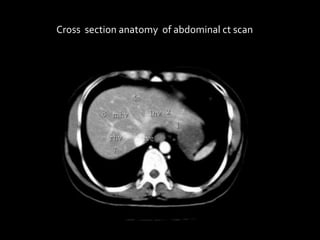
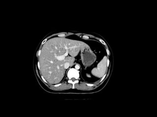
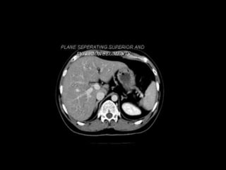
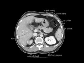
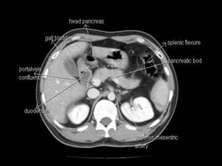
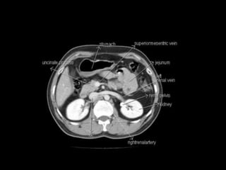
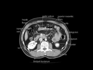
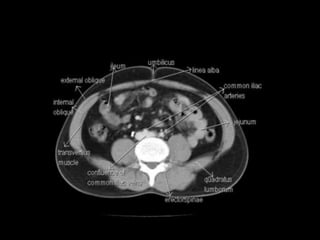
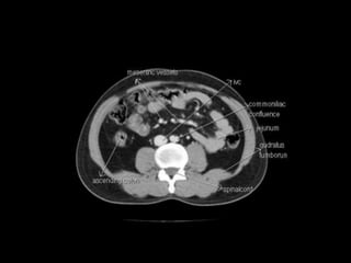
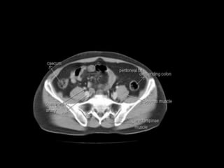
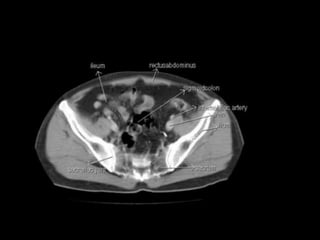
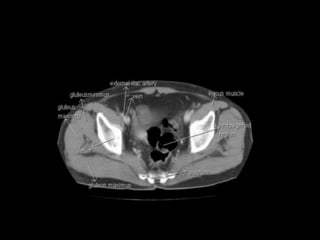
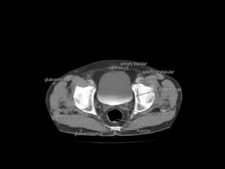
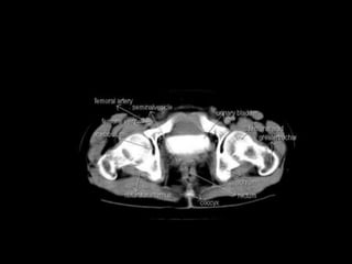
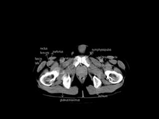
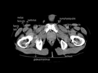
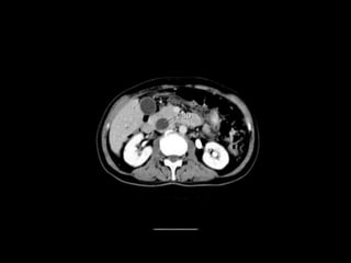
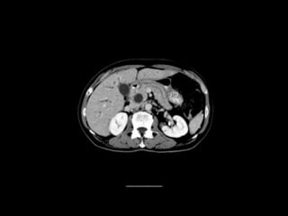
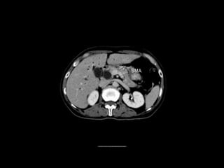
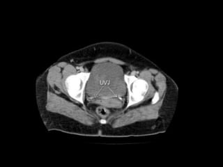
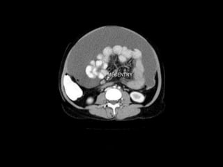
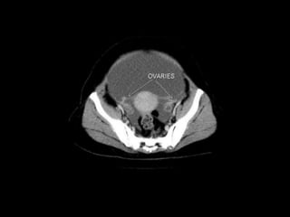
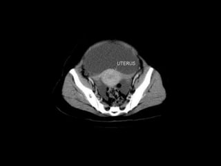
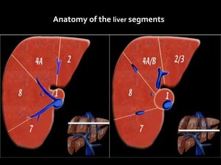
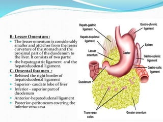
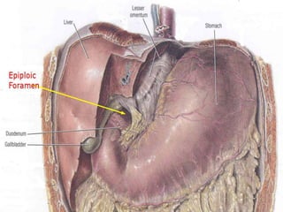
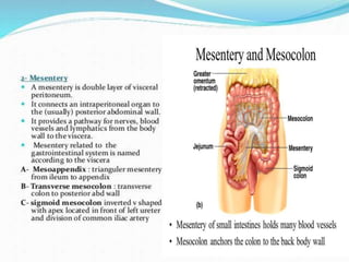
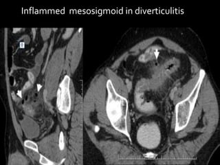
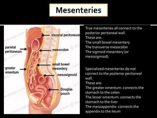
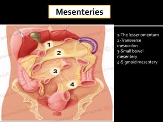
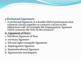
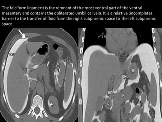

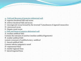
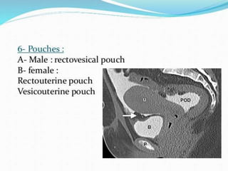
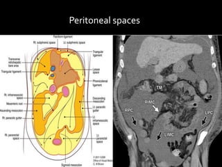
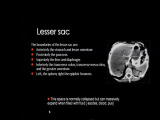
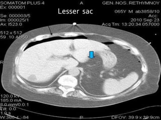
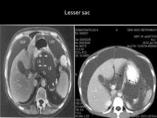
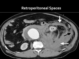

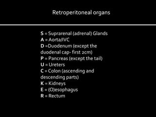


The document provides an anatomical overview of the liver segments and their vascular supplies, as well as details about the peritoneum and its spaces. It describes the organization of the liver into segments based on venous drainage, the classification of mesenteries, and the delineation of peritoneal regions. Additionally, it explains the retroperitoneal space and its divisions, highlighting the importance of these anatomical structures in medical imaging.


