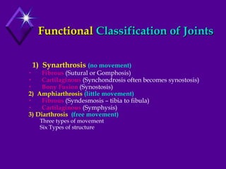
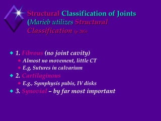
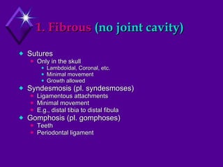
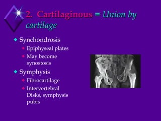
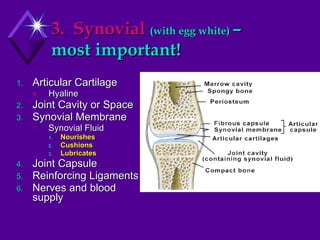
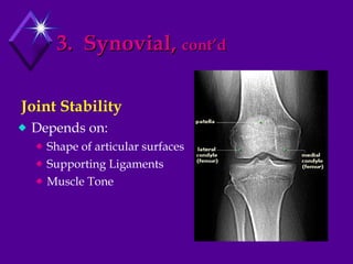
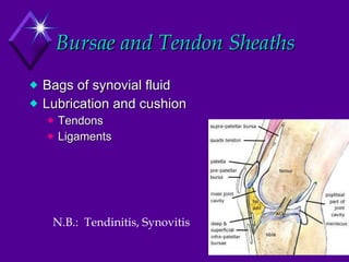
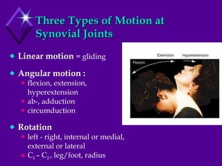
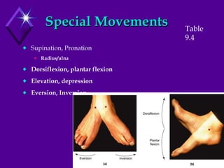
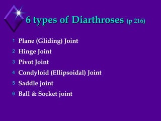
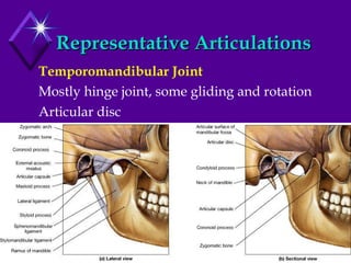
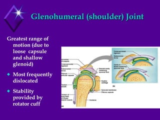
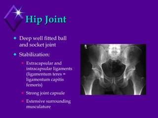
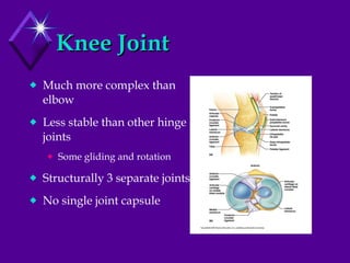
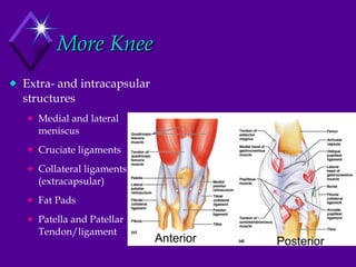
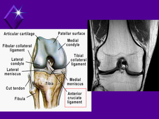
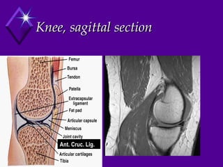
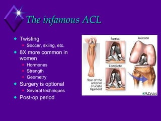
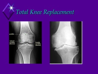
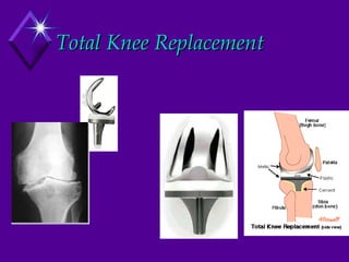
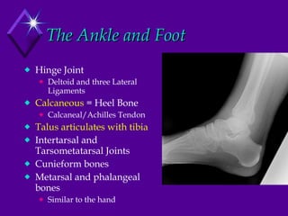
Buy your lab coat and gloves!
Chapter 9 Joints = Articulations Where ANY bones come together
Functional  Classification of Joints 1)  Synarthrosis   (no movement) Fibrous  (Sutural or Gomphosis) Cartilaginous  (Synchondrosis often becomes synostosis) Bony Fusion  (Synostosis) 2)  Amphiarthrosis   (little movement) Fibrous  (Syndesmosis – tibia to fibula)  Cartilaginous  (Symphysis) 3) Diarthrosis  (free movement) Three types of movement Six Types of structure
Structural  Classification of Joints ( Marieb utilizes  Structural Classification  (p 205) 1.  Fibrous  (no joint cavity) Almost no movement, little CT E.g, Sutures in calvarium 2.  Cartilaginous E.g., Symphysis pubis, IV disks 3.  Synovial  – by far most important
1. Fibrous  (no joint cavity) Sutures Only in the skull Lambdoidal, Coronal, etc. Minimal movement Growth allowed Syndesmosis (pl. syndesmoses) Ligamentous attachments Minimal movement E.g., distal tibia to distal fibula Gomphosis (pl. gomphoses) Teeth Periodontal ligament
2.  Cartilaginous  =  Union by cartilage Synchondrosis Epiphyseal plates May become synostosis Symphysis Fibrocartilage Intervertebral Disks, symphysis pubis
3.  Synovial   (with egg white)  –  most important! Articular Cartilage Hyaline Joint Cavity or Space Synovial Membrane Synovial Fluid Nourishes Cushions Lubricates Joint Capsule Reinforcing Ligaments Nerves and blood supply
3.  Synovial,  cont’d Depends on: Shape of articular surfaces Supporting Ligaments Muscle Tone Joint Stability
Bursae and Tendon Sheaths Bags of synovial fluid Lubrication and cushion Tendons Ligaments N.B.:  Tendinitis, Synovitis
Three Types of Motion at Synovial Joints Linear motion  = gliding Angular motion :  flexion, extension, hyperextension ab-, adduction circumduction Rotation left - right, internal or medial, external or lateral C 1  – C 2  , leg/foot, radius
Special Movements Supination, Pronation Radius/ulna Dorsiflexion, plantar flexion Elevation, depression Eversion, Inversion Table 9.4
6 types of Diarthroses  (p 216) Plane (Gliding) Joint Hinge Joint Pivot Joint Condyloid (Ellipsoidal) Joint Saddle joint Ball & Socket joint
Representative Articulations Temporomandibular Joint Mostly hinge joint, some gliding and rotation Articular disc
Intervertebral articulations Gliding joints  between vertebrae (facets) Intervertebral discs:   (Amphiarthroses)  annulus fibrosus:   tough outer layer (fibrocartilage)  nucleus pulposus:   soft, gelatinous core Account for ~25% of vertebral column height – H 2 O loss during aging
Glenohumeral (shoulder) Joint Greatest range of motion (due to loose  capsule and shallow glenoid) Most frequently dislocated Stability provided by rotator cuff
Hip Joint Deep well fitted ball and socket joint Stabilization:  Extracapsular and intracapsular ligaments (ligamentum teres = ligamentum capitis femoris) Strong joint capsule Extensive surrounding musculature
Knee Joint Much more complex than elbow Less stable than other hinge joints Some gliding and rotation Structurally 3 separate joints No single joint capsule
More Knee Extra- and intracapsular structures Medial and lateral meniscus Cruciate ligaments Collateral ligaments (extracapsular) Fat Pads Patella and Patellar Tendon/ligament Anterior Posterior
 
Knee, sagittal section Ant. Cruc. Lig.
The infamous ACL Twisting Soccer, skiing, etc. 8X more common in women Hormones Strength Geometry Surgery is optional Several techniques Post-op period
Total Knee Replacement
The Ankle and Foot Hinge Joint Deltoid and three Lateral Ligaments Calcaneous  = Heel Bone Calcaneal/Achilles Tendon Talus articulates with tibia Intertarsal and Tarsometatarsal Joints Cunieform bones Metarsal and phalangeal bones Similar to the hand


Chapter9 jointsmarieb

  • 1.
    Buy your labcoat and gloves!
  • 2.
    Chapter 9 Joints= Articulations Where ANY bones come together
  • 3.
    Functional Classificationof Joints 1) Synarthrosis (no movement) Fibrous (Sutural or Gomphosis) Cartilaginous (Synchondrosis often becomes synostosis) Bony Fusion (Synostosis) 2) Amphiarthrosis (little movement) Fibrous (Syndesmosis – tibia to fibula) Cartilaginous (Symphysis) 3) Diarthrosis (free movement) Three types of movement Six Types of structure
  • 4.
    Structural Classificationof Joints ( Marieb utilizes Structural Classification (p 205) 1. Fibrous (no joint cavity) Almost no movement, little CT E.g, Sutures in calvarium 2. Cartilaginous E.g., Symphysis pubis, IV disks 3. Synovial – by far most important
  • 5.
    1. Fibrous (no joint cavity) Sutures Only in the skull Lambdoidal, Coronal, etc. Minimal movement Growth allowed Syndesmosis (pl. syndesmoses) Ligamentous attachments Minimal movement E.g., distal tibia to distal fibula Gomphosis (pl. gomphoses) Teeth Periodontal ligament
  • 6.
    2. Cartilaginous = Union by cartilage Synchondrosis Epiphyseal plates May become synostosis Symphysis Fibrocartilage Intervertebral Disks, symphysis pubis
  • 7.
    3. Synovial (with egg white) – most important! Articular Cartilage Hyaline Joint Cavity or Space Synovial Membrane Synovial Fluid Nourishes Cushions Lubricates Joint Capsule Reinforcing Ligaments Nerves and blood supply
  • 8.
    3. Synovial, cont’d Depends on: Shape of articular surfaces Supporting Ligaments Muscle Tone Joint Stability
  • 9.
    Bursae and TendonSheaths Bags of synovial fluid Lubrication and cushion Tendons Ligaments N.B.: Tendinitis, Synovitis
  • 10.
    Three Types ofMotion at Synovial Joints Linear motion = gliding Angular motion : flexion, extension, hyperextension ab-, adduction circumduction Rotation left - right, internal or medial, external or lateral C 1 – C 2 , leg/foot, radius
  • 11.
    Special Movements Supination,Pronation Radius/ulna Dorsiflexion, plantar flexion Elevation, depression Eversion, Inversion Table 9.4
  • 12.
    6 types ofDiarthroses (p 216) Plane (Gliding) Joint Hinge Joint Pivot Joint Condyloid (Ellipsoidal) Joint Saddle joint Ball & Socket joint
  • 13.
    Representative Articulations TemporomandibularJoint Mostly hinge joint, some gliding and rotation Articular disc
  • 14.
    Intervertebral articulations Glidingjoints between vertebrae (facets) Intervertebral discs: (Amphiarthroses)  annulus fibrosus: tough outer layer (fibrocartilage) nucleus pulposus: soft, gelatinous core Account for ~25% of vertebral column height – H 2 O loss during aging
  • 15.
    Glenohumeral (shoulder) JointGreatest range of motion (due to loose capsule and shallow glenoid) Most frequently dislocated Stability provided by rotator cuff
  • 16.
    Hip Joint Deepwell fitted ball and socket joint Stabilization: Extracapsular and intracapsular ligaments (ligamentum teres = ligamentum capitis femoris) Strong joint capsule Extensive surrounding musculature
  • 17.
    Knee Joint Muchmore complex than elbow Less stable than other hinge joints Some gliding and rotation Structurally 3 separate joints No single joint capsule
  • 18.
    More Knee Extra-and intracapsular structures Medial and lateral meniscus Cruciate ligaments Collateral ligaments (extracapsular) Fat Pads Patella and Patellar Tendon/ligament Anterior Posterior
  • 19.
  • 20.
    Knee, sagittal sectionAnt. Cruc. Lig.
  • 21.
    The infamous ACLTwisting Soccer, skiing, etc. 8X more common in women Hormones Strength Geometry Surgery is optional Several techniques Post-op period
  • 22.
  • 23.
  • 24.
    The Ankle andFoot Hinge Joint Deltoid and three Lateral Ligaments Calcaneous = Heel Bone Calcaneal/Achilles Tendon Talus articulates with tibia Intertarsal and Tarsometatarsal Joints Cunieform bones Metarsal and phalangeal bones Similar to the hand
  • 25.
  • 26.