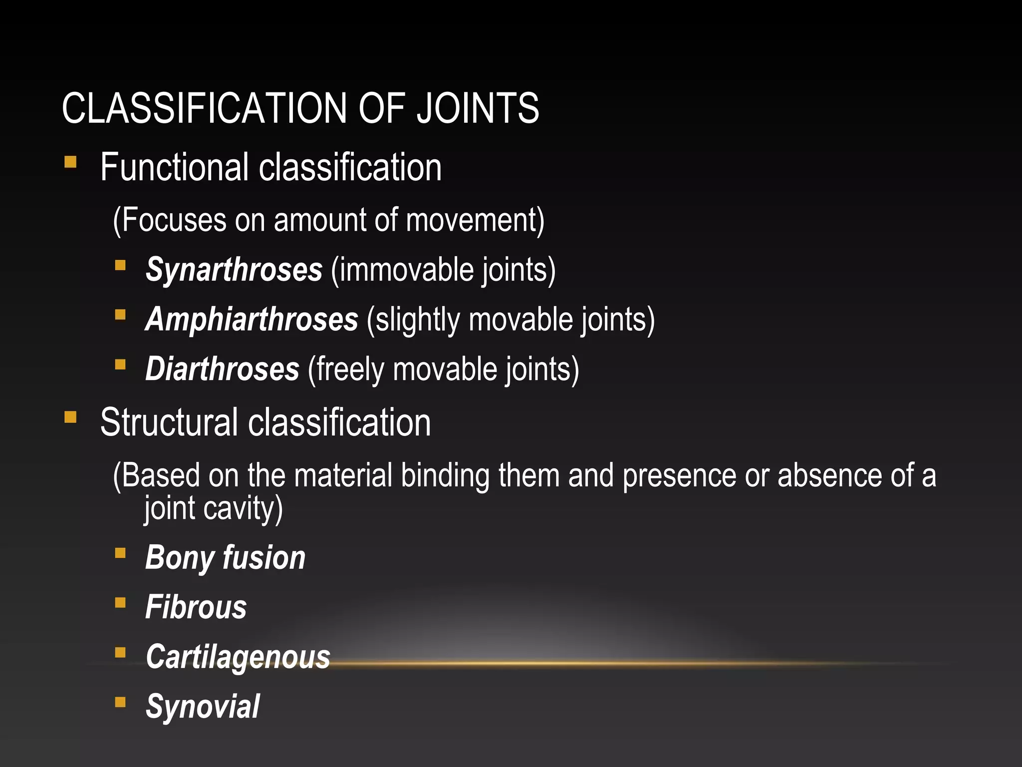
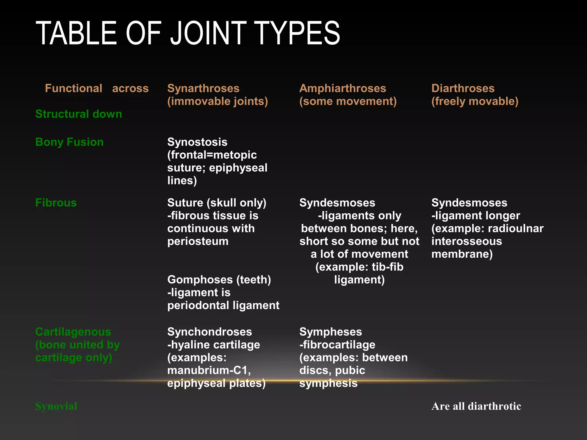
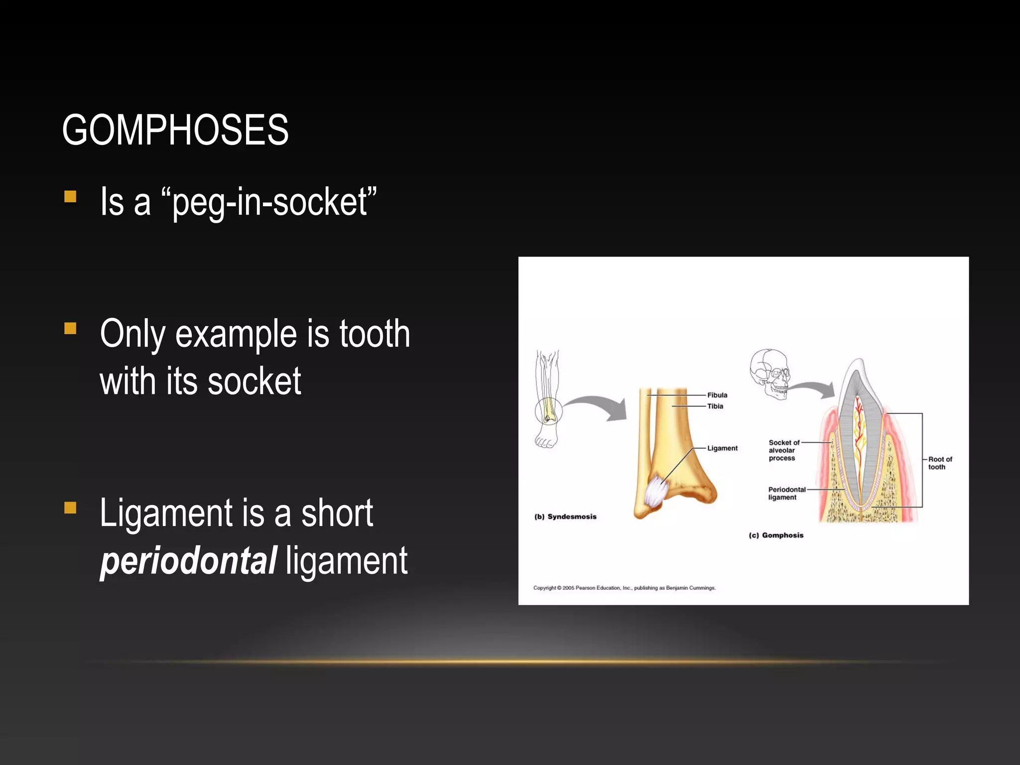
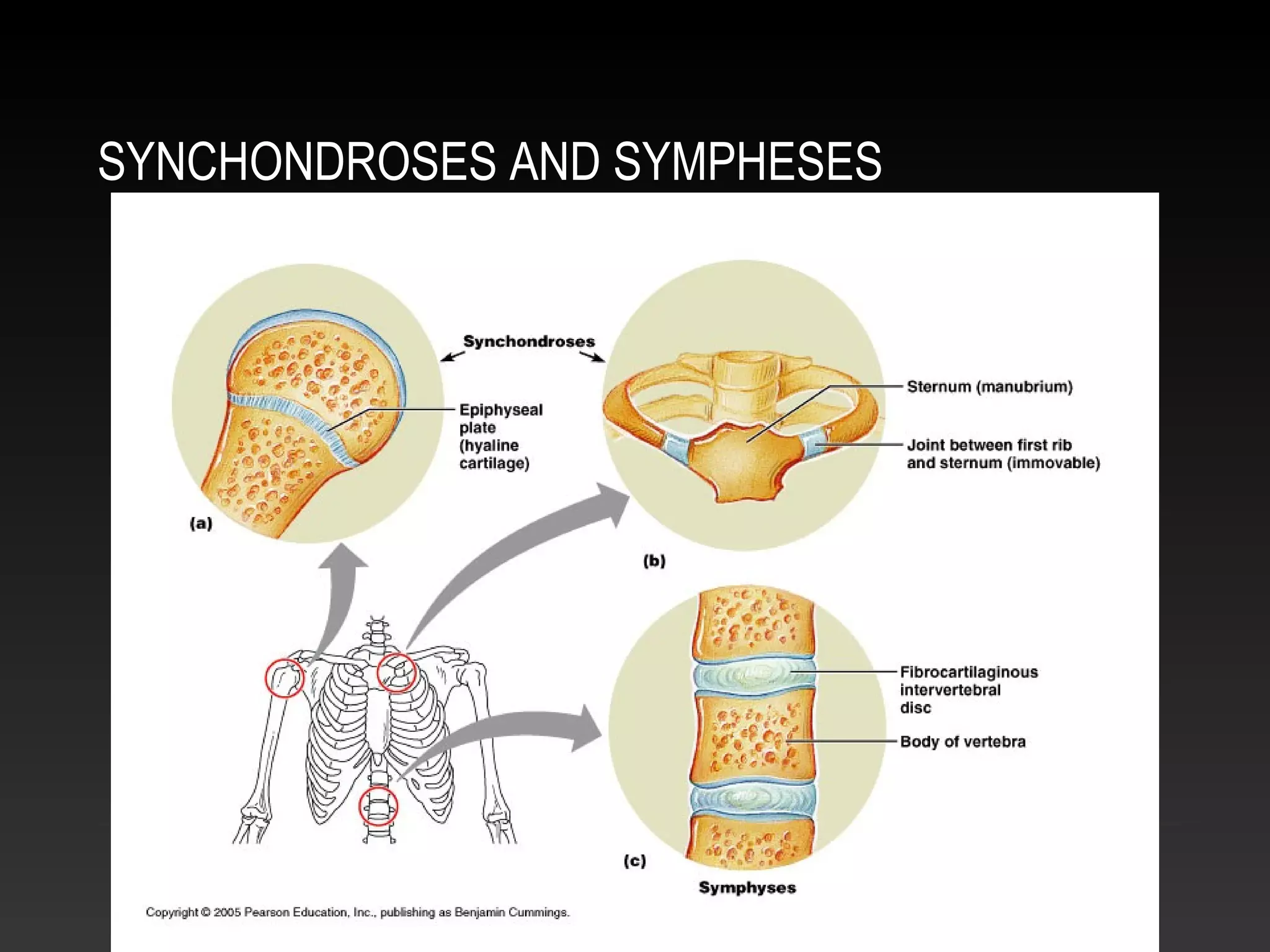
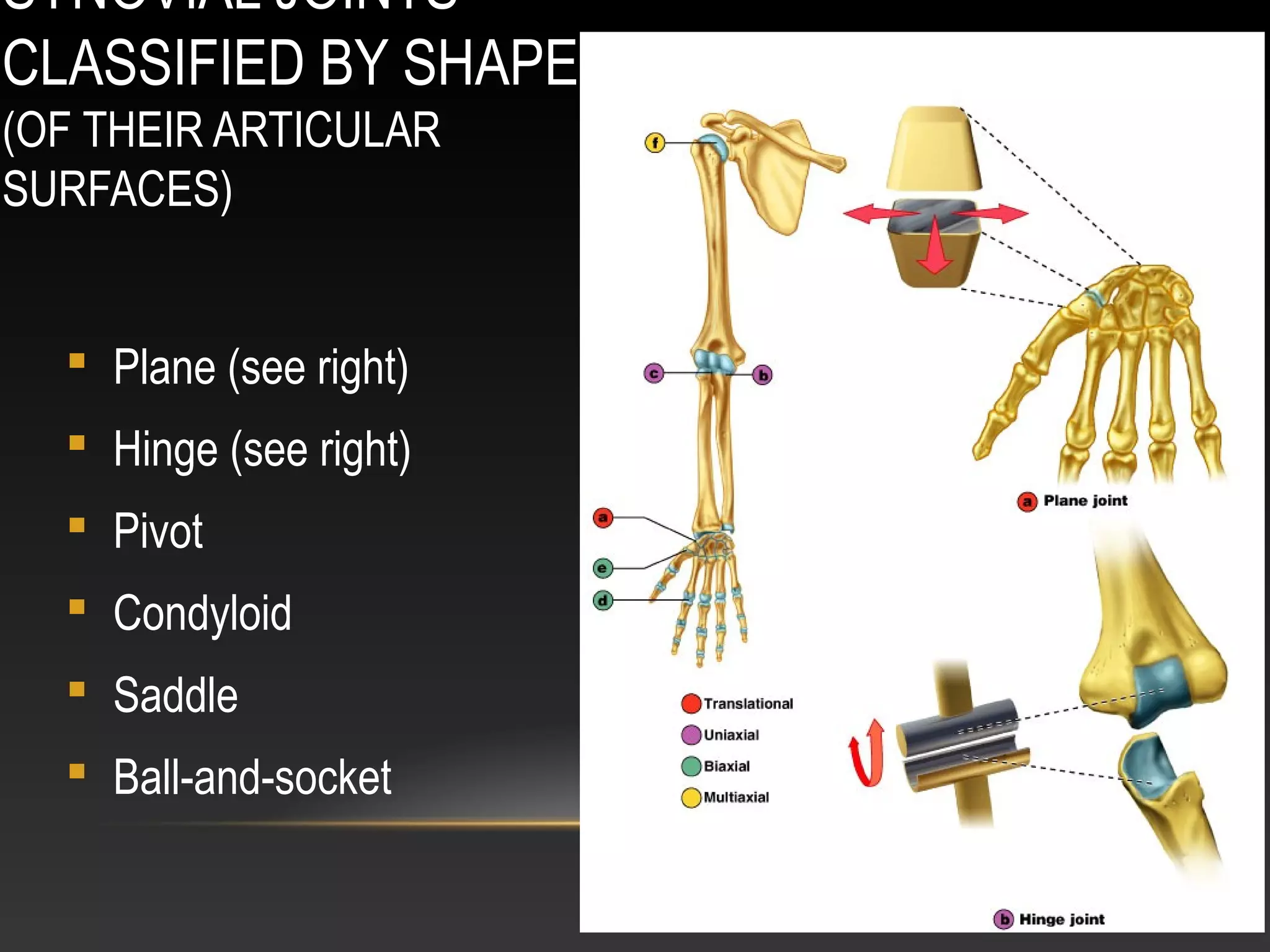
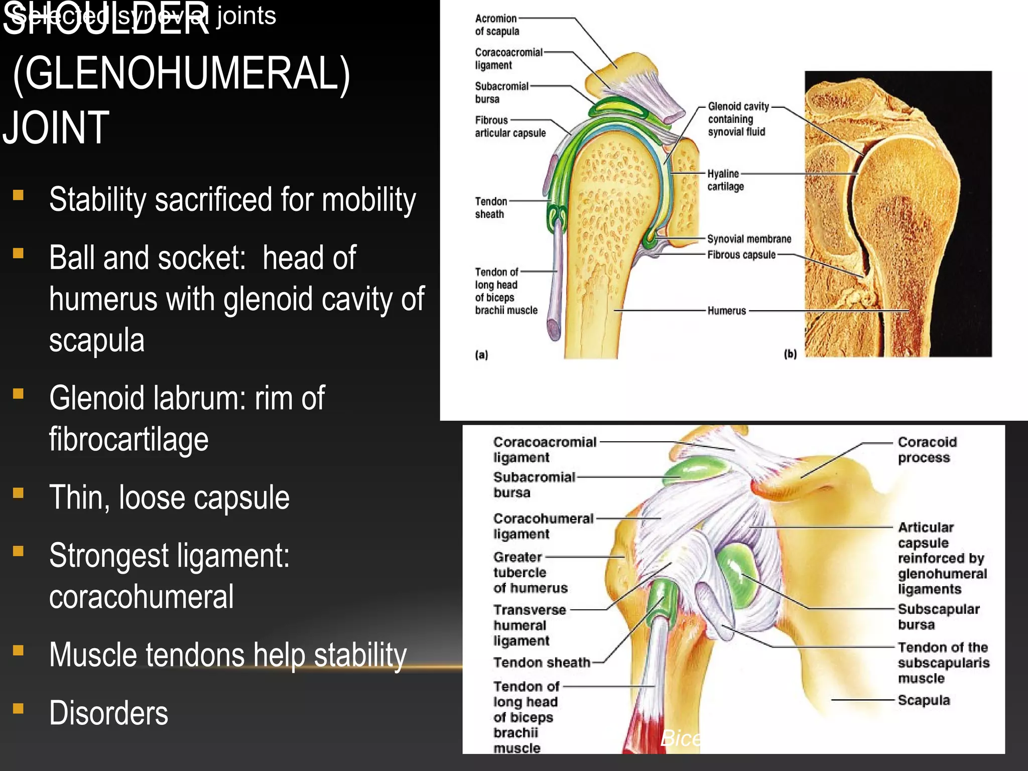
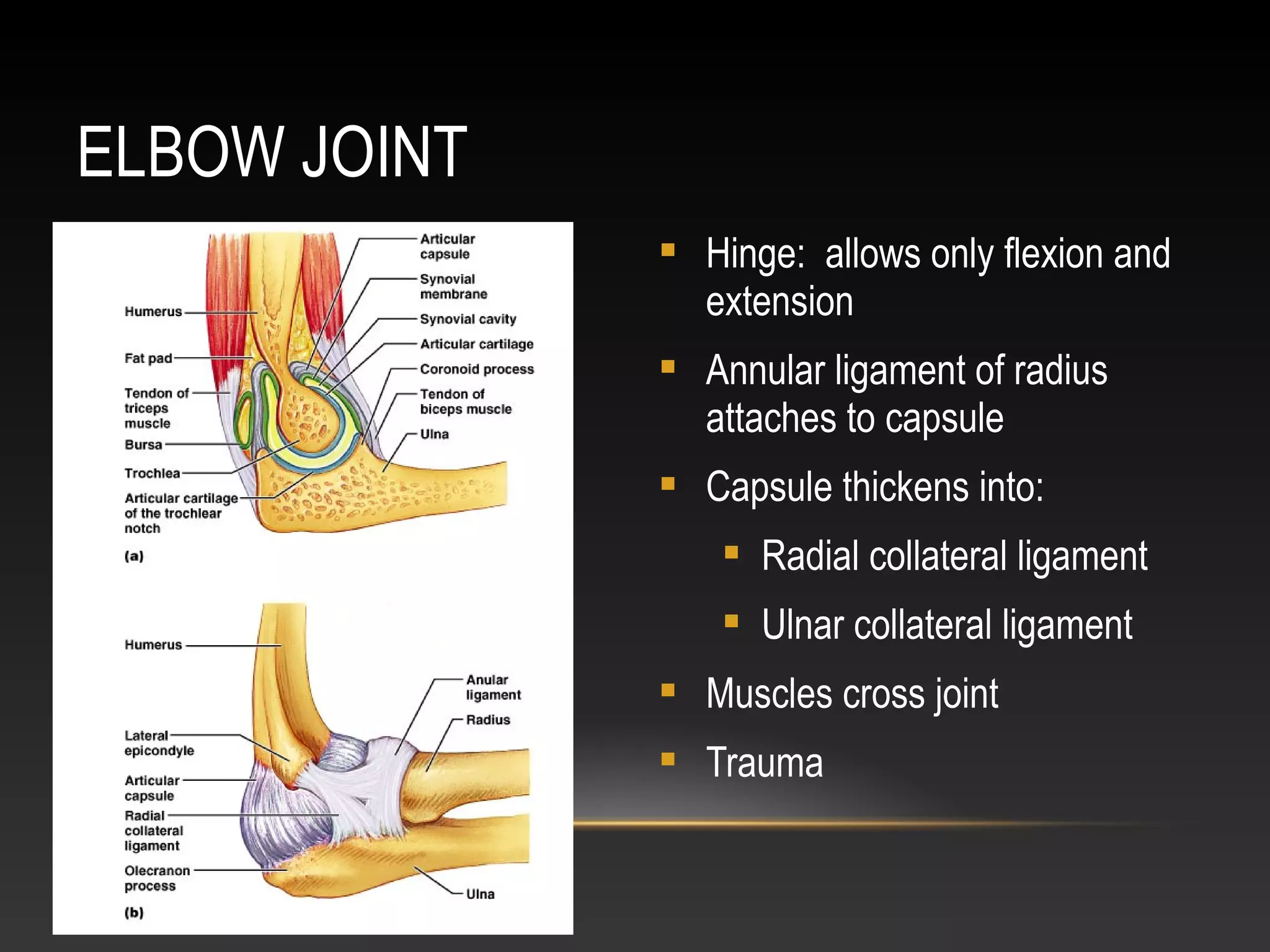
ANATOMY OF JOINTS
By- Dr. Armaan SinghBy- Dr. Armaan Singh
CLASSIFICATION OF JOINTS
 Functional classification
(Focuses on amount of movement)
 Synarthroses (immovable joints)
 Amphiarthroses (slightly movable joints)
 Diarthroses (freely movable joints)
 Structural classification
(Based on the material binding them and presence or absence of a
joint cavity)
 Bony fusion
 Fibrous
 Cartilagenous
 Synovial
TABLE OF JOINT TYPES
Functional across
Structural down
Synarthroses
(immovable joints)
Amphiarthroses
(some movement)
Diarthroses
(freely movable)
Bony Fusion Synostosis
(frontal=metopic
suture; epiphyseal
lines)
Fibrous Suture (skull only)
-fibrous tissue is
continuous with
periosteum
Gomphoses (teeth)
-ligament is
periodontal ligament
Syndesmoses
-ligaments only
between bones; here,
short so some but not
a lot of movement
(example: tib-fib
ligament)
Syndesmoses
-ligament longer
(example: radioulnar
interosseous
membrane)
Cartilagenous
(bone united by
cartilage only)
Synchondroses
-hyaline cartilage
(examples:
manubrium-C1,
epiphyseal plates)
Sympheses
-fibrocartilage
(examples: between
discs, pubic
symphesis
Synovial Are all diarthrotic
FIBROUS JOINTS
 Bones connected by fibrous tissue: dense regular connective tissue
 No joint cavity
 Slightly immovable or
not at all
 Types
 Sutures
 Syndesmoses
 Gomphoses
SUTURES
 Only between bones of skull
 Fibrous tissue continuous with periosteum
 Ossify and fuse in middle age: now
technically called “synostoses”= bony
junctions
SYNDESMOSES
 In Greek: “ligament”
 Bones connected by
ligaments only
 Amount of movement
depends on length of
the fibers: longer than
in sutures
GOMPHOSES
 Is a “peg-in-socket”
 Only example is tooth
with its socket
 Ligament is a short
periodontal ligament
CARTILAGENOUS JOINTS
 Articulating bones united by cartilage
 Lack a joint cavity
 Not highly movable
 Two types
 Synchondroses (singular: synchondrosis)
 Sympheses (singular: symphesis)
SYNCHONDROSES
 Literally: “junction of cartilage”
 Hyaline cartilage unites the bones
 Immovable (synarthroses)
 Examples:
 Epiphyseal plates
 Joint between first rib’s costal cartilage and manubrium of the sternum
SYMPHESES
 Literally “growing together”
 Fibrocartilage unites the bones
 Slightly movable (amphiarthroses)
 Resilient shock absorber
 Provide strength and flexibility
 Hyaline cartilage on articular surfaces of bones to reduce
friction
 Examples
 Intervertebral discs
 Pubic symphysis of the pelvis
SYNCHONDROSES AND SYMPHESES
Also pubic symphsis
SYNOVIAL JOINTS
 Include most of the body’s joints
 All are diarthroses (freely movable)
 All contain fluid-filled joint cavity
GENERAL STRUCTURE OF SYNOVIAL
JOINTS
1. Articular cartilage
 Hyaline
 Spongy cushions absorb
compression
 Protects ends of bones from
being crushed
2. Joint (synovial) cavity
 Potential space
 Small amount of synovial fluid
GENERAL STRUCTURE OF SYNOVIAL JOINTS
(CONT.)
3. Articular (or joint) capsule
 Two layered
 Outer*: fibrous capsule of dense
irregular connective tissue
continuous with periosteum
 Inner*: synovial membrane of
loose connective tissue (makes
synovial fluid)
 Lines all internal joint surfaces not
covered by cartilage*
*
*
*
GENERAL STRUCTURE OF SYNOVIAL JOINTS
(CONT.)
4. Synovial fluid
 Filtrate of blood
 Contains special glycoproteins
 Nourishes cartilage and functions as
slippery lubricant
 “Weeping” lubricatioin
5. Reinforcing ligaments (some joints)
 Capsular (most) – thickened parts of
capsule
 Extracapsular
 Intracapsular
GENERAL STRUCTURE OF SYNOVIAL JOINTS
(CONT.)
6. Nerves
 Detect pain
 Monitor stretch (one of the ways
of sensing posture and body
movements)
7. Blood vessels
 Rich blood supply
 Extensive capillary beds in
synovial membrane (produce the
blood filtrate)
GENERAL STRUCTURE OF SYNOVIAL
JOINTS
SOME JOINTS…
 Articular disc or
meniscus
(literally “crescent”)
 Only some joints
 Those with bone
ends of different
shapes or fitting poorly
 Some to allow two kinds of movement (e.g. jaw)
 Of fibrocartilage
 Examples: knee
TMJ (temporomandibular joint)
BURSAE AND TENDON SHEATHS
 Contain synovial fluid
 Not joints but often associated with them
 Act like ball bearings
 Bursa means “purse” in Latin
 Flattened sac lined by synovial membrane
 Where ligaments, muscles, tendons, or bones overlie each other and rub together
 Tendon sheath
 Only on tendons subjected to friction
BURSAE AND TENDON SHEATHS
JOINT STABILITY
 Articular surfaces
 Shape usually plays only minor role
 Some deep sockets or grooves do provide stability
 Ligaments
 Usually the more, the stronger the joint
 Can stretch only 6% beyond normal length before tear
 Once stretched, stay stretched
 Muscle tone
 Constant, low level of contractile force
 Keeps tension on the ligaments
 Especially important at shoulders, knees, arches of foot
MOVEMENTS ALLOWED BY SYNOVIAL
JOINTS
 Gliding
 Angular movements: or  the angle between two bones
DO TOGETHER
 Flexion
 Extension
 Abduction
 Adduction
 Circumduction
 Rotation
 Special movements
 Pronation
 Supination
 Dorsiflexion
 Plantar flexion
 Inversion
 Eversion
 Protraction
 Retraction
 Elevation
 Depression
 Opposition
SPECIAL MOVEMENTS
JOINT MOVEMENTS PICS
(FROM MARIEB, 4TH
ED.)
SYNOVIAL JOINTS
CLASSIFIED BY SHAPE
(OF THEIR ARTICULAR
SURFACES)
 Plane (see right)
 Hinge (see right)
 Pivot
 Condyloid
 Saddle
 Ball-and-socket
SHOULDER
(GLENOHUMERAL)
JOINT
 Stability sacrificed for mobility
 Ball and socket: head of
humerus with glenoid cavity of
scapula
 Glenoid labrum: rim of
fibrocartilage
 Thin, loose capsule
 Strongest ligament:
coracohumeral
 Muscle tendons help stability
 Disorders
Selected synovial joints
Rotator cuff muscles add to stability
Biceps tendon is intra-articular
ELBOW JOINT
 Hinge: allows only flexion and
extension
 Annular ligament of radius
attaches to capsule
 Capsule thickens into:
 Radial collateral ligament
 Ulnar collateral ligament
 Muscles cross joint
 Trauma
WRIST JOINT
TWO MAJOR JOINT SURFACES
SEVERAL LIGAMENTS
STABILIZE
1. Radiocarpal joint
 Between radius and
proximal carpals
(scaphoid and lunate)
 Condyloid joint
 Flexion extension
adduction, abduction,
circumduction
1. Intercarpal or midcarpal
joint
 Between the proximal and
distal rows of carpals
HIP (COXAL) JOINT
 Ball and socket
 Moves in all axes but
limited by ligaments and
deep socket
 Three ext. ligaments
“screw in” head of femur
when standing
 Iliofemoral
 Pubofemoral
 Ischiofemoral
 Acetabular labrum
diameter smaller than
head of femur
 Dislocations rare
 Ligament of head of femur
supplies artery
 Muscle tendons cross joint
 Hip fractures common in
elderly because of
osteoporosis
Right hip, AP view
KNEE JOINT
 Largest and most complex joint
 Primarily a hinge
 Compound and bicondyloid: femur and tibia both have 2 condyles
 Femoropatellar joint shares joint cavity
 At least a dozen bursae
 Prepatellar
 Suprapatellar
 Lateral and medial menisci
 “torn cartilage”
 Capsule absent anteriorly
 Capsular and extracapsular
ligaments
 Taut when knee extended to
prevent hyperextension
 Patellar ligament
 Continuation of quad
tendon
 Medial and lateral
retinacula
 Fibular and tibial
collateral ligaments
 Called medial and
lateral
 Extracapsular
 Oblique popliteal
 Arcuate popliteal
CRUCIATE LIGAMENTS
 Cross each other (cruciate
means cross)
 Anterior cruciate (ACL)
 Anterior intercondylar area of
tibia to medial side of lateral
condyl of femur
 Posterior cruciate
 Posterior intercondylar area of
tibia to lateral side of medial
condyl
 Restraining straps
 Lock the knee
CRUCIATE LIGAMENTS
KNEE INJURIES
 Flat tibial surface
predisposes to horizontal
injuries
 Lateral blow: multiple tears
 ACL injuries
 Stop and twist
 Commoner in women
athletes
 Heal poorly
 Require surgery
NKLE JOINT
 Hinge joint
 Distal tibia and fibula to talus
 Dorsiflexion and plantar flexion only
 Medial deltoid ligament
 Lateral ligaments: 3 bands
 Anterior talofibular
 Posterior talofibular
 Calcaneofibular
 Anterior and posterior tibiofibular
(syndesmosis)
RIGHT ANKLE, LATERAL VIEW
TEMPOROMANDIBULA
R JOINT (TMJ)
 Head of mandible articulates with
temporal bone
 Disc protects thin mandibular
fossa of temporal bone
 Many movements
Demonstrate movements together
 Disorders common
STERNOCLAVICULAR
JOINT
 Saddle joint
 Only other example is trapezium and
metacarpal 1 (thumb), allowing
opposion
 Sternum and 1st
costal (rib) cartilage
articulate with clavicle
 Very stable: clavicle usually breaks
before dislocation of joint
 Only bony attachment of axial skeleton
to pectoral girdle
Demonstrate movements together
DISORDERS OF JOINTS
 Injuries
 Sprains
 Dislocatios
 Torn cartilage
 Inflammatory and degenerative conditions
 Bursitis
 Tendinitis
 Arthritis
 Osteoarthritis (“DJD” – degenerative joint disease)
 Rheumatoid arthritis (one of many “autoimmune” arthritites)
 Gout (crystal arthropathy)

Joints by dr. armaan singh

  • 1.
    ANATOMY OF JOINTS By-Dr. Armaan SinghBy- Dr. Armaan Singh
  • 2.
    CLASSIFICATION OF JOINTS Functional classification (Focuses on amount of movement)  Synarthroses (immovable joints)  Amphiarthroses (slightly movable joints)  Diarthroses (freely movable joints)  Structural classification (Based on the material binding them and presence or absence of a joint cavity)  Bony fusion  Fibrous  Cartilagenous  Synovial
  • 3.
    TABLE OF JOINTTYPES Functional across Structural down Synarthroses (immovable joints) Amphiarthroses (some movement) Diarthroses (freely movable) Bony Fusion Synostosis (frontal=metopic suture; epiphyseal lines) Fibrous Suture (skull only) -fibrous tissue is continuous with periosteum Gomphoses (teeth) -ligament is periodontal ligament Syndesmoses -ligaments only between bones; here, short so some but not a lot of movement (example: tib-fib ligament) Syndesmoses -ligament longer (example: radioulnar interosseous membrane) Cartilagenous (bone united by cartilage only) Synchondroses -hyaline cartilage (examples: manubrium-C1, epiphyseal plates) Sympheses -fibrocartilage (examples: between discs, pubic symphesis Synovial Are all diarthrotic
  • 4.
    FIBROUS JOINTS  Bonesconnected by fibrous tissue: dense regular connective tissue  No joint cavity  Slightly immovable or not at all  Types  Sutures  Syndesmoses  Gomphoses
  • 5.
    SUTURES  Only betweenbones of skull  Fibrous tissue continuous with periosteum  Ossify and fuse in middle age: now technically called “synostoses”= bony junctions
  • 6.
    SYNDESMOSES  In Greek:“ligament”  Bones connected by ligaments only  Amount of movement depends on length of the fibers: longer than in sutures
  • 7.
    GOMPHOSES  Is a“peg-in-socket”  Only example is tooth with its socket  Ligament is a short periodontal ligament
  • 8.
    CARTILAGENOUS JOINTS  Articulatingbones united by cartilage  Lack a joint cavity  Not highly movable  Two types  Synchondroses (singular: synchondrosis)  Sympheses (singular: symphesis)
  • 9.
    SYNCHONDROSES  Literally: “junctionof cartilage”  Hyaline cartilage unites the bones  Immovable (synarthroses)  Examples:  Epiphyseal plates  Joint between first rib’s costal cartilage and manubrium of the sternum
  • 10.
    SYMPHESES  Literally “growingtogether”  Fibrocartilage unites the bones  Slightly movable (amphiarthroses)  Resilient shock absorber  Provide strength and flexibility  Hyaline cartilage on articular surfaces of bones to reduce friction  Examples  Intervertebral discs  Pubic symphysis of the pelvis
  • 11.
  • 12.
    SYNOVIAL JOINTS  Includemost of the body’s joints  All are diarthroses (freely movable)  All contain fluid-filled joint cavity
  • 13.
    GENERAL STRUCTURE OFSYNOVIAL JOINTS 1. Articular cartilage  Hyaline  Spongy cushions absorb compression  Protects ends of bones from being crushed 2. Joint (synovial) cavity  Potential space  Small amount of synovial fluid
  • 14.
    GENERAL STRUCTURE OFSYNOVIAL JOINTS (CONT.) 3. Articular (or joint) capsule  Two layered  Outer*: fibrous capsule of dense irregular connective tissue continuous with periosteum  Inner*: synovial membrane of loose connective tissue (makes synovial fluid)  Lines all internal joint surfaces not covered by cartilage* * * *
  • 15.
    GENERAL STRUCTURE OFSYNOVIAL JOINTS (CONT.) 4. Synovial fluid  Filtrate of blood  Contains special glycoproteins  Nourishes cartilage and functions as slippery lubricant  “Weeping” lubricatioin 5. Reinforcing ligaments (some joints)  Capsular (most) – thickened parts of capsule  Extracapsular  Intracapsular
  • 16.
    GENERAL STRUCTURE OFSYNOVIAL JOINTS (CONT.) 6. Nerves  Detect pain  Monitor stretch (one of the ways of sensing posture and body movements) 7. Blood vessels  Rich blood supply  Extensive capillary beds in synovial membrane (produce the blood filtrate)
  • 17.
    GENERAL STRUCTURE OFSYNOVIAL JOINTS
  • 18.
    SOME JOINTS…  Articulardisc or meniscus (literally “crescent”)  Only some joints  Those with bone ends of different shapes or fitting poorly  Some to allow two kinds of movement (e.g. jaw)  Of fibrocartilage  Examples: knee TMJ (temporomandibular joint)
  • 19.
    BURSAE AND TENDONSHEATHS  Contain synovial fluid  Not joints but often associated with them  Act like ball bearings  Bursa means “purse” in Latin  Flattened sac lined by synovial membrane  Where ligaments, muscles, tendons, or bones overlie each other and rub together  Tendon sheath  Only on tendons subjected to friction
  • 20.
  • 21.
    JOINT STABILITY  Articularsurfaces  Shape usually plays only minor role  Some deep sockets or grooves do provide stability  Ligaments  Usually the more, the stronger the joint  Can stretch only 6% beyond normal length before tear  Once stretched, stay stretched  Muscle tone  Constant, low level of contractile force  Keeps tension on the ligaments  Especially important at shoulders, knees, arches of foot
  • 22.
    MOVEMENTS ALLOWED BYSYNOVIAL JOINTS  Gliding  Angular movements: or  the angle between two bones DO TOGETHER  Flexion  Extension  Abduction  Adduction  Circumduction  Rotation  Special movements
  • 23.
     Pronation  Supination Dorsiflexion  Plantar flexion  Inversion  Eversion  Protraction  Retraction  Elevation  Depression  Opposition SPECIAL MOVEMENTS
  • 24.
  • 30.
    SYNOVIAL JOINTS CLASSIFIED BYSHAPE (OF THEIR ARTICULAR SURFACES)  Plane (see right)  Hinge (see right)  Pivot  Condyloid  Saddle  Ball-and-socket
  • 32.
    SHOULDER (GLENOHUMERAL) JOINT  Stability sacrificedfor mobility  Ball and socket: head of humerus with glenoid cavity of scapula  Glenoid labrum: rim of fibrocartilage  Thin, loose capsule  Strongest ligament: coracohumeral  Muscle tendons help stability  Disorders Selected synovial joints Rotator cuff muscles add to stability Biceps tendon is intra-articular
  • 33.
    ELBOW JOINT  Hinge:allows only flexion and extension  Annular ligament of radius attaches to capsule  Capsule thickens into:  Radial collateral ligament  Ulnar collateral ligament  Muscles cross joint  Trauma
  • 34.
    WRIST JOINT TWO MAJORJOINT SURFACES SEVERAL LIGAMENTS STABILIZE 1. Radiocarpal joint  Between radius and proximal carpals (scaphoid and lunate)  Condyloid joint  Flexion extension adduction, abduction, circumduction 1. Intercarpal or midcarpal joint  Between the proximal and distal rows of carpals
  • 35.
    HIP (COXAL) JOINT Ball and socket  Moves in all axes but limited by ligaments and deep socket  Three ext. ligaments “screw in” head of femur when standing  Iliofemoral  Pubofemoral  Ischiofemoral
  • 36.
     Acetabular labrum diametersmaller than head of femur  Dislocations rare  Ligament of head of femur supplies artery  Muscle tendons cross joint  Hip fractures common in elderly because of osteoporosis
  • 37.
  • 38.
    KNEE JOINT  Largestand most complex joint  Primarily a hinge  Compound and bicondyloid: femur and tibia both have 2 condyles  Femoropatellar joint shares joint cavity  At least a dozen bursae  Prepatellar  Suprapatellar
  • 39.
     Lateral andmedial menisci  “torn cartilage”  Capsule absent anteriorly  Capsular and extracapsular ligaments  Taut when knee extended to prevent hyperextension
  • 40.
     Patellar ligament Continuation of quad tendon  Medial and lateral retinacula  Fibular and tibial collateral ligaments  Called medial and lateral  Extracapsular  Oblique popliteal  Arcuate popliteal
  • 41.
    CRUCIATE LIGAMENTS  Crosseach other (cruciate means cross)  Anterior cruciate (ACL)  Anterior intercondylar area of tibia to medial side of lateral condyl of femur  Posterior cruciate  Posterior intercondylar area of tibia to lateral side of medial condyl  Restraining straps  Lock the knee
  • 42.
  • 43.
    KNEE INJURIES  Flattibial surface predisposes to horizontal injuries  Lateral blow: multiple tears  ACL injuries  Stop and twist  Commoner in women athletes  Heal poorly  Require surgery
  • 44.
    NKLE JOINT  Hingejoint  Distal tibia and fibula to talus  Dorsiflexion and plantar flexion only  Medial deltoid ligament  Lateral ligaments: 3 bands  Anterior talofibular  Posterior talofibular  Calcaneofibular  Anterior and posterior tibiofibular (syndesmosis)
  • 45.
  • 48.
    TEMPOROMANDIBULA R JOINT (TMJ) Head of mandible articulates with temporal bone  Disc protects thin mandibular fossa of temporal bone  Many movements Demonstrate movements together  Disorders common
  • 49.
    STERNOCLAVICULAR JOINT  Saddle joint Only other example is trapezium and metacarpal 1 (thumb), allowing opposion  Sternum and 1st costal (rib) cartilage articulate with clavicle  Very stable: clavicle usually breaks before dislocation of joint  Only bony attachment of axial skeleton to pectoral girdle Demonstrate movements together
  • 50.
    DISORDERS OF JOINTS Injuries  Sprains  Dislocatios  Torn cartilage  Inflammatory and degenerative conditions  Bursitis  Tendinitis  Arthritis  Osteoarthritis (“DJD” – degenerative joint disease)  Rheumatoid arthritis (one of many “autoimmune” arthritites)  Gout (crystal arthropathy)