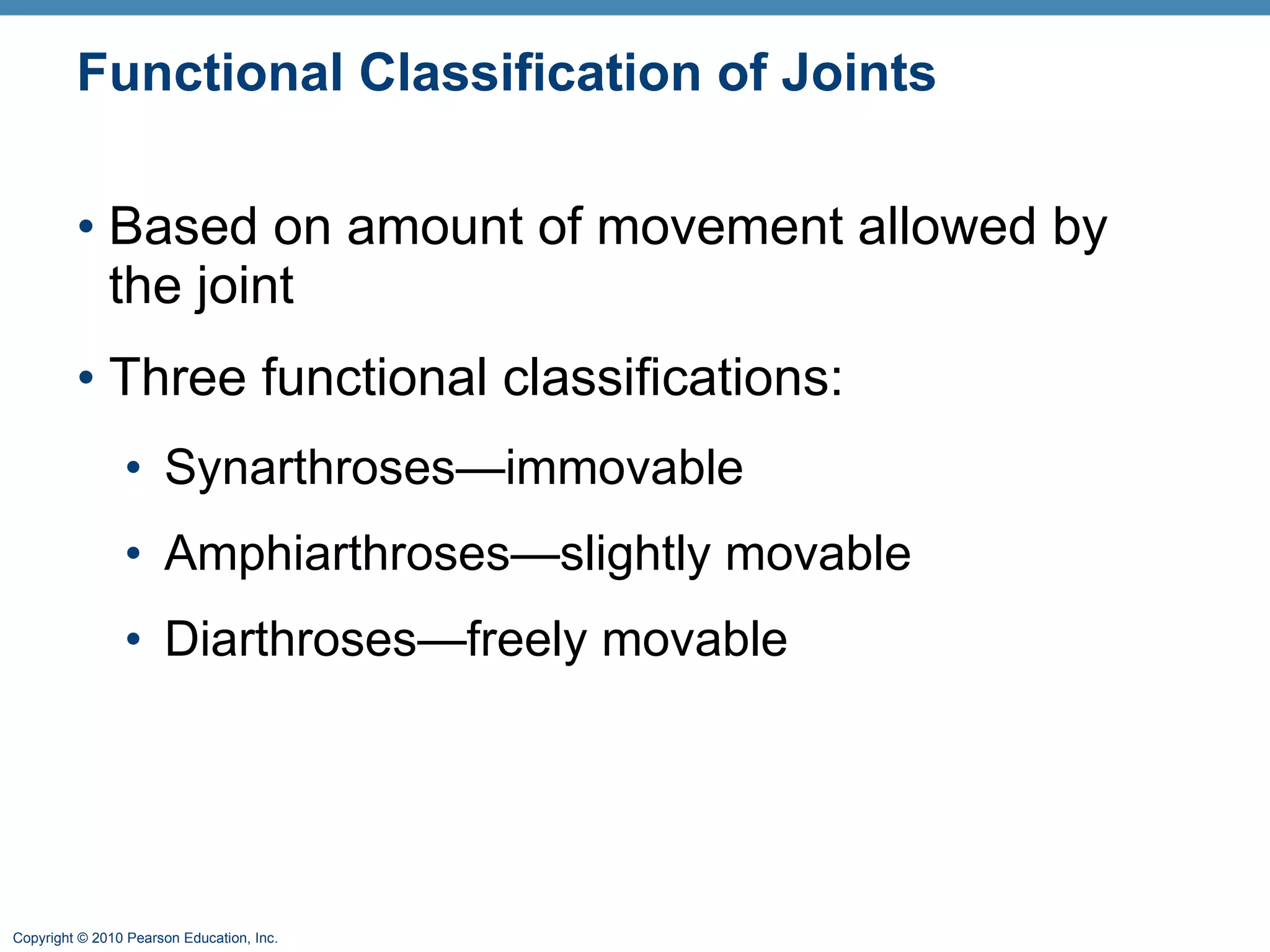
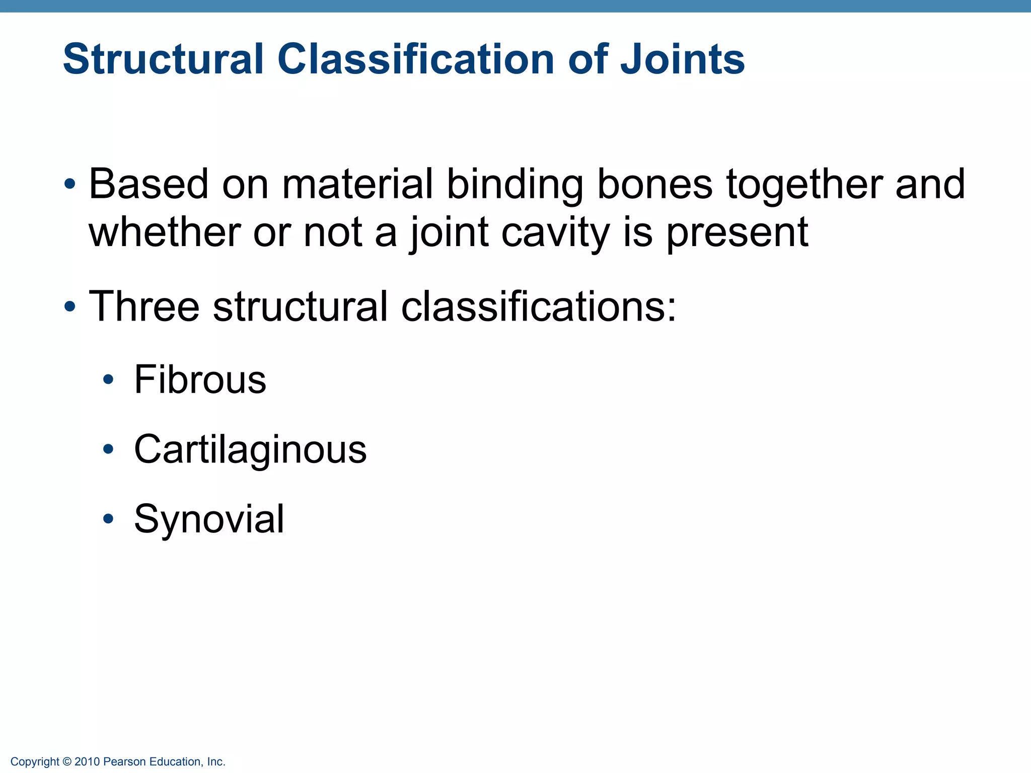
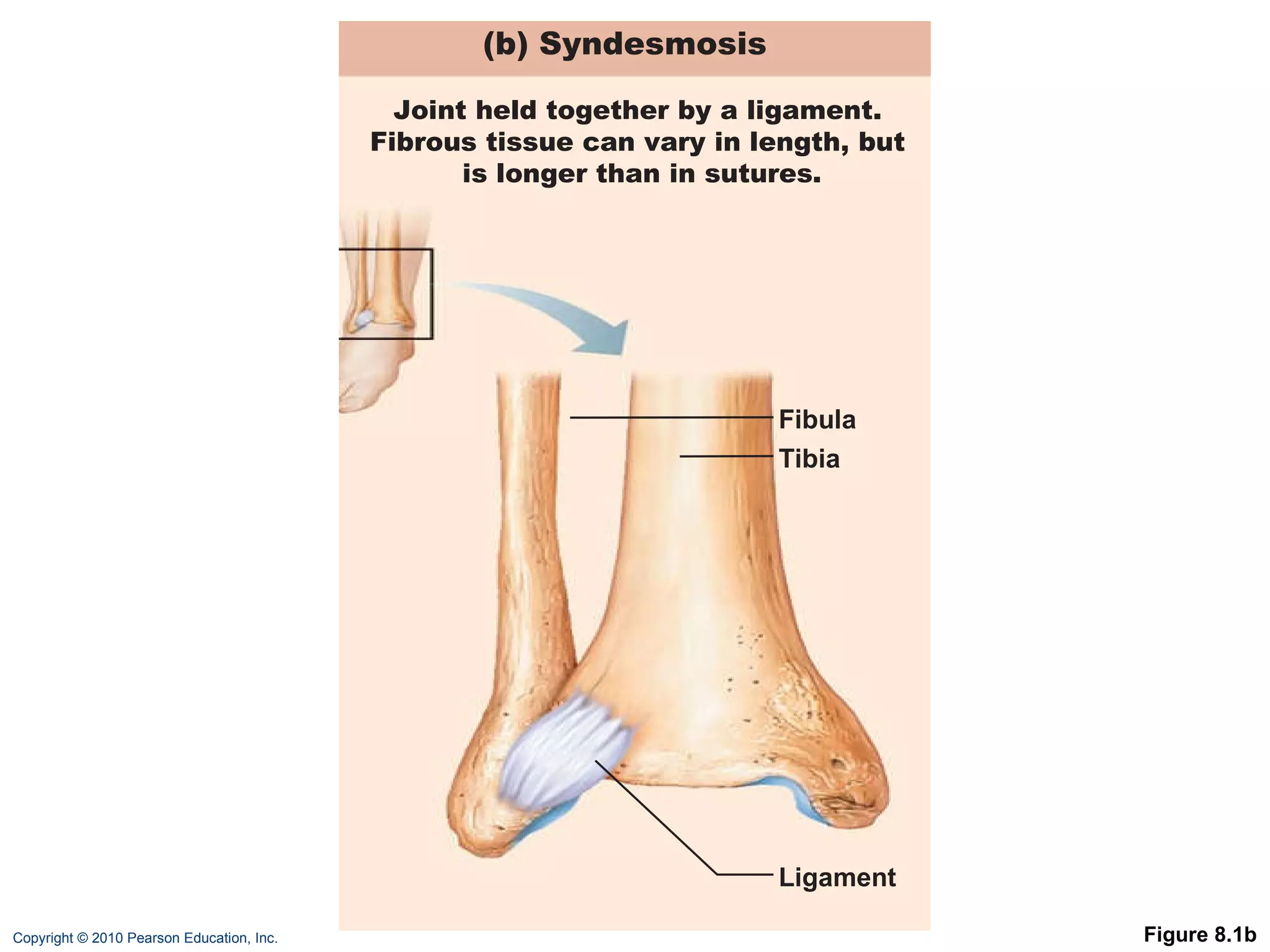
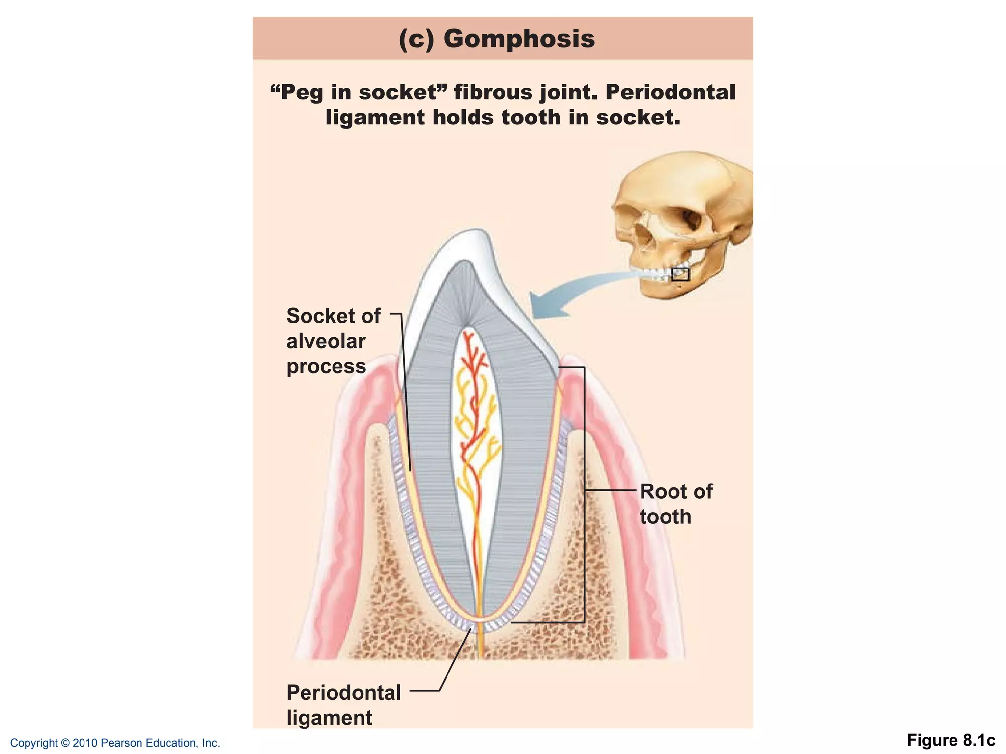
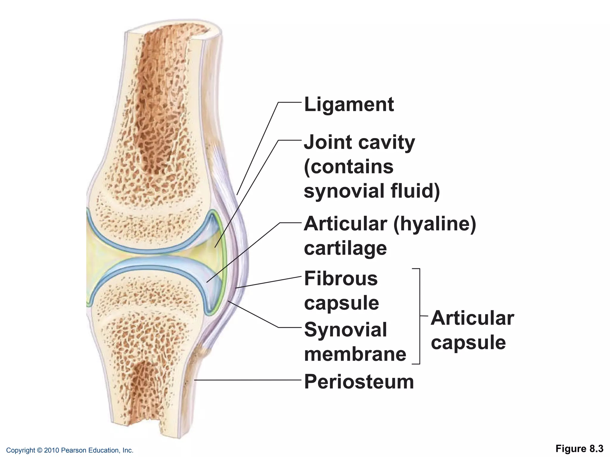
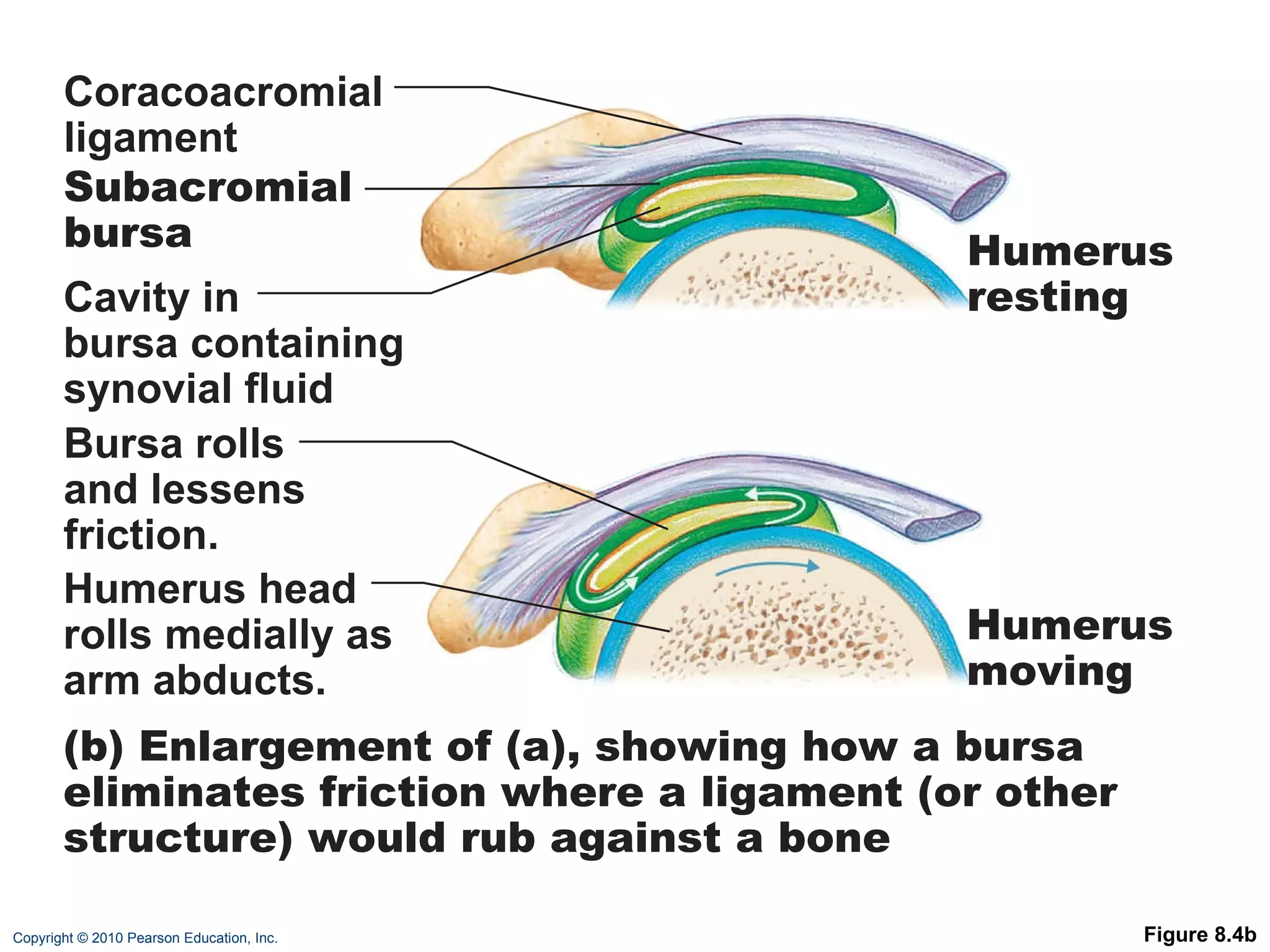
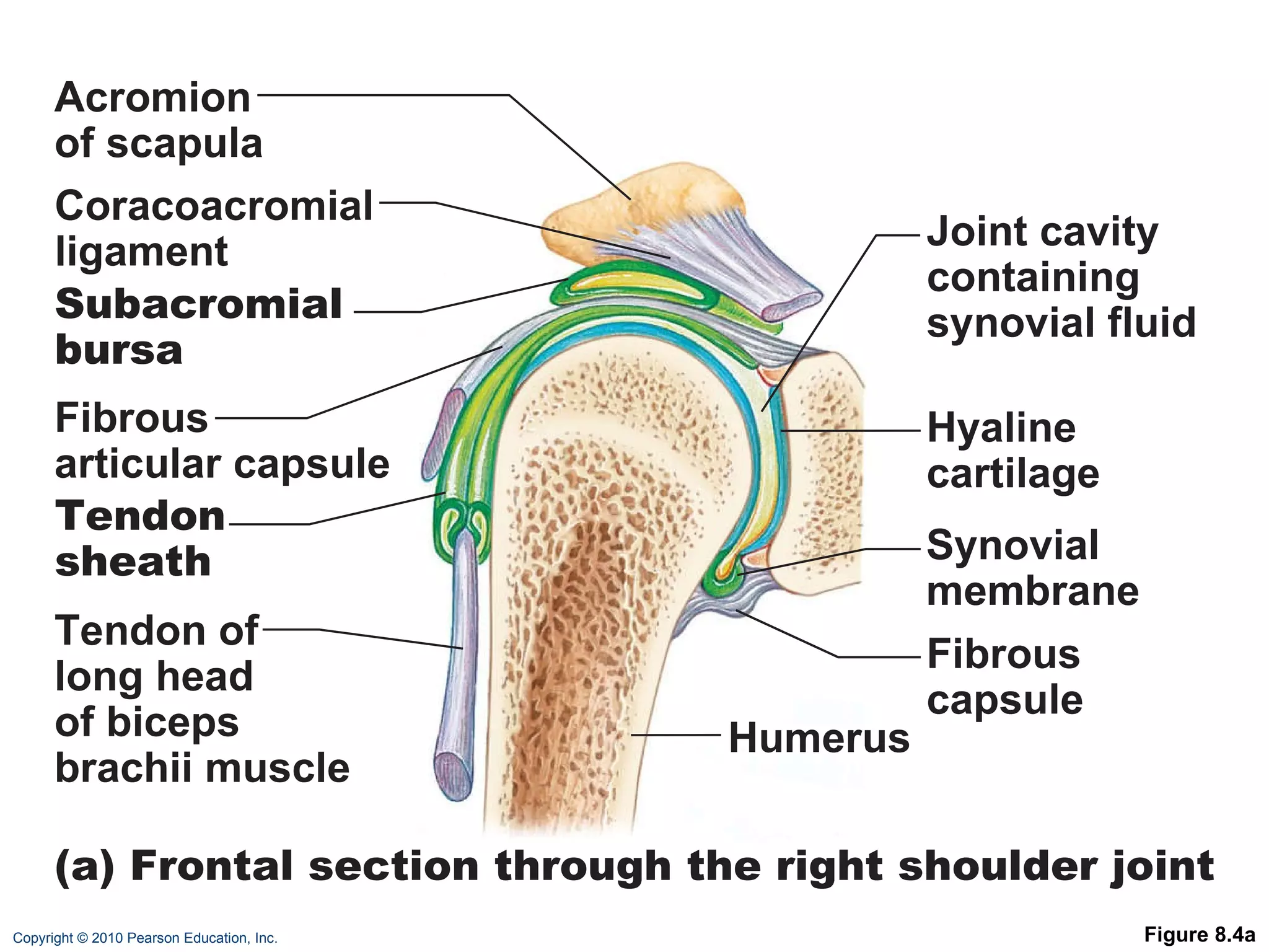
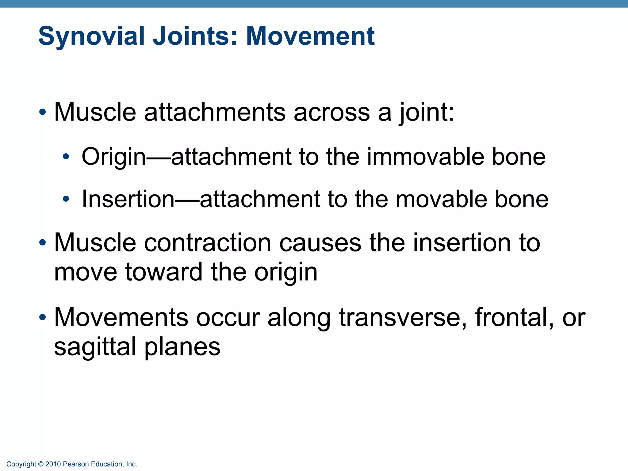
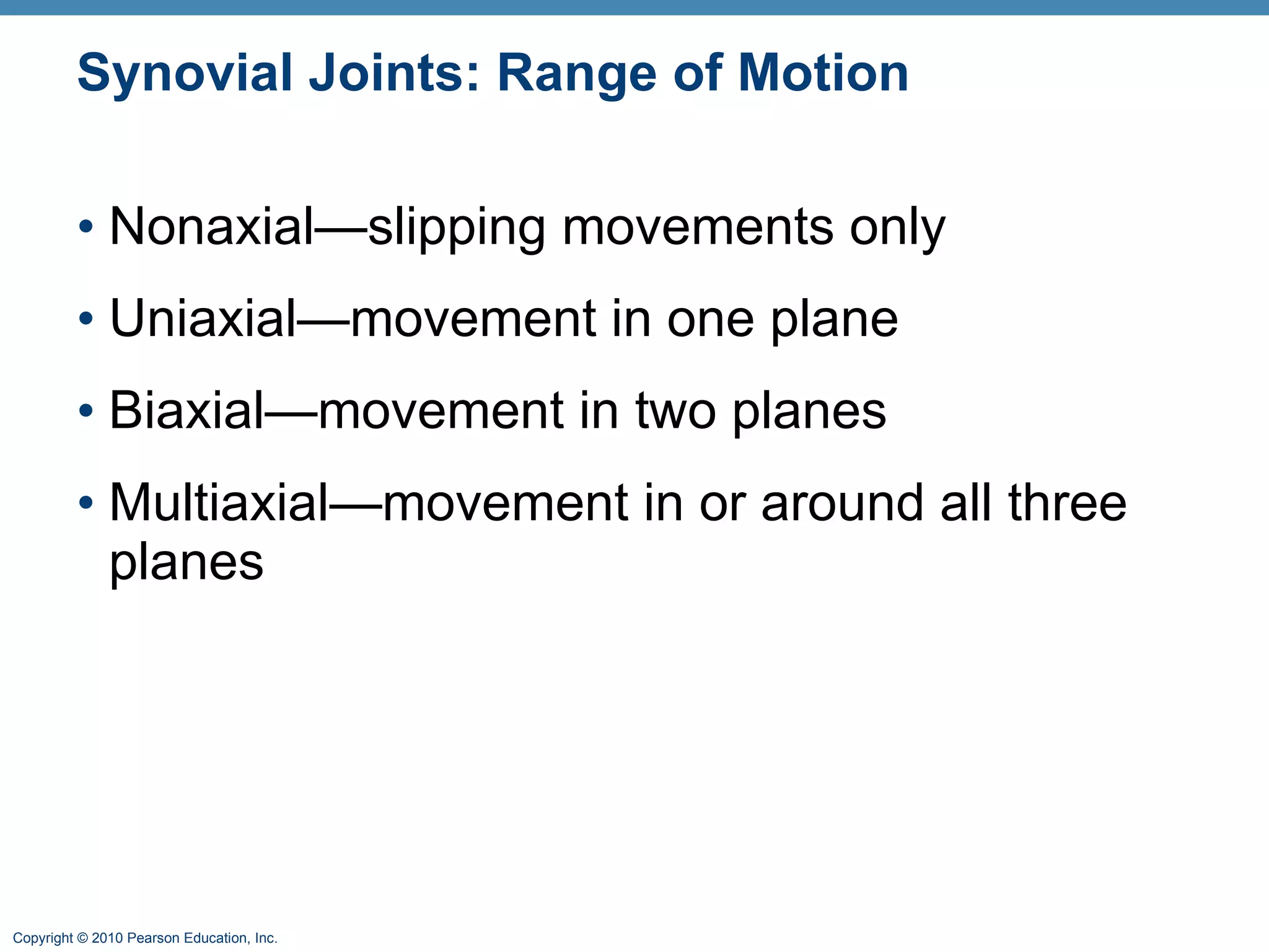
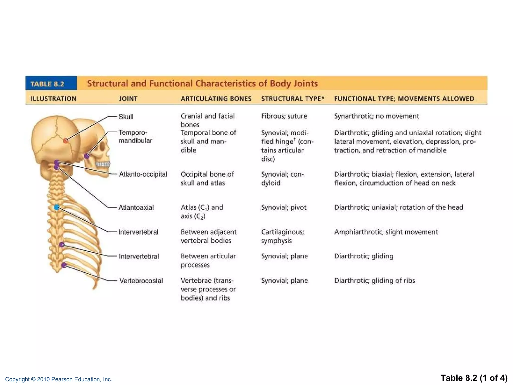
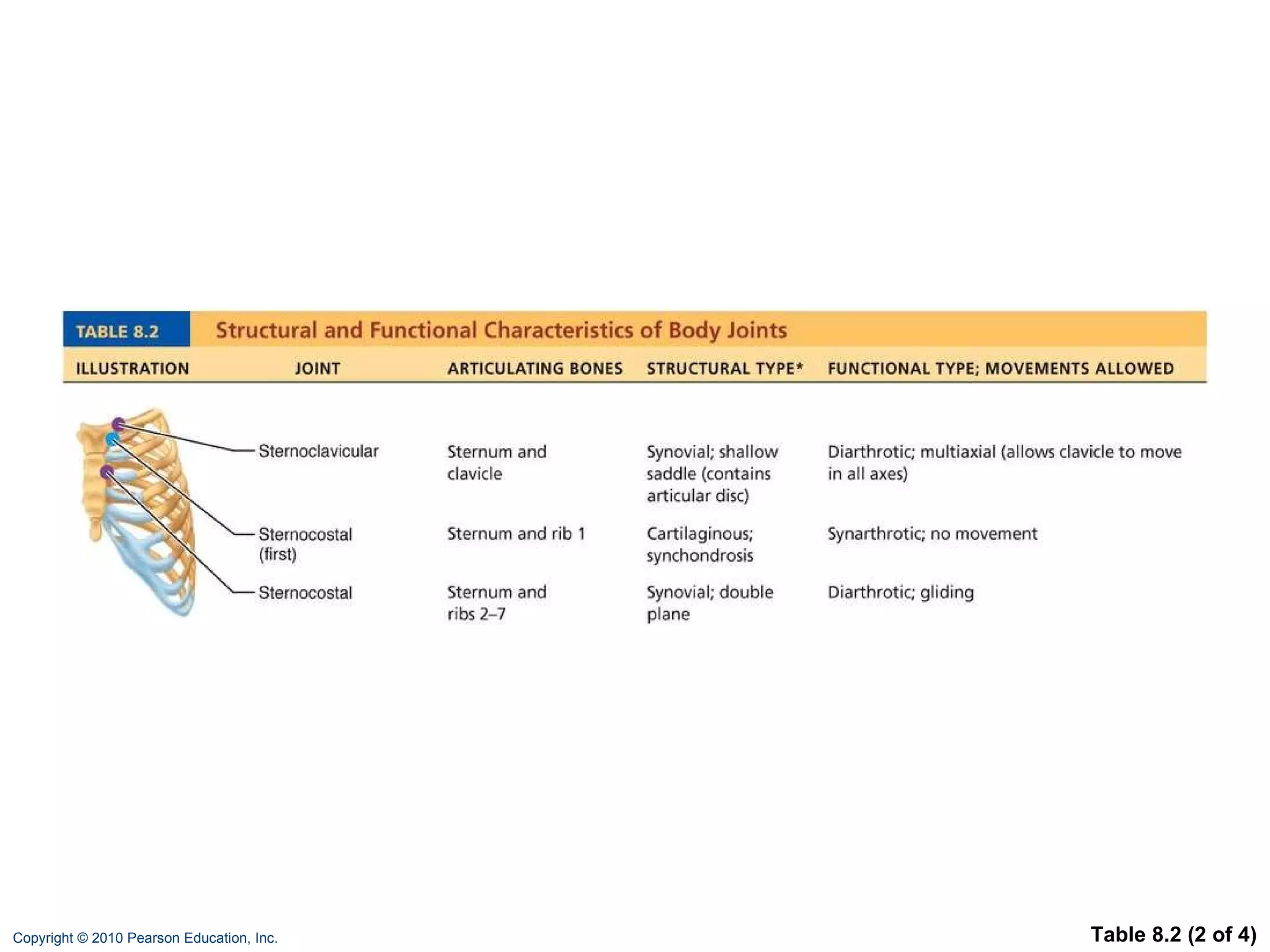
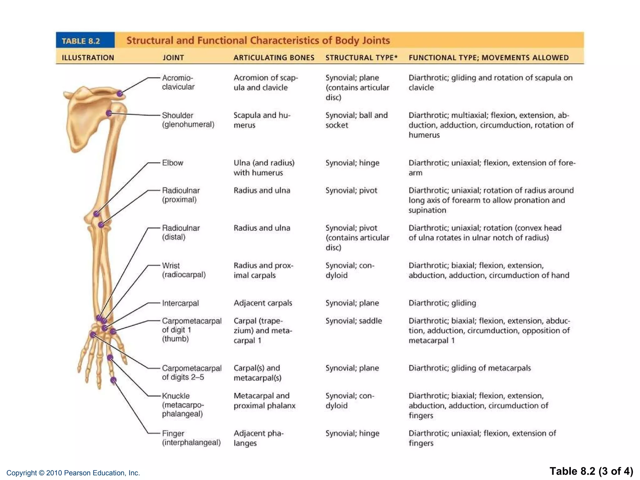
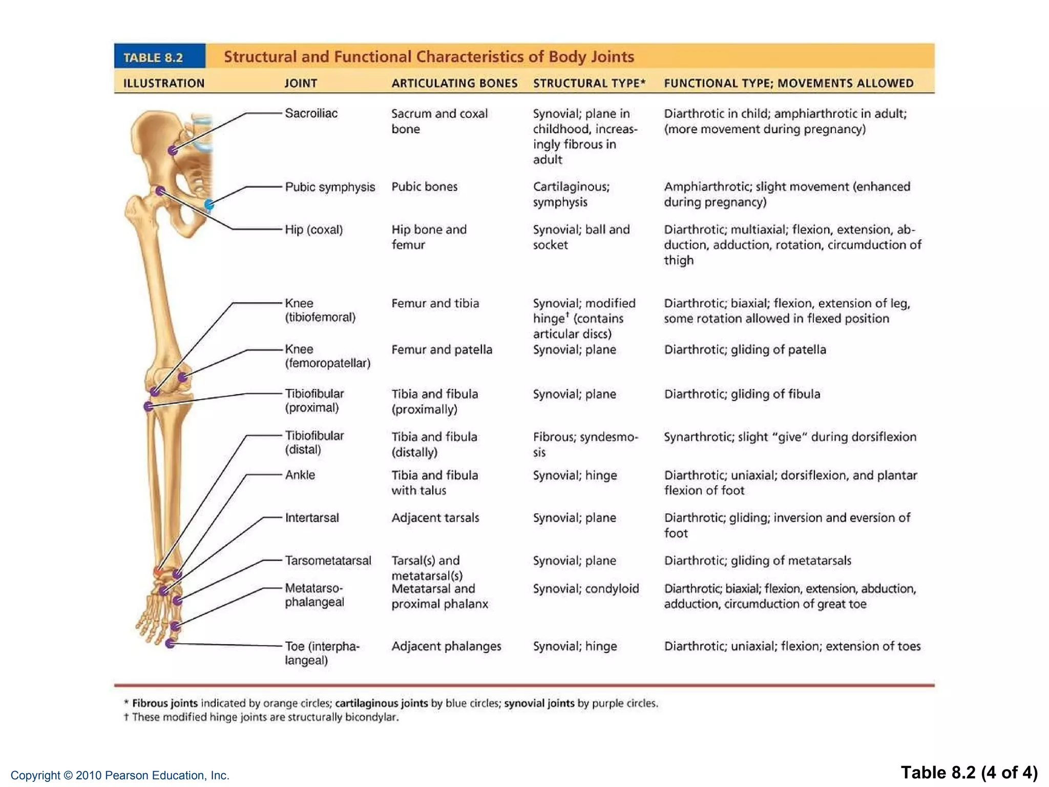
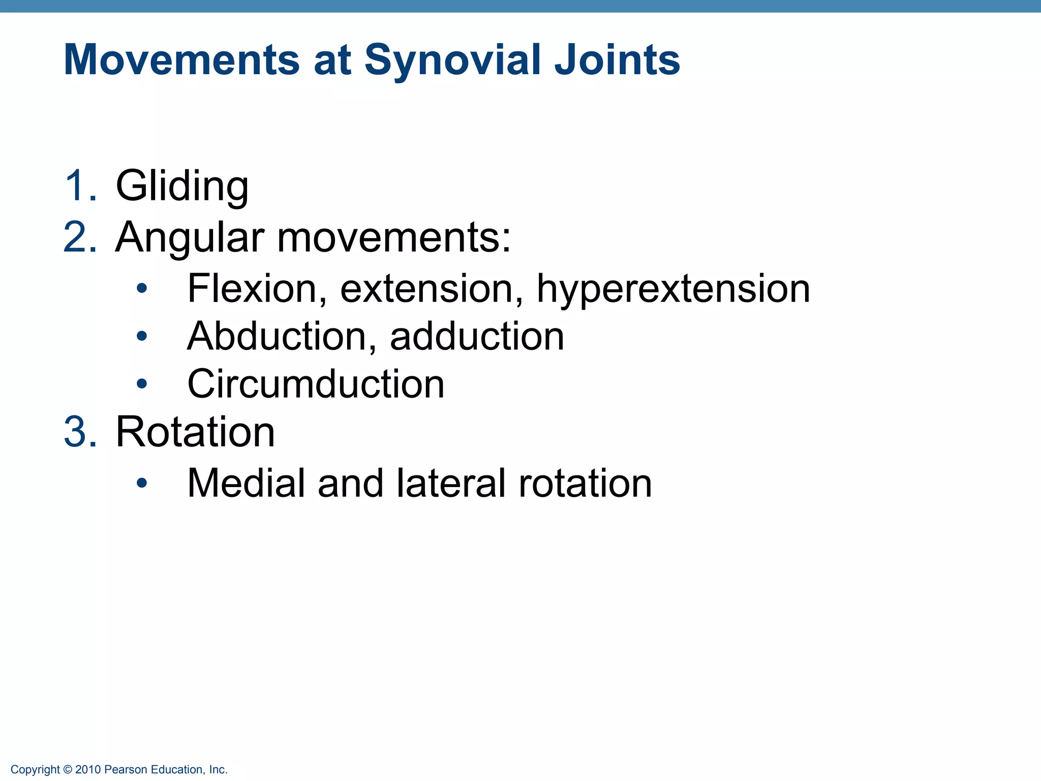
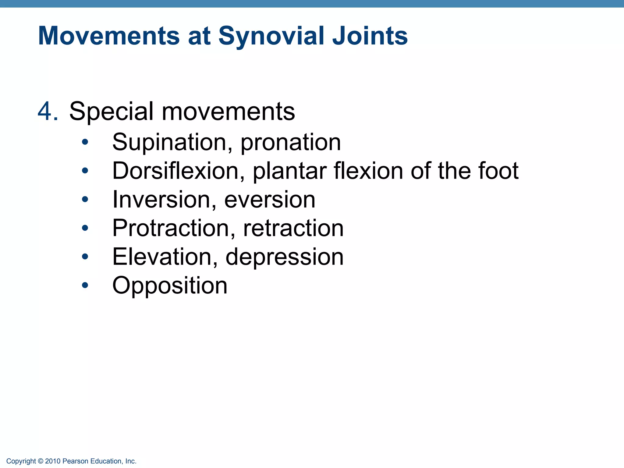
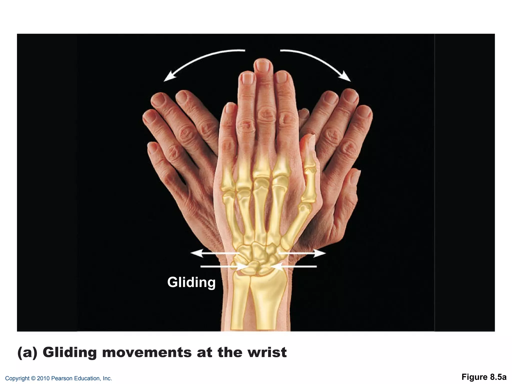
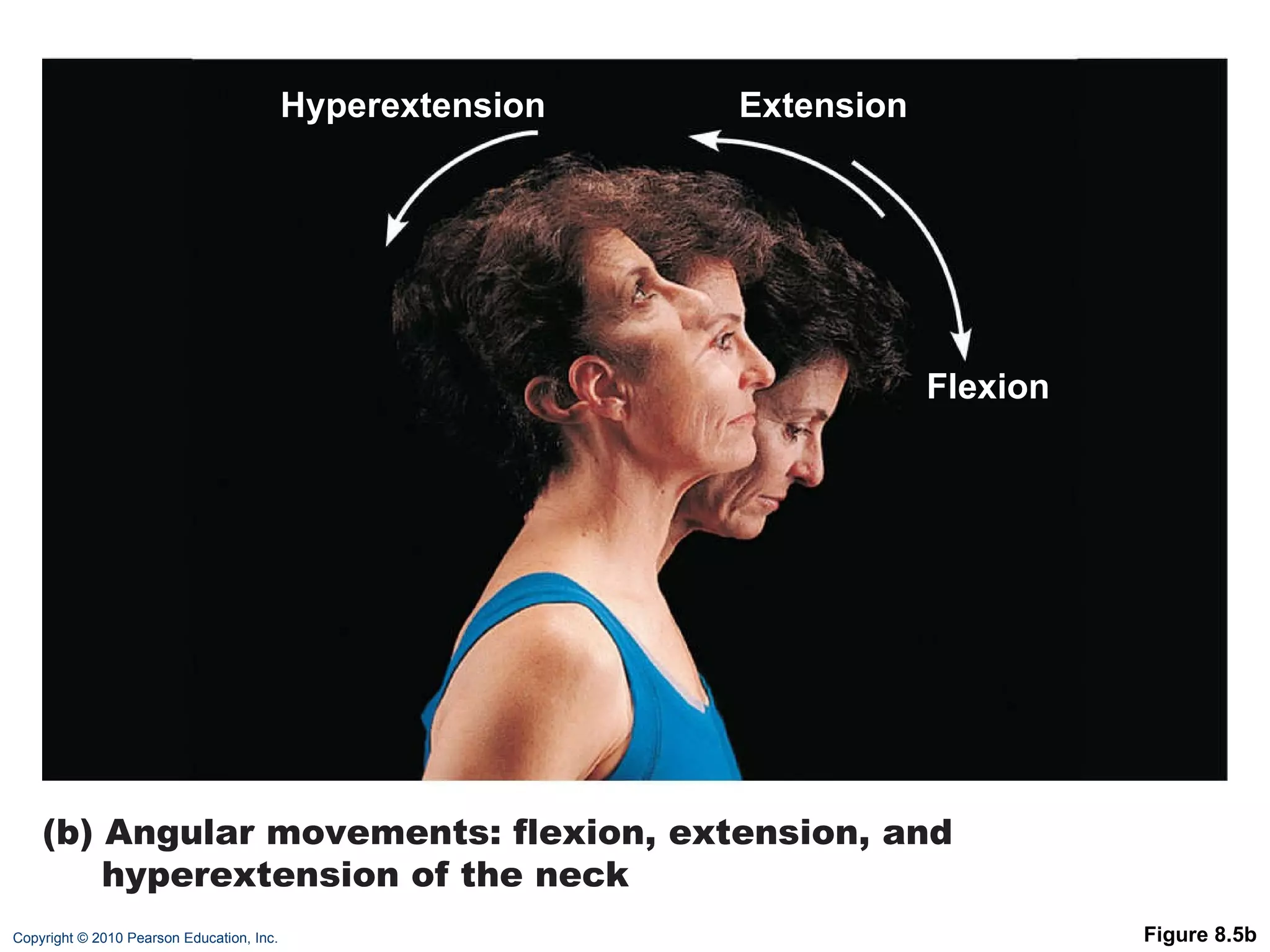
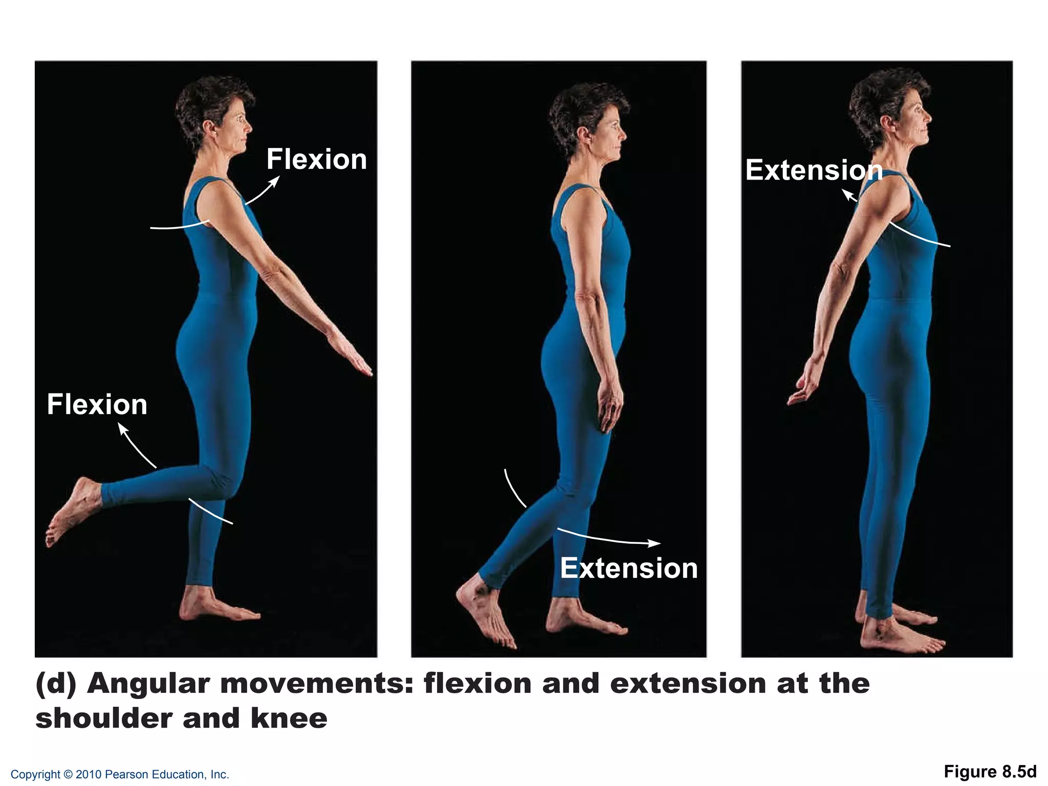
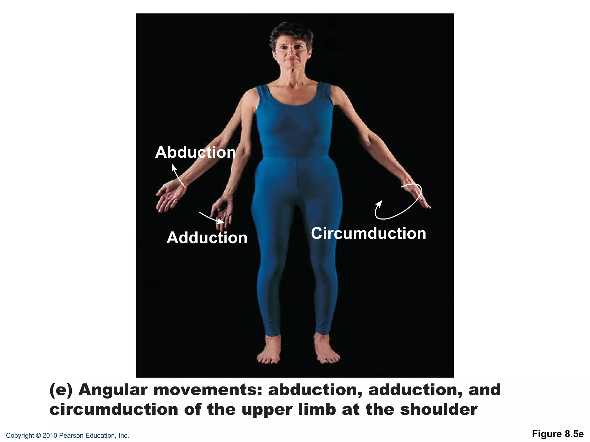
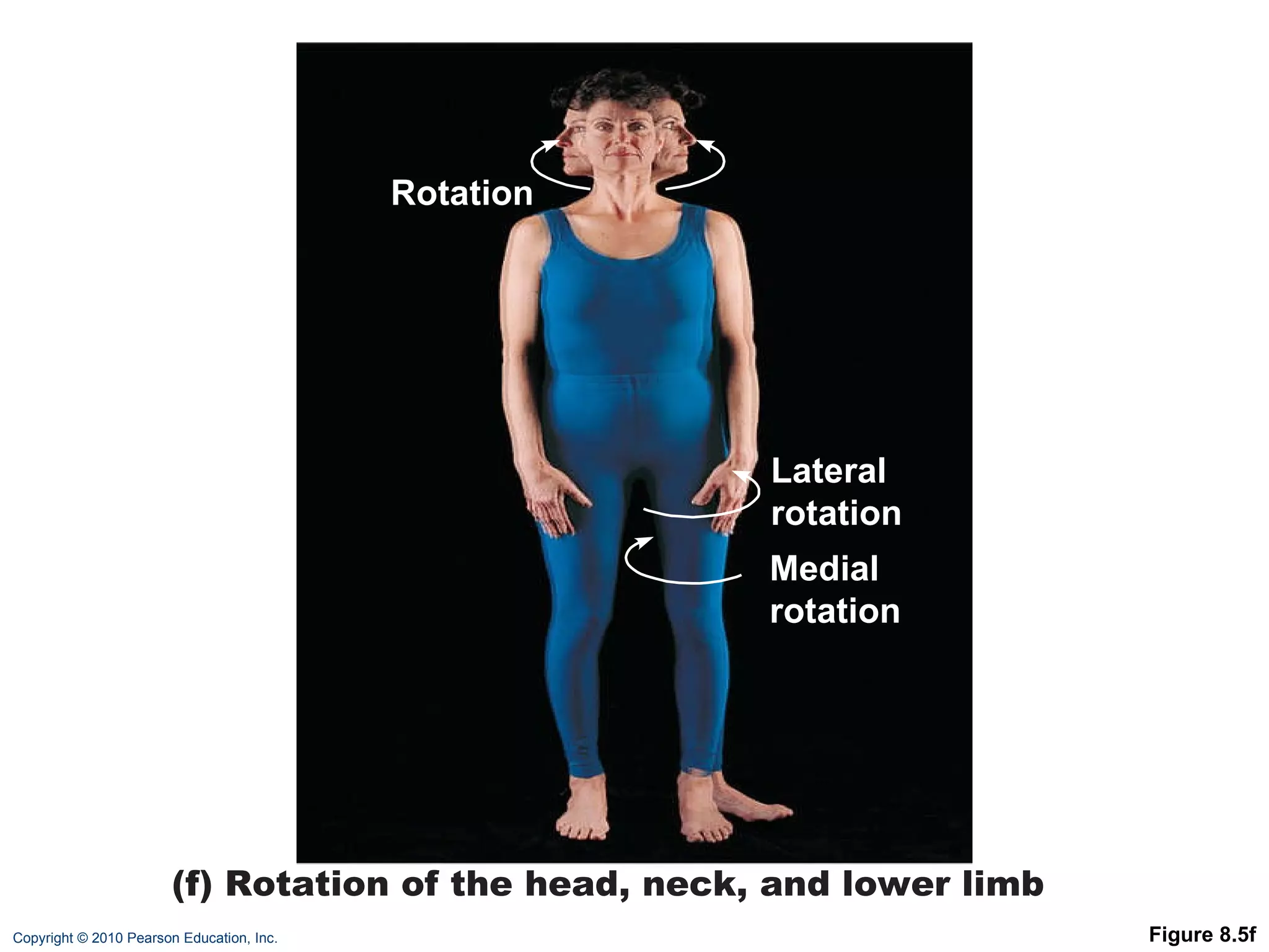
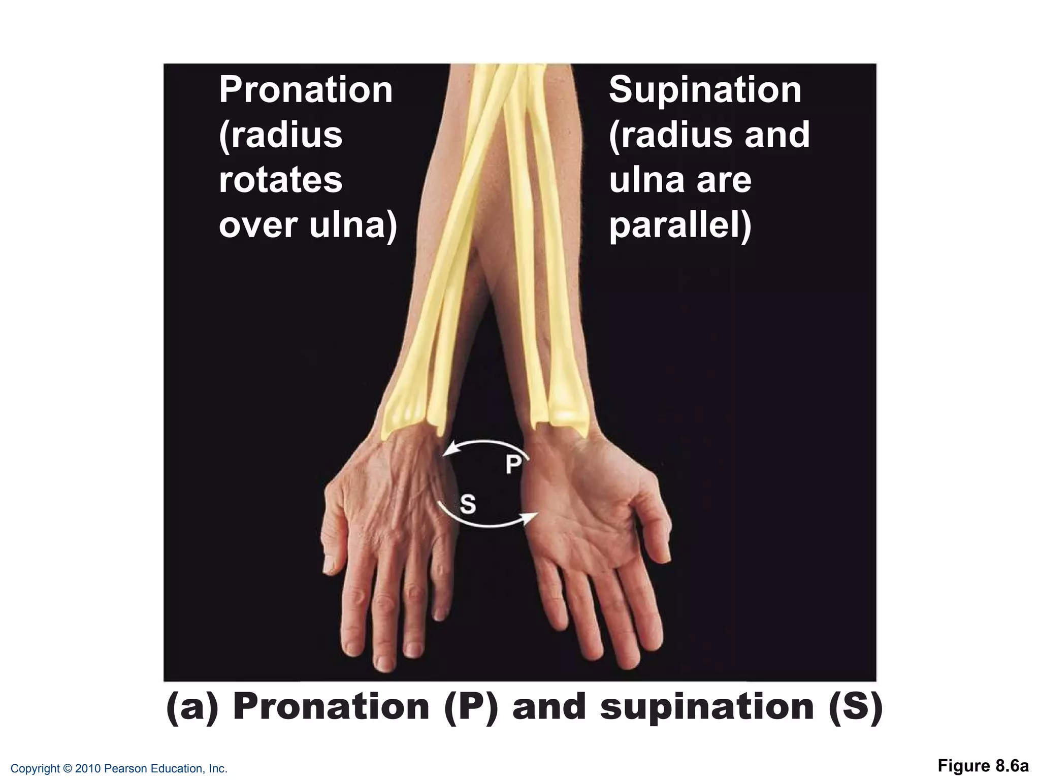
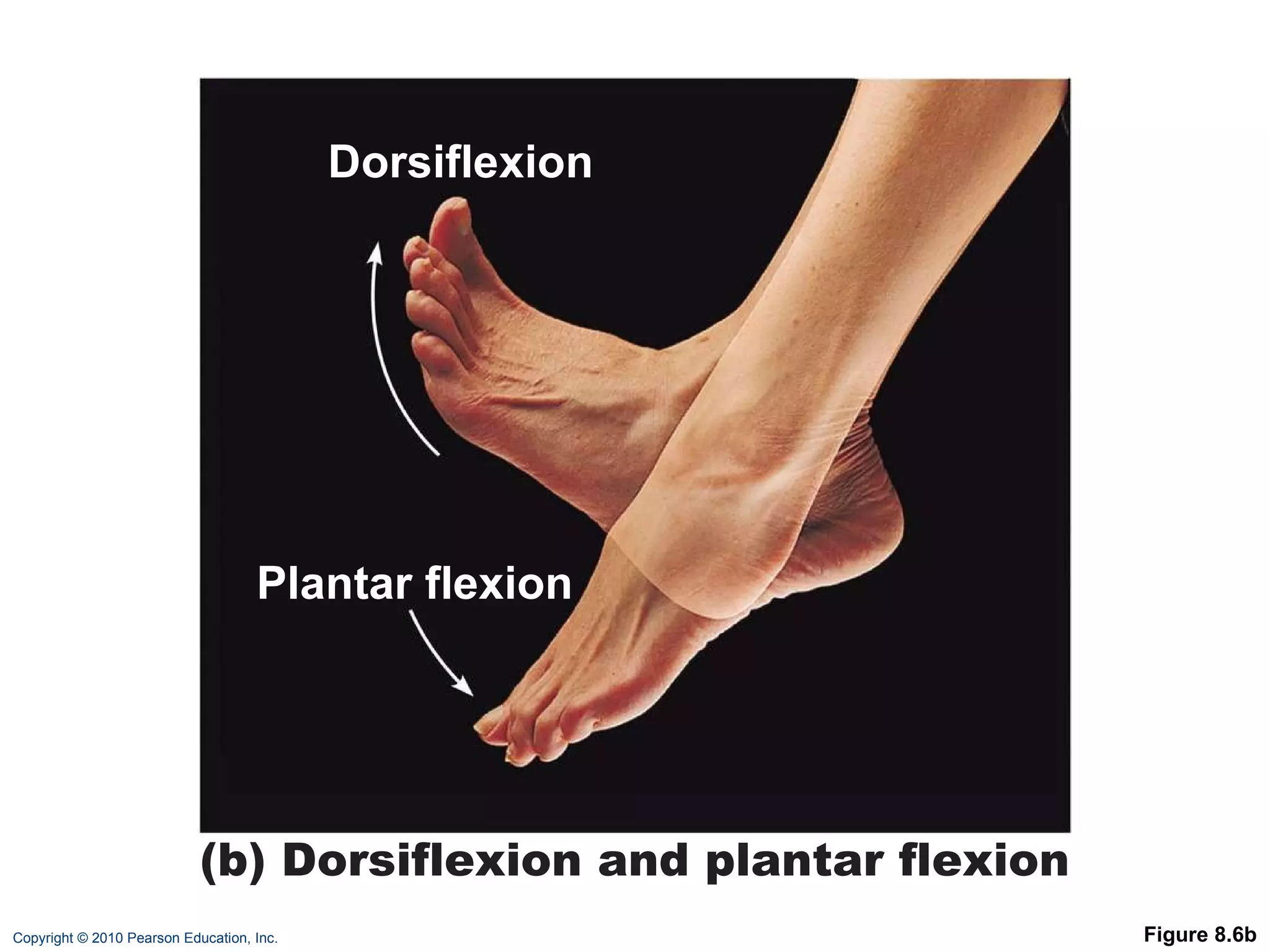
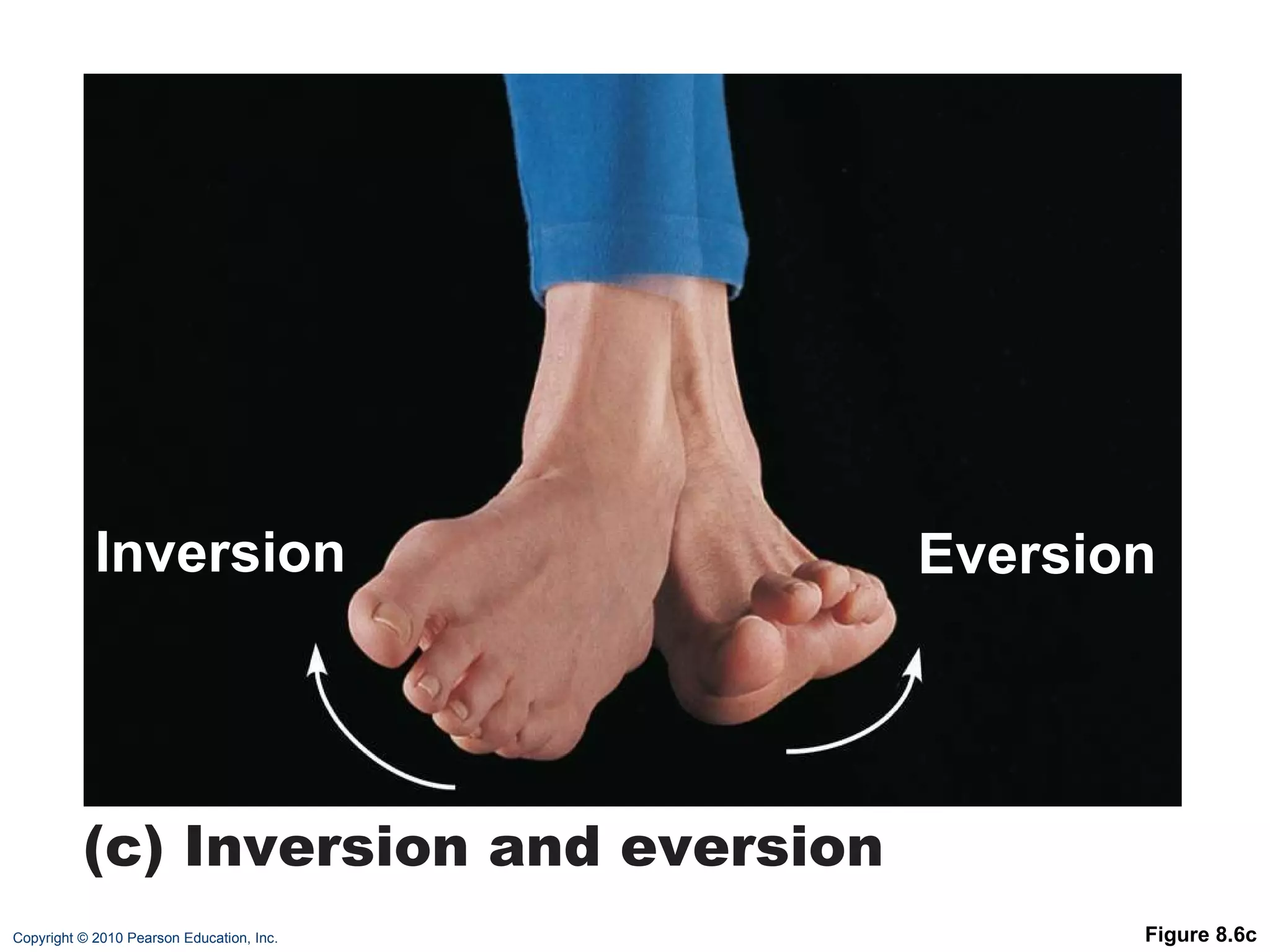
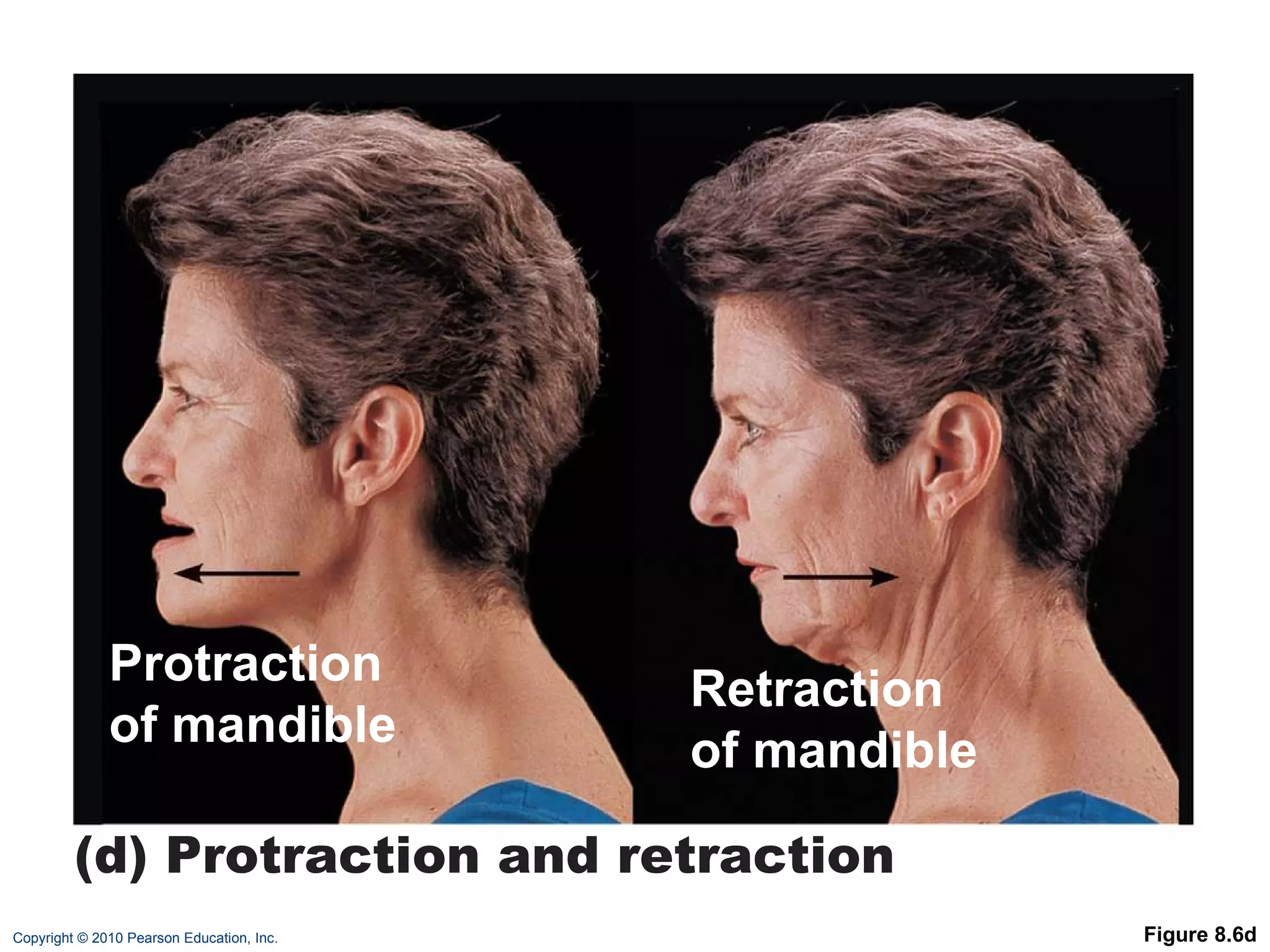
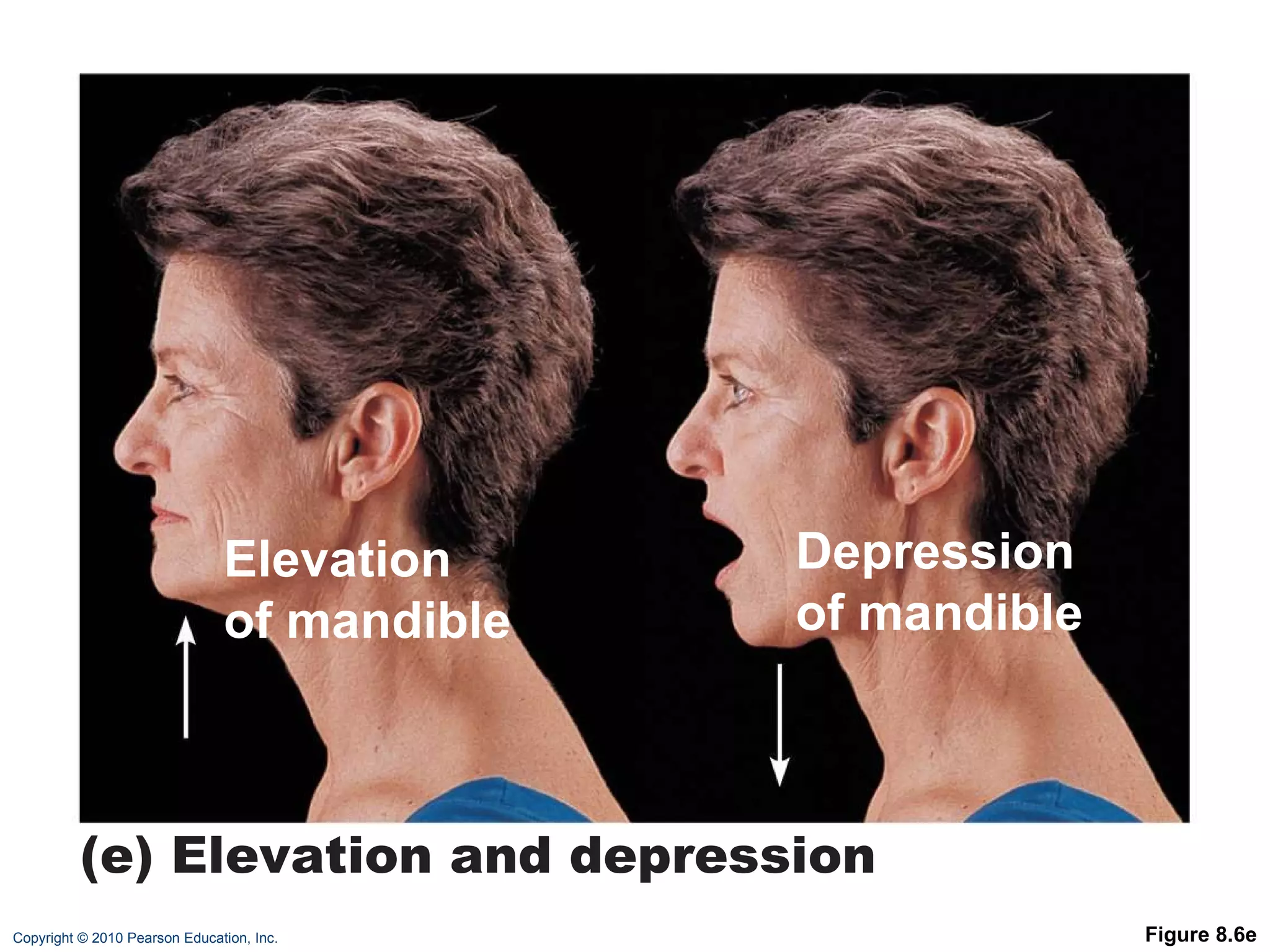
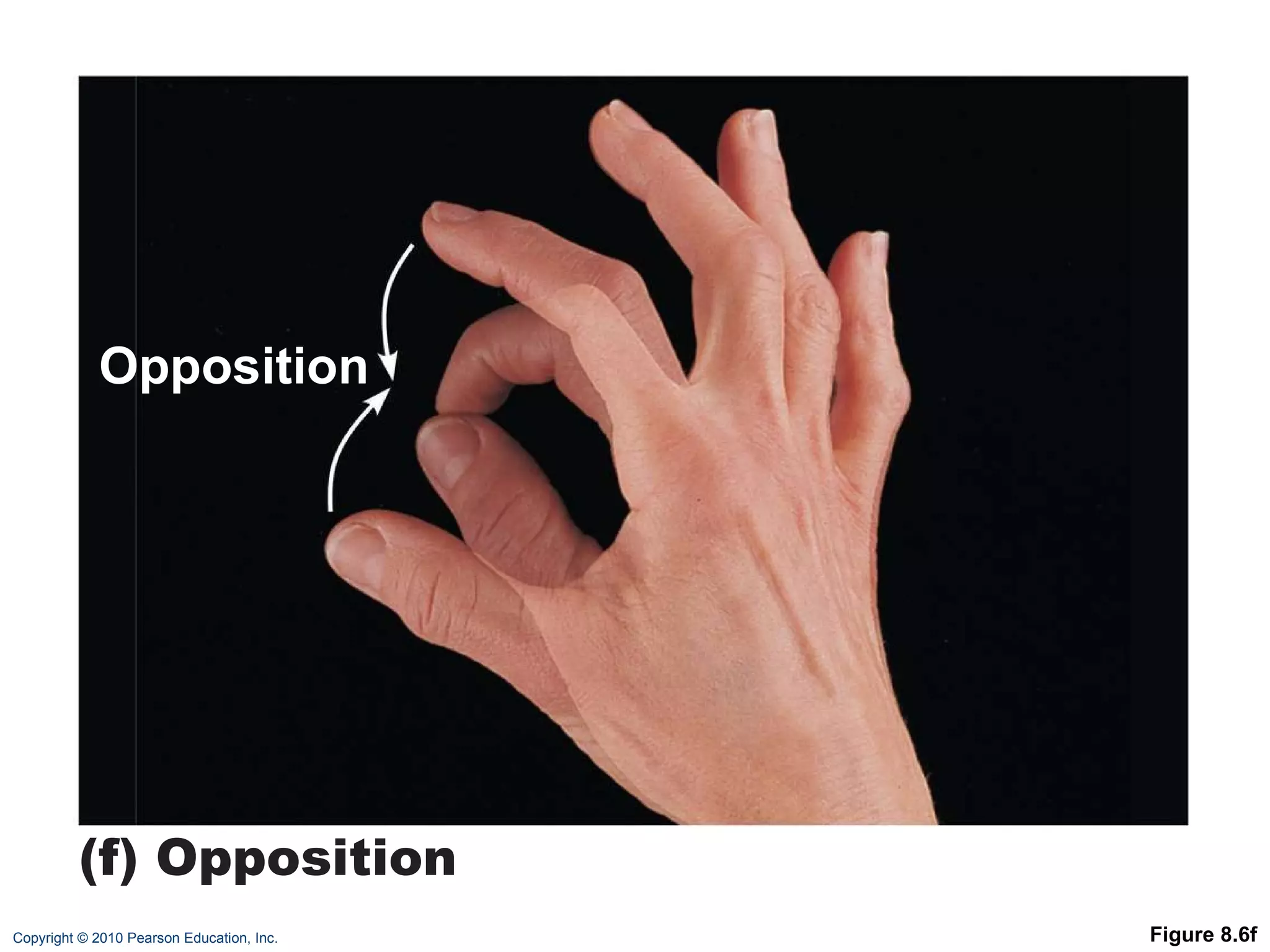
8  Joints: Part A
Joints (Articulations) Articulation—site where two or more bones meet Functions of joints: Give skeleton mobility Hold skeleton together
Functional Classification of Joints  Based on amount of movement allowed by the joint Three functional classifications: Synarthroses—immovable  Amphiarthroses—slightly movable  Diarthroses—freely movable
Structural Classification of Joints  Based on material binding bones together and whether or not a joint cavity is present Three structural classifications: Fibrous Cartilaginous  Synovial
Fibrous Joints Bones joined by dense fibrous connective tissue No joint cavity Most are synarthrotic (immovable) Three types: Sutures Syndesmoses Gomphoses
Fibrous Joints: Sutures Rigid, interlocking joints containing short connective tissue fibers Allow for growth during youth In middle age, sutures ossify and are called synostoses
Figure 8.1a Dense fibrous connective tissue Suture line (a)  Suture   Joint held together with very short, interconnecting fibers, and bone edges interlock. Found only in the skull.
Fibrous Joints: Syndesmoses Bones connected by ligaments (bands of fibrous tissue) Movement varies from immovable to slightly movable Examples:  Synarthrotic distal tibiofibular joint Diarthrotic interosseous connection between radius and ulna
Figure 8.1b Fibula Tibia Ligament (b) Syndesmosis  Joint held together by a ligament. Fibrous tissue can vary in length, but is longer than in sutures.
Fibrous Joints: Gomphoses Peg-in-socket joints of teeth in alveolar sockets Fibrous connection is the periodontal ligament
Figure 8.1c Root of tooth Socket of alveolar process Periodontal ligament (c) Gomphosis  “ Peg in socket” fibrous joint. Periodontal ligament holds tooth in socket.
Cartilaginous Joints Bones united by cartilage No joint cavity Two types: Synchondroses Symphyses
Cartilaginous Joints: Synchondroses A bar or plate of hyaline cartilage unites the bones All are synarthrotic
Figure 8.2a Epiphyseal plate (temporary hyaline cartilage joint) Sternum (manubrium) Joint between first rib and sternum (immovable) (a)  Synchondroses Bones united by hyaline cartilage
Cartilaginous Joints: Symphyses Hyaline cartilage covers the articulating surfaces and is fused to an intervening pad of fibrocartilage Strong, flexible amphiarthroses
Figure 8.2b Fibrocartilaginous intervertebral disc Pubic symphysis Body of vertebra Hyaline cartilage (b)  Symphyses Bones united by fibrocartilage
Synovial Joints All are diarthrotic Include all limb joints; most joints of the body
Synovial Joints Distinguishing features: Articular cartilage: hyaline cartilage Joint (synovial) cavity: small potential space
Synovial Joints Distinguishing features: 3. Articular (joint) capsule:  Outer fibrous capsule of dense irregular connective tissue Inner synovial membrane of loose connective tissue
Synovial Joints Distinguishing features: 4. Synovial fluid:  Viscous slippery filtrate of plasma + hyaluronic acid  Lubricates and nourishes articular cartilage
Figure 8.3 Periosteum Ligament Fibrous capsule Synovial membrane Joint cavity (contains synovial fluid) Articular (hyaline) cartilage Articular capsule
Synovial Joints Distinguishing features: 5. Three possible types of reinforcing ligaments: Capsular (intrinsic)—part of the fibrous capsule Extracapsular—outside the capsule Intracapsular—deep to capsule; covered by synovial membrane
Synovial Joints Distinguishing features: 6. Rich nerve and blood vessel supply: Nerve fibers detect pain, monitor joint position and stretch Capillary beds produce filtrate for synovial fluid
Synovial Joints: Friction-Reducing Structures Bursae: Flattened, fibrous sacs lined with synovial membranes  Contain synovial fluid Commonly act as “ball bearings” where ligaments, muscles, skin, tendons, or bones rub together
Figure 8.4b Coracoacromial ligament Subacromial bursa Cavity in bursa containing synovial fluid Bursa rolls and lessens friction. Humerus head rolls medially as arm abducts. (b) Enlargement of (a), showing how a bursa eliminates friction where a ligament (or other structure) would rub against a bone Humerus resting  Humerus moving
Synovial Joints: Friction-Reducing Structures Tendon sheath: Elongated bursa that wraps completely around a tendon
Figure 8.4a Acromion of scapula Joint cavity containing synovial fluid Synovial membrane Fibrous capsule Humerus Hyaline cartilage Coracoacromial ligament Subacromial bursa Fibrous articular capsule Tendon sheath Tendon of long head of biceps brachii muscle (a) Frontal section through the right shoulder joint
Stabilizing Factors at Synovial Joints Shapes of articular surfaces (minor role) Ligament number and location (limited role) Muscle tone, which keeps tendons that cross the joint taut Extremely important in reinforcing shoulder and knee joints and arches of the foot
Synovial Joints: Movement Muscle attachments across a joint: Origin—attachment to the immovable bone Insertion—attachment to the movable bone Muscle contraction causes the insertion to move toward the origin Movements occur along transverse, frontal, or sagittal planes
Synovial Joints: Range of Motion Nonaxial—slipping movements only Uniaxial—movement in one plane Biaxial—movement in two planes Multiaxial—movement in or around all three planes
Summary of Characteristics of Body Joints Consult Table 8.2 for: Joint names Articulating bones Structural classification Functional classification Movements allowed
Table 8.2 (1 of 4)
Table 8.2 (2 of 4)
Table 8.2 (3 of 4)
Table 8.2 (4 of 4)
Movements at Synovial Joints Gliding Angular movements: Flexion, extension, hyperextension Abduction, adduction Circumduction Rotation Medial and lateral rotation
Movements at Synovial Joints 4. Special movements Supination, pronation Dorsiflexion, plantar flexion of the foot Inversion, eversion Protraction, retraction Elevation, depression Opposition
Gliding Movements One flat bone surface glides or slips over another similar surface  Examples: Intercarpal joints Intertarsal joints Between articular processes of vertebrae
Figure 8.5a Gliding (a) Gliding movements at the wrist
Angular Movements Movements that occur along the sagittal plane: Flexion—decreases the angle of the joint Extension— increases the angle of the joint Hyperextension—excessive extension beyond normal range of motion
Figure 8.5b (b) Angular movements: flexion, extension, and hyperextension of the neck Hyperextension Extension Flexion
Figure 8.5c Hyperextension Flexion Extension (c) Angular movements: flexion, extension, and hyperextension of the vertebral column
Figure 8.5d Extension Extension Flexion Flexion (d) Angular movements: flexion and extension at the shoulder and knee
Angular Movements Movements that occur along the frontal plane: Abduction—movement away from the midline Adduction—movement toward the midline Circumduction—flexion + abduction + extension + adduction of a limb so as to describe a cone in space
Figure 8.5e Abduction Adduction (e) Angular movements: abduction, adduction, and circumduction of the upper limb at the shoulder Circumduction
Rotation The turning of a bone around its own long axis Examples: Between C 1  and C 2  vertebrae Rotation of humerus and femur
Figure 8.5f Lateral rotation Medial rotation Rotation (f) Rotation of the head, neck, and lower limb
Special Movements Movements of radius around ulna: Supination (turning hand backward) Pronation (turning hand forward)
Figure 8.6a Supination (radius and  ulna are  parallel) (a) Pronation (P) and supination (S) Pronation (radius  rotates over ulna)
Special Movements Movements of the foot: Dorsiflexion (upward movement)  Plantar flexion (downward movement)
Figure 8.6b Dorsiflexion Plantar flexion Dorsiflexion Plantar flexion (b) Dorsiflexion and plantar flexion
Special Movements Movements of the foot: Inversion (turn sole medially) Eversion (turn sole laterally)
Figure 8.6c Eversion Inversion (c) Inversion and eversion
Special Movements Movements in a transverse plane: Protraction (anterior movement) Retraction (posterior movement)
Figure 8.6d Protraction of mandible Retraction of mandible (d) Protraction and retraction
Special Movements Elevation (lifting a body part superiorly) Depression (moving a body part inferiorly)
Figure 8.6e Elevation of mandible Depression of mandible (e) Elevation and depression
Special Movements Opposition of the thumb Movement in the saddle joint so that the thumb touches the tips of the other fingers
Figure 8.6f (f) Opposition Opposition

Ch 08 lecture_outline_a

  • 1.
    8 Joints:Part A
  • 2.
    Joints (Articulations) Articulation—sitewhere two or more bones meet Functions of joints: Give skeleton mobility Hold skeleton together
  • 3.
    Functional Classification ofJoints Based on amount of movement allowed by the joint Three functional classifications: Synarthroses—immovable Amphiarthroses—slightly movable Diarthroses—freely movable
  • 4.
    Structural Classification ofJoints Based on material binding bones together and whether or not a joint cavity is present Three structural classifications: Fibrous Cartilaginous Synovial
  • 5.
    Fibrous Joints Bonesjoined by dense fibrous connective tissue No joint cavity Most are synarthrotic (immovable) Three types: Sutures Syndesmoses Gomphoses
  • 6.
    Fibrous Joints: SuturesRigid, interlocking joints containing short connective tissue fibers Allow for growth during youth In middle age, sutures ossify and are called synostoses
  • 7.
    Figure 8.1a Densefibrous connective tissue Suture line (a) Suture Joint held together with very short, interconnecting fibers, and bone edges interlock. Found only in the skull.
  • 8.
    Fibrous Joints: SyndesmosesBones connected by ligaments (bands of fibrous tissue) Movement varies from immovable to slightly movable Examples: Synarthrotic distal tibiofibular joint Diarthrotic interosseous connection between radius and ulna
  • 9.
    Figure 8.1b FibulaTibia Ligament (b) Syndesmosis Joint held together by a ligament. Fibrous tissue can vary in length, but is longer than in sutures.
  • 10.
    Fibrous Joints: GomphosesPeg-in-socket joints of teeth in alveolar sockets Fibrous connection is the periodontal ligament
  • 11.
    Figure 8.1c Rootof tooth Socket of alveolar process Periodontal ligament (c) Gomphosis “ Peg in socket” fibrous joint. Periodontal ligament holds tooth in socket.
  • 12.
    Cartilaginous Joints Bonesunited by cartilage No joint cavity Two types: Synchondroses Symphyses
  • 13.
    Cartilaginous Joints: SynchondrosesA bar or plate of hyaline cartilage unites the bones All are synarthrotic
  • 14.
    Figure 8.2a Epiphysealplate (temporary hyaline cartilage joint) Sternum (manubrium) Joint between first rib and sternum (immovable) (a) Synchondroses Bones united by hyaline cartilage
  • 15.
    Cartilaginous Joints: SymphysesHyaline cartilage covers the articulating surfaces and is fused to an intervening pad of fibrocartilage Strong, flexible amphiarthroses
  • 16.
    Figure 8.2b Fibrocartilaginousintervertebral disc Pubic symphysis Body of vertebra Hyaline cartilage (b) Symphyses Bones united by fibrocartilage
  • 17.
    Synovial Joints Allare diarthrotic Include all limb joints; most joints of the body
  • 18.
    Synovial Joints Distinguishingfeatures: Articular cartilage: hyaline cartilage Joint (synovial) cavity: small potential space
  • 19.
    Synovial Joints Distinguishingfeatures: 3. Articular (joint) capsule: Outer fibrous capsule of dense irregular connective tissue Inner synovial membrane of loose connective tissue
  • 20.
    Synovial Joints Distinguishingfeatures: 4. Synovial fluid: Viscous slippery filtrate of plasma + hyaluronic acid Lubricates and nourishes articular cartilage
  • 21.
    Figure 8.3 PeriosteumLigament Fibrous capsule Synovial membrane Joint cavity (contains synovial fluid) Articular (hyaline) cartilage Articular capsule
  • 22.
    Synovial Joints Distinguishingfeatures: 5. Three possible types of reinforcing ligaments: Capsular (intrinsic)—part of the fibrous capsule Extracapsular—outside the capsule Intracapsular—deep to capsule; covered by synovial membrane
  • 23.
    Synovial Joints Distinguishingfeatures: 6. Rich nerve and blood vessel supply: Nerve fibers detect pain, monitor joint position and stretch Capillary beds produce filtrate for synovial fluid
  • 24.
    Synovial Joints: Friction-ReducingStructures Bursae: Flattened, fibrous sacs lined with synovial membranes Contain synovial fluid Commonly act as “ball bearings” where ligaments, muscles, skin, tendons, or bones rub together
  • 25.
    Figure 8.4b Coracoacromialligament Subacromial bursa Cavity in bursa containing synovial fluid Bursa rolls and lessens friction. Humerus head rolls medially as arm abducts. (b) Enlargement of (a), showing how a bursa eliminates friction where a ligament (or other structure) would rub against a bone Humerus resting Humerus moving
  • 26.
    Synovial Joints: Friction-ReducingStructures Tendon sheath: Elongated bursa that wraps completely around a tendon
  • 27.
    Figure 8.4a Acromionof scapula Joint cavity containing synovial fluid Synovial membrane Fibrous capsule Humerus Hyaline cartilage Coracoacromial ligament Subacromial bursa Fibrous articular capsule Tendon sheath Tendon of long head of biceps brachii muscle (a) Frontal section through the right shoulder joint
  • 28.
    Stabilizing Factors atSynovial Joints Shapes of articular surfaces (minor role) Ligament number and location (limited role) Muscle tone, which keeps tendons that cross the joint taut Extremely important in reinforcing shoulder and knee joints and arches of the foot
  • 29.
    Synovial Joints: MovementMuscle attachments across a joint: Origin—attachment to the immovable bone Insertion—attachment to the movable bone Muscle contraction causes the insertion to move toward the origin Movements occur along transverse, frontal, or sagittal planes
  • 30.
    Synovial Joints: Rangeof Motion Nonaxial—slipping movements only Uniaxial—movement in one plane Biaxial—movement in two planes Multiaxial—movement in or around all three planes
  • 31.
    Summary of Characteristicsof Body Joints Consult Table 8.2 for: Joint names Articulating bones Structural classification Functional classification Movements allowed
  • 32.
  • 33.
  • 34.
  • 35.
  • 36.
    Movements at SynovialJoints Gliding Angular movements: Flexion, extension, hyperextension Abduction, adduction Circumduction Rotation Medial and lateral rotation
  • 37.
    Movements at SynovialJoints 4. Special movements Supination, pronation Dorsiflexion, plantar flexion of the foot Inversion, eversion Protraction, retraction Elevation, depression Opposition
  • 38.
    Gliding Movements Oneflat bone surface glides or slips over another similar surface Examples: Intercarpal joints Intertarsal joints Between articular processes of vertebrae
  • 39.
    Figure 8.5a Gliding(a) Gliding movements at the wrist
  • 40.
    Angular Movements Movementsthat occur along the sagittal plane: Flexion—decreases the angle of the joint Extension— increases the angle of the joint Hyperextension—excessive extension beyond normal range of motion
  • 41.
    Figure 8.5b (b)Angular movements: flexion, extension, and hyperextension of the neck Hyperextension Extension Flexion
  • 42.
    Figure 8.5c HyperextensionFlexion Extension (c) Angular movements: flexion, extension, and hyperextension of the vertebral column
  • 43.
    Figure 8.5d ExtensionExtension Flexion Flexion (d) Angular movements: flexion and extension at the shoulder and knee
  • 44.
    Angular Movements Movementsthat occur along the frontal plane: Abduction—movement away from the midline Adduction—movement toward the midline Circumduction—flexion + abduction + extension + adduction of a limb so as to describe a cone in space
  • 45.
    Figure 8.5e AbductionAdduction (e) Angular movements: abduction, adduction, and circumduction of the upper limb at the shoulder Circumduction
  • 46.
    Rotation The turningof a bone around its own long axis Examples: Between C 1 and C 2 vertebrae Rotation of humerus and femur
  • 47.
    Figure 8.5f Lateralrotation Medial rotation Rotation (f) Rotation of the head, neck, and lower limb
  • 48.
    Special Movements Movementsof radius around ulna: Supination (turning hand backward) Pronation (turning hand forward)
  • 49.
    Figure 8.6a Supination(radius and ulna are parallel) (a) Pronation (P) and supination (S) Pronation (radius rotates over ulna)
  • 50.
    Special Movements Movementsof the foot: Dorsiflexion (upward movement) Plantar flexion (downward movement)
  • 51.
    Figure 8.6b DorsiflexionPlantar flexion Dorsiflexion Plantar flexion (b) Dorsiflexion and plantar flexion
  • 52.
    Special Movements Movementsof the foot: Inversion (turn sole medially) Eversion (turn sole laterally)
  • 53.
    Figure 8.6c EversionInversion (c) Inversion and eversion
  • 54.
    Special Movements Movementsin a transverse plane: Protraction (anterior movement) Retraction (posterior movement)
  • 55.
    Figure 8.6d Protractionof mandible Retraction of mandible (d) Protraction and retraction
  • 56.
    Special Movements Elevation(lifting a body part superiorly) Depression (moving a body part inferiorly)
  • 57.
    Figure 8.6e Elevationof mandible Depression of mandible (e) Elevation and depression
  • 58.
    Special Movements Oppositionof the thumb Movement in the saddle joint so that the thumb touches the tips of the other fingers
  • 59.
    Figure 8.6f (f)Opposition Opposition