1. Discuss theclassification, types, location, structure and functions,
the blood and nervous supply of bones, joints, cartilages and
muscles;
2. Locate the major cartilages of the adult skeleton.
3. Discuss the functional properties of the types of cartilages.
4. Describe the location, structure and function of skeletal, smooth
and cardiac muscles;
Objectives
4.
Joint
• Are knownas articulations
• Functional junctions between bones
• Bind parts of skeletal system together
• Make bone growth possible
• Permit parts of the skeleton to change shape
during childbirth
• Enable body to move in response to skeletal
muscle contraction
• Three (3) classifications of joints will be considered
5.
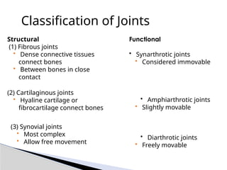
Classification of Joints
Structural
(1)Fibrous joints
• Dense connective tissues
connect bones
• Between bones in close
contact
(2) Cartilaginous joints
• Hyaline cartilage or
fibrocartilage connect bones
(3) Synovial joints
• Most complex
• Allow free movement
Functional
• Synarthrotic joints
• Considered immovable
• Amphiarthrotic joints
• Slightly movable
• Diarthrotic joints
• Freely movable
6.
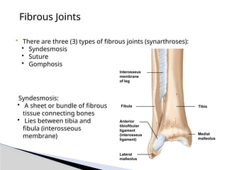
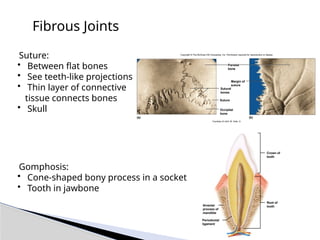
Fibrous Joints
• Thereare three (3) types of fibrous joints (synarthroses):
• Syndesmosis
• Suture
• Gomphosis
• Syndesmosis:
• A sheet or bundle of fibrous
tissue connecting bones
• Lies between tibia and
fibula (interosseous
membrane)
Fibula
Interosseus
membrane
of leg
Tibia
Medial
malleolus
Anterior
tibiofibular
ligament
(interosseus
ligament)
Lateral
malleolus
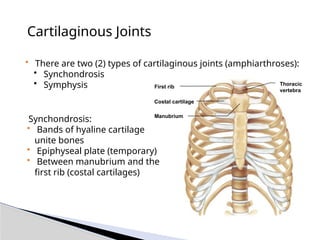
Cartilaginous Joints
• Thereare two (2) types of cartilaginous joints (amphiarthroses):
• Synchondrosis
• Symphysis
• Synchondrosis:
• Bands of hyaline cartilage
unite bones
• Epiphyseal plate (temporary)
• Between manubrium and the
first rib (costal cartilages)
Thoracic
vertebra
Costal cartilage
Manubrium
First rib
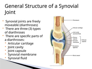
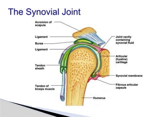
General Structure ofa Synovial
Joint
• Synovial joints are freely
moveable (diarthroses)
• There are three (3) types
of diarthroses
• There are specific parts of
a diarthroses:
• Articular cartilage
• Joint cavity
• Joint capsule
• Synovial membrane
• Synovial fluid
11.
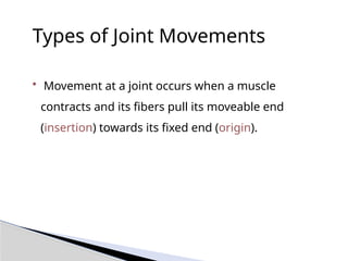
Types of JointMovements
• Movement at a joint occurs when a muscle
contracts and its fibers pull its moveable end
(insertion) towards its fixed end (origin).
12.
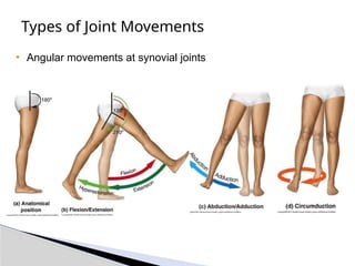
Types of JointMovements
• Angular movements at synovial joints
Type Description Example
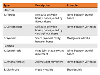
Structure
1.Fibrous No space between
bones; bones joined by
fibrous tissue
Joints between cranial
bones
2. Cartilaginous No space between
bones; bones joined by
cartilaginous tissue
Joints between vertebrae
3. Synovial Space (synovial cavity)
between bones
Most joints in limbs
Function
1. Synarthrosis Fixed joint that allows no
movement
Joints between cranial
bones
2. Amphiarthrosis Allows slight movement Joints between vertebrae
3. Diarthrosis Freely movable Shoulder, hip
15.
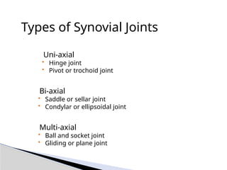
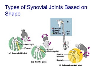
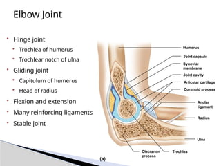
Types of SynovialJoints
• Uni-axial
• Hinge joint
• Pivot or trochoid joint
• Bi-axial
• Saddle or sellar joint
• Condylar or ellipsoidal joint
• Multi-axial
• Ball and socket joint
• Gliding or plane joint
16.
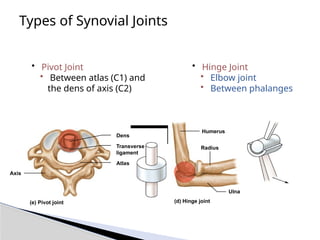
Types of SynovialJoints
• Pivot Joint
• Between atlas (C1) and
the dens of axis (C2)
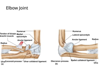
• Hinge Joint
• Elbow joint
• Between phalanges
(e) Pivot joint
Dens
Transverse
ligament
Atlas
Axis
(d) Hinge joint
Humerus
Ulna
Radius
17.
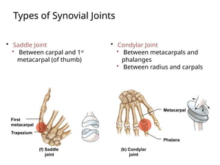
Types of SynovialJoints
• Saddle Joint
• Between carpal and 1st
metacarpal (of thumb)
• Condylar Joint
• Between metacarpals and
phalanges
• Between radius and carpals
Metacarpal
Phalanx
(b) Condylar
joint
(f) Saddle
joint
First
metacarpal
Trapezium
18.
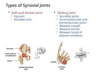
Types of SynovialJoints
• Ball-and-Socket Joint
• Hip joint
• Shoulder joint
• Gliding Joint
• Sacroiliac joints
• Acromioclavicular and
sternoclavicular joints
• Between carpals
• Between tarsals
• Between facets of
adjacent vertebrae
Hip bone
(a) Ball-and-socket
joint
Head of femur
in acetabulum
Femur
(c) Plane
joint
Carpals
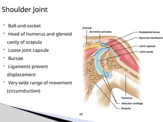
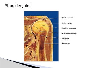
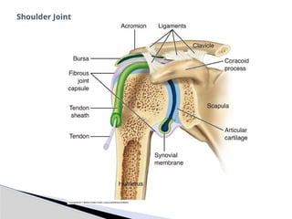
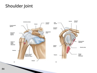
Shoulder Joint
• Ball-and-socket
•Head of humerus and glenoid
cavity of scapula
• Loose joint capsule
• Bursae
• Ligaments prevent
displacement
• Very wide range of movement
(circumduction)
Humerus
Articular cartilage
Scapula
Clavicle
Acromion process Subdeltoid bursa
Synovial membrane
Joint capsule
Joint cavity
(a)
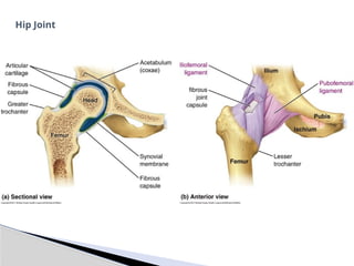
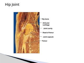
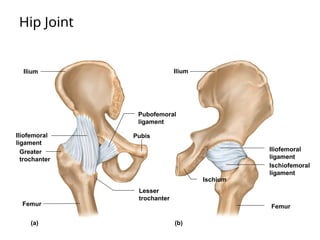
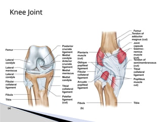
Hip Joint
• Ball-and-socketjoint
• Head of femur and
acetabulum of coxa
• Heavy joint capsule
• Many reinforcing ligaments
• Less freedom of movement
than shoulder joint
• Circumduction
Hip bone
Joint cavity
Articular cartilage
Synovial membrane
Joint capsule
Ligamentum capitis
Femur
(a)
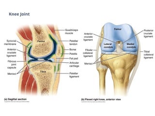
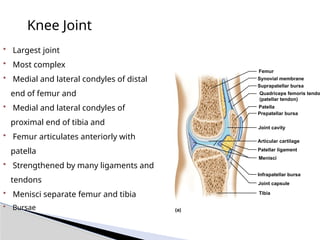
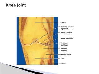
Knee Joint
• Largestjoint
• Most complex
• Medial and lateral condyles of distal
end of femur and
• Medial and lateral condyles of
proximal end of tibia and
• Femur articulates anteriorly with
patella
• Strengthened by many ligaments and
tendons
• Menisci separate femur and tibia
• Bursae
Femur
Quadriceps femoris tendo
(patellar tendon)
Synovial membrane
Suprapatellar bursa
Patella
Prepatellar bursa
Joint cavity
Articular cartilage
Menisci
Patellar ligament
Infrapatellar bursa
Joint capsule
Tibia
(a)
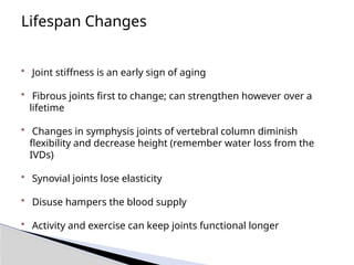
Lifespan Changes
• Jointstiffness is an early sign of aging
• Fibrous joints first to change; can strengthen however over a
lifetime
• Changes in symphysis joints of vertebral column diminish
flexibility and decrease height (remember water loss from the
IVDs)
• Synovial joints lose elasticity
• Disuse hampers the blood supply
• Activity and exercise can keep joints functional longer
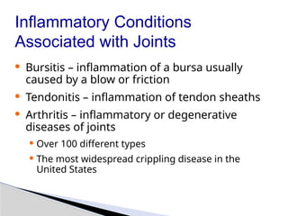
Inflammatory Conditions
Associated withJoints
· Bursitis – inflammation of a bursa usually
caused by a blow or friction
· Tendonitis – inflammation of tendon sheaths
· Arthritis – inflammatory or degenerative
diseases of joints
· Over 100 different types
· The most widespread crippling disease in the
United States
52.
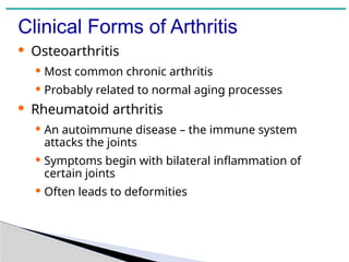
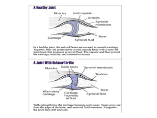
Clinical Forms ofArthritis
· Osteoarthritis
· Most common chronic arthritis
· Probably related to normal aging processes
· Rheumatoid arthritis
· An autoimmune disease – the immune system
attacks the joints
· Symptoms begin with bilateral inflammation of
certain joints
· Often leads to deformities
55.
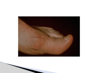
Clinical Forms ofArthritis
· Gouty Arthritis
·Inflammation of joints is caused by a
deposition of urate crystals from the blood
·Can usually be controlled with diet
57.
McConnell, T.H. & Hull, K. L. (2011). Human form and function:
Essentials of anatomy& physiology. Philadelphia: Wolters
Kluwer, Lippincott Williams & Wilkins.
Shier, D., Lewis, R. & Butler, J. (2002). Hole’s human anatomy &
physiology. New York: McGraw Hill.
Tortora, Gerard J. & Derrickson, Bryan H. (2011). Principles of
anatomy and physiology. Somerset, New Jersey: John Wiley &
Sons.
Recommended Reading
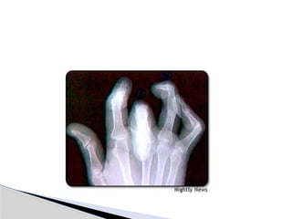
#7 Gomphosis – only found where the peridental ligament joins to the mandible or maxilla
#10 synovial membrane lines the interior of the joint capsule and secretes synovial fluid into the joint cavity.
This fluid lubricates, cushions shocks, prevents abrasion, and supports the chondrocytes of the articular cartilages through nutrient distribution.
Even in a large joint such as the knee, the total quantity of synovial fluid in a joint is normally less than 3 mL.
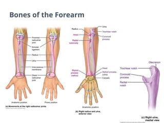
#33 The forearm consists of the radius and ulna.
A. The radius and ulna are uncrossed in anatomical position, but cross over when the arm is pronated.
B. Right radius and ulna, anterior view in the anatomical position.
C. Right ulna, medial view.
Question: Which bone has the coronoid process?
Answer: Ulna
#35 Question: Which structure is continuous with the synovial membrane – the tendon sheath or the ligaments?
Answer: Tendon sheath
#38 The right elbow is illustrated in the flexed position.
Question: Name the ligament that wraps around the radius.
Answer: Annular ligament
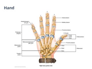
#39 Question: How many phalanges are found in the thumb?
Answer: Two
#40 A. Sectional view
B. Anterior view
Question: What is the name of the socket into which the ball of the femoral head fits?
Answer: Acetabulum
#44 A. Sagittal section.
B. Flexed knee, anterior view. The patella and other anterior structures have been removed.
Question: Which 2 ligaments are found within the synovial cavity?
Answer: Anterior and posterior cruciate ligaments
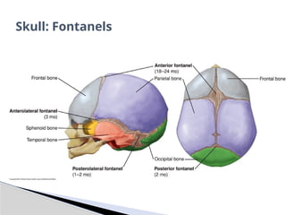
#50 The infant skull at birth, showing the fontanels. The fontanels close at the age shown in the brackets.
Question: Which fontanel closes last?
Answer: Anterior fontanel


![Chapt08 Holes Lecture[1]](https://cdn.slidesharecdn.com/ss_thumbnails/chapt08holeslecture1-091122122447-phpapp02-thumbnail.jpg?width=640&height=640&fit=bounds)


![Polymer [ बहुलक ] Chemistry Notes PDF - Irfanullah Mehar - JJ Sir Chemistry.pdf](https://cdn.slidesharecdn.com/ss_thumbnails/polymerchemistrynotespdf-irfanullahmehar-jjsirchemistry-260210172118-3f9b37f7-thumbnail.jpg?width=640&height=640&fit=bounds)


