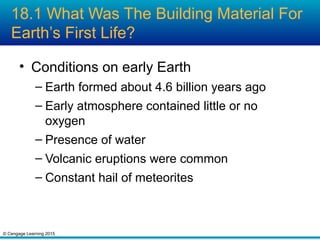
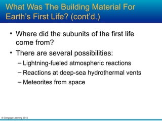
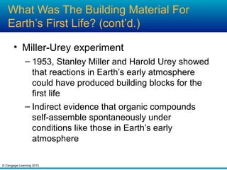
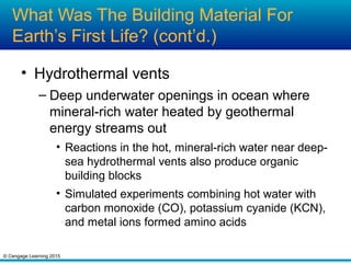
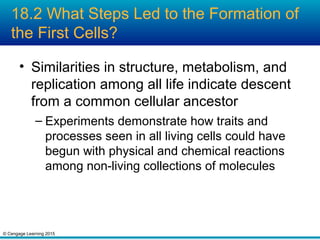
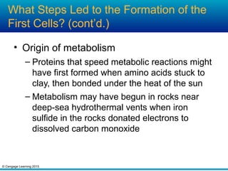
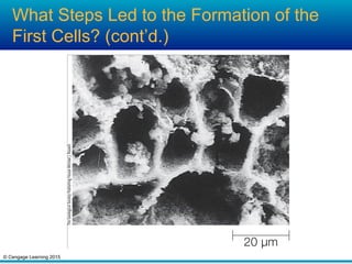
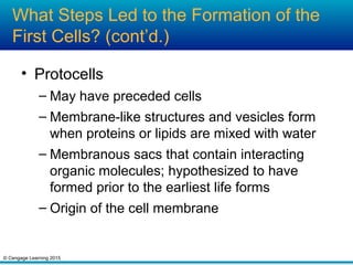
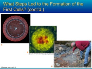
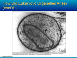
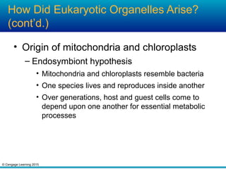
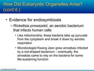
The document discusses the origins and early evolution of life on Earth. It describes some of the conditions on the early Earth and how simple organic compounds may have formed through atmospheric reactions, hydrothermal vents, or delivery by meteorites. It also discusses experiments that demonstrate how self-assembly of these molecules could have led to the first protocells and metabolism, as well as the possible emergence of RNA prior to DNA as the genetic material. The document then covers the divergence of bacteria and archaea, the rise of oxygen due to cyanobacteria, the appearance of eukaryotes, and current hypotheses about the endosymbiotic origins of organelles like mitochondria and chloroplasts.
Figure 18.3 Cell-sized chambers in iron-sulfide-rich rocks formed by simulations of conditions near hydrothermal vents. Similar chambers could have served as protected environments in which the first metabolic reactions took place.
Figure 18.4 Protocells. Scientists test hypotheses about protocell formation through laboratory simulations and field experiments
A Computer model of a protocell with a bilayer membrane of fatty acids around strands of RNA.
B Laboratory-formed protocell consisting of RNA-coated clay (red) surrounded by fatty acids and alcohols.
C Field-testing a hypothesis about protocell formation. David Deamer pours a mix of small organic molecules and phosphates into a hot acidic pool in Russia.
Figure 18.5 Possible microfossils from western Australia
A Micrograph of a filament that may be a 3.5-billion-year-old fossilized chain of photosynthetic bacteria.
B Micrograph showing structures that may be fossilized 3.4-billion-year-old sulfur-reducing bacteria.
Figure 18.6 Fossil stromatolite. It consists of layered remains of countless photosynthetic bacteria and sediment they captured.
Figure 18.8 {Animated} One hypothesis for the steps in organelle evolution. Membrane in-folding produced the endomembrane system. Then, an early endosymbiosis produced mitochondria, which occur in almost all eukaryotes. Later, photosynthetic bacteria entered a eukaryotic cell and, over many generations, evolved into chloroplasts.
Figure 18.9 Nucleus-like structure in a prokaryote. The DNA of Gemmata obscuriglobus, a species of bacteria, is enclosed by a double lipid bilayer membrane (indicated by the arrow).
Figure 18.10 Modern relatives of mitochondria
Rickettsias are tiny bacteria that invade eukaryotic
cells and replicate inside them. The species shown
here, Rickettsia prowazeki, is of special interest to
scientists for two reasons. First, it causes typhus,
a deadly human disease spread by lice and fleas.
Second, its genome is very similar to that of a
mitochondrion. R. prowazeki is one of the closest
living relatives of mitochondria. Some free-living
bacteria that float in the ocean’s surface waters
are also genetically similar to both rickettsias and
mitochondria. We do not know exactly how these
modern groups are related, but by one hypothesis,
all three—mitochondria, rickettsias, and the
modern marine bacteria—descended from
an ancient free-living marine species.
Figure 18.12 The Mars-like landscape of Chile’s Atacama Desert. Scientist Jay Quade, visible in the distance at the right, was a member of a team that found bacteria living beneath this desert soil.