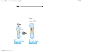
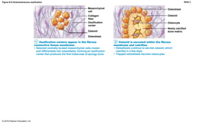
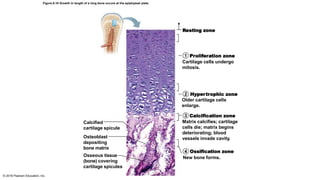
SlideShare a Scribd company logo
6.5 Bone Development
• Ossification (osteogenesis) is the process of bone tissue
formation
– Formation of bony skeleton begins in month 2 of development
– Postnatal bone growth occurs until early adulthood
– Bone remodeling and repair are lifelong
© 2016 Pearson Education, Inc.
Formation of the Bony Skeleton
• Up to about week 8, fibrous membranes and hyaline cartilage of
fetal skeleton are replaced with bone tissue
• Endochondral ossification
– Bone forms by replacing hyaline cartilage
– Bones are called cartilage (endochondral) bones
– Form most of skeleton
• Intramembranous ossification
– Bone develops from fibrous membrane
– Bones are called membrane bones
© 2016 Pearson Education, Inc.
Formation of the Bony Skeleton (cont.)
• Endochondral ossification
– Forms essentially all bones inferior to base of skull, except clavicles
– Begins late in month 2 of development
– Uses previously formed hyaline cartilage models
– Requires breakdown of hyaline cartilage prior to ossification
– Begins at primary ossification center in center of shaft
• Blood vessels infiltrate perichondrium, converting it to periosteum
• Mesenchymal cells specialize into osteoblasts
© 2016 Pearson Education, Inc.
Formation of the Bony Skeleton (cont.)
• Five main steps in the process of ossification:
1. Bone collar forms around diaphysis of cartilage model
2. Central cartilage in diaphysis calcifies, then develops cavities
3. Periosteal bud invades cavities, leading to formation of spongy
bone
• Bud is made up of blood vessels, nerves, red marrow, osteogenic cells, and
osteoclasts
© 2016 Pearson Education, Inc.
Formation of the Bony Skeleton (cont.)
• Five main steps in the process of ossification (cont.):
4. Diaphysis elongates, and medullary cavity forms
• Secondary ossification centers appear in epiphyses
5. Epiphyses ossify
• Hyaline cartilage remains only in epiphyseal plates and articular cartilages
© 2016 Pearson Education, Inc.
Figure 6.8 Endochondral ossification in a long bone.
© 2016 Pearson Education, Inc.
Week 9
Hyaline
cartilage
Primary
ossification
center
Bone
collar
1 Bone collar forms
around the diaphysis
of the hyaline
cartilage model.
1
Slide 2
Figure 6.8 Endochondral ossification in a long bone.
© 2016 Pearson Education, Inc.
Week 9
Area of
deteriorating
cartilage matrix
Hyaline
cartilage
Primary
ossification
center
Bone
collar
1 Bone collar forms
around the diaphysis
of the hyaline
cartilage model.
Cartilage in the
center of the diaphysis
calcifies and then
develops cavities.
1 2
Slide 3
Figure 6.8 Endochondral ossification in a long bone.
© 2016 Pearson Education, Inc.
Month 3Week 9
Area of
deteriorating
cartilage matrix
Hyaline
cartilage
Spongy
bone
formation
Blood
vessel of
periosteal
bud
Primary
ossification
center
Bone
collar
1 Bone collar forms
around the diaphysis
of the hyaline
cartilage model.
Cartilage in the
center of the diaphysis
calcifies and then
develops cavities.
The periosteal
bud invades the
internal cavities
and spongy bone
forms.
1 2 3
Slide 4
Figure 6.8 Endochondral ossification in a long bone.
© 2016 Pearson Education, Inc.
Month 3Week 9 Birth
Secondary
ossification
center
Epiphyseal
blood vesselArea of
deteriorating
cartilage matrix
Hyaline
cartilage Medullary
cavity
Spongy
bone
formation
Blood
vessel of
periosteal
bud
Primary
ossification
center
Bone
collar
1 Bone collar forms
around the diaphysis
of the hyaline
cartilage model.
Cartilage in the
center of the diaphysis
calcifies and then
develops cavities.
The periosteal
bud invades the
internal cavities
and spongy bone
forms.
1 2 3 The diaphysis
elongates and a
medullary cavity forms.
Secondary ossification
centers appear in the
epiphyses.
4
Slide 5
Figure 6.8 Endochondral ossification in a long bone.
© 2016 Pearson Education, Inc.
Month 3Week 9 Birth Childhood to adolescence
Articular
cartilage
Secondary
ossification
center
Spongy
bone
Epiphyseal
blood vesselArea of
deteriorating
cartilage matrix
Epiphyseal
plate
cartilage
Hyaline
cartilage Medullary
cavity
Spongy
bone
formation
Blood
vessel of
periosteal
bud
Primary
ossification
center
Bone
collar
1 Bone collar forms
around the diaphysis
of the hyaline
cartilage model.
Cartilage in the
center of the diaphysis
calcifies and then
develops cavities.
The periosteal
bud invades the
internal cavities
and spongy bone
forms.
1 2 3 The epiphyses ossify.
When completed, hyaline
cartilage remains only in
the epiphyseal plates and
articular cartilages.
5The diaphysis
elongates and a
medullary cavity forms.
Secondary ossification
centers appear in the
epiphyses.
4
Slide 6
Formation of the Bony Skeleton (cont.)
• Intramembranous ossification: begins within fibrous connective
tissue membranes formed by mesenchymal cells
– Forms frontal, parietal, occipital, temporal, and clavicle bones
© 2016 Pearson Education, Inc.
Formation of the Bony Skeleton (cont.)
• Four major steps are involved:
1. Ossification centers are formed when mesenchymal cells cluster
and become osteoblasts
2. Osteoid is secreted, then calcified
3. Woven bone is formed when osteoid is laid down around blood
vessels, resulting in trabeculae
• Outer layer of woven bone forms periosteum
4. Lamellar bone replaces woven bone, and red marrow appears
© 2016 Pearson Education, Inc.
Mesenchymal
cell
Osteoid
Osteoblast
Ossification centers appear in the fibrous
connective tissue membrane.
• Selected centrally located mesenchymal cells cluster
and differentiate into osteoblasts, forming an ossification
center that produces the first trabeculae of spongy bone.
Ossification
center
Collagen
fiber
Figure 6.9 Intramembranous ossification.
© 2016 Pearson Education, Inc.
1
Slide 2
Mesenchymal
cell
Osteoblast
Osteoid
Osteocyte
Newly calcified
bone matrixOsteoid
Osteoblast
Ossification centers appear in the fibrous
connective tissue membrane.
Osteoid is secreted within the fibrous
membrane and calcifies.
• Selected centrally located mesenchymal cells cluster
and differentiate into osteoblasts, forming an ossification
center that produces the first trabeculae of spongy bone.
• Osteoblasts continue to secrete osteoid, which
calcifies in a few days.
• Trapped osteoblasts become osteocytes.
Ossification
center
Collagen
fiber
Figure 6.9 Intramembranous ossification.
© 2016 Pearson Education, Inc.
1 2
Slide 3
Mesenchymal
cell
Osteoblast
Osteoid
Osteocyte
Newly calcified
bone matrixOsteoid
Osteoblast
Ossification centers appear in the fibrous
connective tissue membrane.
Osteoid is secreted within the fibrous
membrane and calcifies.
• Selected centrally located mesenchymal cells cluster
and differentiate into osteoblasts, forming an ossification
center that produces the first trabeculae of spongy bone.
• Osteoblasts continue to secrete osteoid, which
calcifies in a few days.
• Trapped osteoblasts become osteocytes.
Mesenchyme
condensing
to form the
periosteum
Trabeculae of
woven bone
Blood vessel
Woven bone and periosteum form.
• Accumulating osteoid is laid down between embryonic
blood vessels in a manner that results in a network
(instead of concentric lamellae) of trabeculae called
woven bone.
• Vascularized mesenchyme condenses on the external
face of the woven bone and becomes the periosteum.
Ossification
center
Collagen
fiber
Figure 6.9 Intramembranous ossification.
© 2016 Pearson Education, Inc.
1 2
3
Slide 4
Mesenchymal
cell
Osteoblast
Osteoid
Osteocyte
Newly calcified
bone matrixOsteoid
Osteoblast
Ossification centers appear in the fibrous
connective tissue membrane.
Osteoid is secreted within the fibrous
membrane and calcifies.
• Selected centrally located mesenchymal cells cluster
and differentiate into osteoblasts, forming an ossification
center that produces the first trabeculae of spongy bone.
• Osteoblasts continue to secrete osteoid, which
calcifies in a few days.
• Trapped osteoblasts become osteocytes.
Mesenchyme
condensing
to form the
periosteum
Fibrous
periosteum
Plate of
compact bone
Trabeculae of
woven bone
Diploë (spongy
bone) cavities
contain red
marrow
Blood vessel
Woven bone and periosteum form.
• Accumulating osteoid is laid down between embryonic
blood vessels in a manner that results in a network
(instead of concentric lamellae) of trabeculae called
woven bone.
• Vascularized mesenchyme condenses on the external
face of the woven bone and becomes the periosteum.
• Trabeculae just deep to the periosteum thicken. Mature
lamellar bone replaces them, forming compact bone
plates.
• Spongy bone (diploë), consisting of distinct trabeculae,
persists internally and its vascular tissue becomes red
marrow.
Ossification
center
Collagen
fiber
Osteoblast
Lamellar bone replaces woven bone, just deep
to the periosteum. Red marrow appears.
Figure 6.9 Intramembranous ossification.
© 2016 Pearson Education, Inc.
1 2
3 4
Slide 5
Postnatal Bone Growth
• Long bones grow lengthwise by interstitial (longitudinal) growth of
epiphyseal plate
• Bones increase thickness through appositional growth
• Bones stop growing during adolescence
– Some facial bones continue to grow slowly through life
© 2016 Pearson Education, Inc.
Growth in Length of Long Bones
• Interstitial growth requires presence of epiphyseal cartilage in the
epiphyseal plate
• Epiphyseal plate maintains constant thickness
– Rate of cartilage growth on one side balanced by bone
replacement on other
• Epiphyseal plate consists of five zones:
1. Resting (quiescent) zone
2. Proliferation (growth) zone
3. Hypertrophic zone
4. Calcification zone
5. Ossification (osteogenic) zone
© 2016 Pearson Education, Inc.
Growth in Length of Long Bones (cont.)
1. Resting (quiescent) zone
– Area of cartilage on epiphyseal side of epiphyseal plate that is
relatively inactive
2. Proliferation (growth) zone
– Area of cartilage on diaphysis side of epiphyseal plate that is
rapidly dividing
– New cells formed move upward, pushing epiphysis away from
diaphysis, causing lengthening
© 2016 Pearson Education, Inc.
Growth in Length of Long Bones (cont.)
3. Hypertrophic zone
– Area with older chondrocytes closer to diaphysis
– Cartilage lacunae enlarge and erode, forming interconnecting
spaces
4. Calcification zone
– Surrounding cartilage matrix calcifies; chondrocytes die and
deteriorate
© 2016 Pearson Education, Inc.
Growth in Length of Long Bones (cont.)
5. Ossification zone
– Chondrocyte deterioration leaves long spicules of calcified cartilage
at epiphysis-diaphysis junction
– Spicules are then eroded by osteoclasts and are covered with new
bone by osteoblasts
– Ultimately replaced with spongy bone
– Medullary cavity enlarges as spicules are eroded
© 2016 Pearson Education, Inc.
Resting zone
Proliferation zone
Cartilage cells undergo
mitosis.
Hypertrophic zone
Older cartilage cells
enlarge.
Calcification zone
Matrix calcifies; cartilage
cells die; matrix begins
deteriorating; blood
vessels invade cavity.
Calcified
cartilage spicule
Ossification zone
New bone forms.Osseous tissue
(bone) covering
cartilage spicules
Osteoblast
depositing
bone matrix
Figure 6.10 Growth in length of a long bone occurs at the epiphyseal plate.
© 2016 Pearson Education, Inc.
1
2
3
4
Growth in Length of Long Bones (cont.)
• Near end of adolescence, chondroblasts divide less often
• Epiphyseal plate thins, then is replaced by bone
• Epiphyseal plate closure occurs when epiphysis and diaphysis
fuse
• Bone lengthening ceases
– Females: occurs around 18 years of age
– Males: occurs around 21 years of age
© 2016 Pearson Education, Inc.
Growth in Width (Thickness)
• Growing bones widen as they lengthen through appositional
growth
– Can occur throughout life
• Bones thicken in response to increased stress from muscle
activity or added weight
• Osteoblasts beneath periosteum secrete bone matrix on external
bone
• Osteoclasts remove bone on endosteal surface
• Usually more building up than breaking down which leads to
thicker, stronger bone that is not too heavy
© 2016 Pearson Education, Inc.
Figure 6.11 Long bone growth and remodeling during youth.
© 2016 Pearson Education, Inc.
Bone growth Bone remodeling
Articular cartilage
Cartilage
grows here.
Epiphyseal plate
Bone that was
here has been
resorbed.
Bone replaces
cartilage here.
Bone that was
here has been
resorbed.
Appositional
growth adds
bone here.
Cartilage
grows here.
Bone
replaces
cartilage
here.
Hormonal Regulation of Bone Growth
• Growth hormone: most important hormone in stimulating
epiphyseal plate activity in infancy and childhood
• Thyroid hormone: modulates activity of growth hormone,
ensuring proper proportions
• Testosterone (males) and estrogens (females) at puberty:
promote adolescent growth spurts
– End growth by inducing epiphyseal plate closure
• Excesses or deficits of any hormones cause abnormal skeletal
growth
© 2016 Pearson Education, Inc.

More Related Content

What's hot

Muscle - Dr Sanjana ravindra
Muscle - Dr Sanjana ravindraMuscle - Dr Sanjana ravindra
Muscle - Dr Sanjana ravindra
Dr. Sanjana Ravindra
 
Skeletal Muscle Physiology Basics
Skeletal Muscle Physiology BasicsSkeletal Muscle Physiology Basics
Skeletal Muscle Physiology Basics
Armani Ahmad
 
Ossification
OssificationOssification
Ossification
Aditya Samantaray
 
Joints anatomy and physiology
Joints anatomy and physiologyJoints anatomy and physiology
Joints anatomy and physiology
Manthan Kapadiya
 
The muscular system
The muscular systemThe muscular system
The muscular system
tazuddin
 
ossification
ossificationossification
ossificationtheo2010
 
1 Bone Structure and Composition
1 Bone Structure and Composition1 Bone Structure and Composition
1 Bone Structure and Composition
Lisa Benson
 
Bone structure and clinical importance
Bone structure and clinical importanceBone structure and clinical importance
Bone structure and clinical importance
GIRIDHAR BOYAPATI
 
Classification of joints
Classification of jointsClassification of joints
Classification of jointsFarhan Ali
 
osteology of clavicle
osteology of clavicleosteology of clavicle
osteology of clavicle
Kd Sudarsan
 
BONE
BONEBONE
Lecture 2. (fibrous & cartilagenous joint)
Lecture 2. (fibrous & cartilagenous joint)Lecture 2. (fibrous & cartilagenous joint)
Lecture 2. (fibrous & cartilagenous joint)
Ayub Abdi
 
Chap07 Bone Tissue
Chap07 Bone TissueChap07 Bone Tissue
Chap07 Bone Tissue
kevperrino
 
Bones and ossification
Bones and ossificationBones and ossification
Bones and ossification
Ashok Kumar
 
Hip bone (Gross Anatomy)
Hip bone (Gross Anatomy)Hip bone (Gross Anatomy)
Hip bone (Gross Anatomy)
AtifRaza11
 
Nerve muscle physiology 1
Nerve muscle physiology  1Nerve muscle physiology  1
Nerve muscle physiology 1
Shilpasree Saha
 
Muscular system
 Muscular system Muscular system
Muscular system
Mohammed Alsailani
 
Growth of bone
Growth of boneGrowth of bone
Growth of boneLloyd Dean
 

What's hot (20)

Muscle - Dr Sanjana ravindra
Muscle - Dr Sanjana ravindraMuscle - Dr Sanjana ravindra
Muscle - Dr Sanjana ravindra
 
Skeletal Muscle Physiology Basics
Skeletal Muscle Physiology BasicsSkeletal Muscle Physiology Basics
Skeletal Muscle Physiology Basics
 
Ossification
OssificationOssification
Ossification
 
Joints anatomy and physiology
Joints anatomy and physiologyJoints anatomy and physiology
Joints anatomy and physiology
 
Lec25
Lec25Lec25
Lec25
 
The muscular system
The muscular systemThe muscular system
The muscular system
 
ossification
ossificationossification
ossification
 
1 Bone Structure and Composition
1 Bone Structure and Composition1 Bone Structure and Composition
1 Bone Structure and Composition
 
Bone development
Bone  developmentBone  development
Bone development
 
Bone structure and clinical importance
Bone structure and clinical importanceBone structure and clinical importance
Bone structure and clinical importance
 
Classification of joints
Classification of jointsClassification of joints
Classification of joints
 
osteology of clavicle
osteology of clavicleosteology of clavicle
osteology of clavicle
 
BONE
BONEBONE
BONE
 
Lecture 2. (fibrous & cartilagenous joint)
Lecture 2. (fibrous & cartilagenous joint)Lecture 2. (fibrous & cartilagenous joint)
Lecture 2. (fibrous & cartilagenous joint)
 
Chap07 Bone Tissue
Chap07 Bone TissueChap07 Bone Tissue
Chap07 Bone Tissue
 
Bones and ossification
Bones and ossificationBones and ossification
Bones and ossification
 
Hip bone (Gross Anatomy)
Hip bone (Gross Anatomy)Hip bone (Gross Anatomy)
Hip bone (Gross Anatomy)
 
Nerve muscle physiology 1
Nerve muscle physiology  1Nerve muscle physiology  1
Nerve muscle physiology 1
 
Muscular system
 Muscular system Muscular system
Muscular system
 
Growth of bone
Growth of boneGrowth of bone
Growth of bone
 

Viewers also liked

Skeletal system
Skeletal systemSkeletal system
Skeletal systemAbay Alem
 
Ch06 b.bones
Ch06 b.bonesCh06 b.bones
Ch06 b.bonesTheSlaps
 
Development of the skeletal system by fathima trunkwala
Development of the skeletal system by fathima trunkwalaDevelopment of the skeletal system by fathima trunkwala
Development of the skeletal system by fathima trunkwala
Cyril Skaria
 
Lecture posterior fusion for ossification of the posterior longitudinal liga...
Lecture  posterior fusion for ossification of the posterior longitudinal liga...Lecture  posterior fusion for ossification of the posterior longitudinal liga...
Lecture posterior fusion for ossification of the posterior longitudinal liga...
Spiro Antoniades
 
G06 fracture classification
G06 fracture classificationG06 fracture classification
G06 fracture classification
Claudiu Cucu
 
Skeletal System
Skeletal SystemSkeletal System
Skeletal System
RONALDO QUITCO
 
Classification of bones (anatomy)
Classification of bones (anatomy)Classification of bones (anatomy)
Classification of bones (anatomy)
Saadiyah Naeemi
 
Bone classification
Bone classificationBone classification
Bone classification
Kamal Deen
 
Ossification
Ossification Ossification
Ossification
KemUnited
 
Anatomy of bone and cartilage 1
Anatomy of bone and cartilage 1Anatomy of bone and cartilage 1
Anatomy of bone and cartilage 1
Vinay Jain
 
Classification of fractures in general
Classification of fractures in generalClassification of fractures in general
Classification of fractures in general
Sukhvinder Basran
 
Dr. B Ch 05_lecture_presentation
Dr. B Ch 05_lecture_presentationDr. B Ch 05_lecture_presentation
Dr. B Ch 05_lecture_presentation
TheSlaps
 
bone formation and resorption
bone formation and resorptionbone formation and resorption
bone formation and resorption
Dandu Prasad Reddy
 
Pelvic fracture classification
Pelvic fracture classificationPelvic fracture classification
Pelvic fracture classification
Zahid Askar
 
Jaundice presentation
Jaundice presentationJaundice presentation
Jaundice presentationmbishara
 
Classification of Fractures & Compound Fracture Managment
Classification of Fractures & Compound Fracture ManagmentClassification of Fractures & Compound Fracture Managment
Classification of Fractures & Compound Fracture Managment
Kevin Ambadan
 

Viewers also liked (16)

Skeletal system
Skeletal systemSkeletal system
Skeletal system
 
Ch06 b.bones
Ch06 b.bonesCh06 b.bones
Ch06 b.bones
 
Development of the skeletal system by fathima trunkwala
Development of the skeletal system by fathima trunkwalaDevelopment of the skeletal system by fathima trunkwala
Development of the skeletal system by fathima trunkwala
 
Lecture posterior fusion for ossification of the posterior longitudinal liga...
Lecture  posterior fusion for ossification of the posterior longitudinal liga...Lecture  posterior fusion for ossification of the posterior longitudinal liga...
Lecture posterior fusion for ossification of the posterior longitudinal liga...
 
G06 fracture classification
G06 fracture classificationG06 fracture classification
G06 fracture classification
 
Skeletal System
Skeletal SystemSkeletal System
Skeletal System
 
Classification of bones (anatomy)
Classification of bones (anatomy)Classification of bones (anatomy)
Classification of bones (anatomy)
 
Bone classification
Bone classificationBone classification
Bone classification
 
Ossification
Ossification Ossification
Ossification
 
Anatomy of bone and cartilage 1
Anatomy of bone and cartilage 1Anatomy of bone and cartilage 1
Anatomy of bone and cartilage 1
 
Classification of fractures in general
Classification of fractures in generalClassification of fractures in general
Classification of fractures in general
 
Dr. B Ch 05_lecture_presentation
Dr. B Ch 05_lecture_presentationDr. B Ch 05_lecture_presentation
Dr. B Ch 05_lecture_presentation
 
bone formation and resorption
bone formation and resorptionbone formation and resorption
bone formation and resorption
 
Pelvic fracture classification
Pelvic fracture classificationPelvic fracture classification
Pelvic fracture classification
 
Jaundice presentation
Jaundice presentationJaundice presentation
Jaundice presentation
 
Classification of Fractures & Compound Fracture Managment
Classification of Fractures & Compound Fracture ManagmentClassification of Fractures & Compound Fracture Managment
Classification of Fractures & Compound Fracture Managment
 

Similar to 6.5

Lab manual (bone)2 (3).pptx
Lab manual (bone)2 (3).pptxLab manual (bone)2 (3).pptx
Lab manual (bone)2 (3).pptx
FyslZargary
 
Bone_lecture_medical_2013.ppt
Bone_lecture_medical_2013.pptBone_lecture_medical_2013.ppt
Bone_lecture_medical_2013.ppt
Gurumurthy B R
 
6_histo_bone.ppt
6_histo_bone.ppt6_histo_bone.ppt
6_histo_bone.ppt
PaulMarkPilar2
 
Anatomy And Physiology of cartilage
Anatomy And Physiology of cartilage Anatomy And Physiology of cartilage
Anatomy And Physiology of cartilage
C L GUPTA EYE INSTITUTE MORADABAD UTTER PRADESH
 
bone formation _Osteogenesis Flowchart.ppt
bone formation _Osteogenesis Flowchart.pptbone formation _Osteogenesis Flowchart.ppt
bone formation _Osteogenesis Flowchart.ppt
ssuser7ec6af
 
Bone development & growth
Bone development & growthBone development & growth
Bone development & growth
Sweta Padhiar
 
section 1, chapter 7: skeletal system
section 1, chapter 7: skeletal systemsection 1, chapter 7: skeletal system
section 1, chapter 7: skeletal system
Michael Walls
 
OSIFICACIÓN ÓSEA.pdf
OSIFICACIÓN ÓSEA.pdfOSIFICACIÓN ÓSEA.pdf
OSIFICACIÓN ÓSEA.pdf
DulcemariaSarmiento
 
osteogenesis-150319145427-conversion-gate01.pdf
osteogenesis-150319145427-conversion-gate01.pdfosteogenesis-150319145427-conversion-gate01.pdf
osteogenesis-150319145427-conversion-gate01.pdf
ssuserf488eb
 
Lecture 3 (bone marking and development)
Lecture 3 (bone marking and development)Lecture 3 (bone marking and development)
Lecture 3 (bone marking and development)
Ayub Abdi
 
9. Bone tissue.pptx
9. Bone tissue.pptx9. Bone tissue.pptx
9. Bone tissue.pptx
JonathanKanyenge
 
Bones
BonesBones
Ossification
OssificationOssification
Ossificationfatinabby
 
Artifact #2 skeletal physiology
Artifact #2 skeletal physiologyArtifact #2 skeletal physiology
Artifact #2 skeletal physiologycz0634bn
 
Human Bone Tissue
Human Bone TissueHuman Bone Tissue
Human Bone Tissue
Ana Rita Ramos
 
Alveolar bone dr rahul
Alveolar bone dr rahulAlveolar bone dr rahul
Alveolar bone dr rahul
Rahul Roy
 

Similar to 6.5 (20)

Lab manual (bone)2 (3).pptx
Lab manual (bone)2 (3).pptxLab manual (bone)2 (3).pptx
Lab manual (bone)2 (3).pptx
 
Bone_lecture_medical_2013.ppt
Bone_lecture_medical_2013.pptBone_lecture_medical_2013.ppt
Bone_lecture_medical_2013.ppt
 
6_histo_bone.ppt
6_histo_bone.ppt6_histo_bone.ppt
6_histo_bone.ppt
 
Anatomy And Physiology of cartilage
Anatomy And Physiology of cartilage Anatomy And Physiology of cartilage
Anatomy And Physiology of cartilage
 
bone formation _Osteogenesis Flowchart.ppt
bone formation _Osteogenesis Flowchart.pptbone formation _Osteogenesis Flowchart.ppt
bone formation _Osteogenesis Flowchart.ppt
 
Bone development & growth
Bone development & growthBone development & growth
Bone development & growth
 
section 1, chapter 7: skeletal system
section 1, chapter 7: skeletal systemsection 1, chapter 7: skeletal system
section 1, chapter 7: skeletal system
 
bones
bonesbones
bones
 
Osteogenesis
OsteogenesisOsteogenesis
Osteogenesis
 
OSIFICACIÓN ÓSEA.pdf
OSIFICACIÓN ÓSEA.pdfOSIFICACIÓN ÓSEA.pdf
OSIFICACIÓN ÓSEA.pdf
 
osteogenesis-150319145427-conversion-gate01.pdf
osteogenesis-150319145427-conversion-gate01.pdfosteogenesis-150319145427-conversion-gate01.pdf
osteogenesis-150319145427-conversion-gate01.pdf
 
Osteogenesis
OsteogenesisOsteogenesis
Osteogenesis
 
Lecture 3 (bone marking and development)
Lecture 3 (bone marking and development)Lecture 3 (bone marking and development)
Lecture 3 (bone marking and development)
 
9. Bone tissue.pptx
9. Bone tissue.pptx9. Bone tissue.pptx
9. Bone tissue.pptx
 
Bones
BonesBones
Bones
 
A and P Osseous tissue
A and P Osseous tissueA and P Osseous tissue
A and P Osseous tissue
 
Ossification
OssificationOssification
Ossification
 
Artifact #2 skeletal physiology
Artifact #2 skeletal physiologyArtifact #2 skeletal physiology
Artifact #2 skeletal physiology
 
Human Bone Tissue
Human Bone TissueHuman Bone Tissue
Human Bone Tissue
 
Alveolar bone dr rahul
Alveolar bone dr rahulAlveolar bone dr rahul
Alveolar bone dr rahul
 

More from Amo Oliverio

liters to quarts conversion.pptx
liters to quarts conversion.pptxliters to quarts conversion.pptx
liters to quarts conversion.pptx
Amo Oliverio
 
GSC109 01 lecture outline
GSC109 01 lecture outlineGSC109 01 lecture outline
GSC109 01 lecture outline
Amo Oliverio
 
Chapter 17 blood q and q part 2
Chapter 17 blood q and q part 2Chapter 17 blood q and q part 2
Chapter 17 blood q and q part 2
Amo Oliverio
 
Chapter 17 blood q and a part 1
Chapter 17 blood q and a part 1Chapter 17 blood q and a part 1
Chapter 17 blood q and a part 1
Amo Oliverio
 
Chapter18
Chapter18Chapter18
Chapter18
Amo Oliverio
 
8.6
8.68.6
8.4
8.48.4
8.3
8.38.3
8.2
8.28.2
8.1
8.18.1
8.5 no vids
8.5 no vids8.5 no vids
8.5 no vids
Amo Oliverio
 
8.5
8.58.5
8.4
8.48.4
6.7
6.76.7
6.6
6.66.6
6.4
6.46.4
6.8
6.86.8
6.3
6.36.3
6.2
6.26.2
6.1
6.16.1

More from Amo Oliverio (20)

liters to quarts conversion.pptx
liters to quarts conversion.pptxliters to quarts conversion.pptx
liters to quarts conversion.pptx
 
GSC109 01 lecture outline
GSC109 01 lecture outlineGSC109 01 lecture outline
GSC109 01 lecture outline
 
Chapter 17 blood q and q part 2
Chapter 17 blood q and q part 2Chapter 17 blood q and q part 2
Chapter 17 blood q and q part 2
 
Chapter 17 blood q and a part 1
Chapter 17 blood q and a part 1Chapter 17 blood q and a part 1
Chapter 17 blood q and a part 1
 
Chapter18
Chapter18Chapter18
Chapter18
 
8.6
8.68.6
8.6
 
8.4
8.48.4
8.4
 
8.3
8.38.3
8.3
 
8.2
8.28.2
8.2
 
8.1
8.18.1
8.1
 
8.5 no vids
8.5 no vids8.5 no vids
8.5 no vids
 
8.5
8.58.5
8.5
 
8.4
8.48.4
8.4
 
6.7
6.76.7
6.7
 
6.6
6.66.6
6.6
 
6.4
6.46.4
6.4
 
6.8
6.86.8
6.8
 
6.3
6.36.3
6.3
 
6.2
6.26.2
6.2
 
6.1
6.16.1
6.1
 

Recently uploaded

Sha'Carri Richardson Presentation 202345
Sha'Carri Richardson Presentation 202345Sha'Carri Richardson Presentation 202345
Sha'Carri Richardson Presentation 202345
beazzy04
 
678020731-Sumas-y-Restas-Para-Colorear.pdf
678020731-Sumas-y-Restas-Para-Colorear.pdf678020731-Sumas-y-Restas-Para-Colorear.pdf
678020731-Sumas-y-Restas-Para-Colorear.pdf
CarlosHernanMontoyab2
 
Adversarial Attention Modeling for Multi-dimensional Emotion Regression.pdf
Adversarial Attention Modeling for Multi-dimensional Emotion Regression.pdfAdversarial Attention Modeling for Multi-dimensional Emotion Regression.pdf
Adversarial Attention Modeling for Multi-dimensional Emotion Regression.pdf
Po-Chuan Chen
 
Phrasal Verbs.XXXXXXXXXXXXXXXXXXXXXXXXXX
Phrasal Verbs.XXXXXXXXXXXXXXXXXXXXXXXXXXPhrasal Verbs.XXXXXXXXXXXXXXXXXXXXXXXXXX
Phrasal Verbs.XXXXXXXXXXXXXXXXXXXXXXXXXX
MIRIAMSALINAS13
 
How libraries can support authors with open access requirements for UKRI fund...
How libraries can support authors with open access requirements for UKRI fund...How libraries can support authors with open access requirements for UKRI fund...
How libraries can support authors with open access requirements for UKRI fund...
Jisc
 
Mule 4.6 & Java 17 Upgrade | MuleSoft Mysore Meetup #46
Mule 4.6 & Java 17 Upgrade | MuleSoft Mysore Meetup #46Mule 4.6 & Java 17 Upgrade | MuleSoft Mysore Meetup #46
Mule 4.6 & Java 17 Upgrade | MuleSoft Mysore Meetup #46
MysoreMuleSoftMeetup
 
CACJapan - GROUP Presentation 1- Wk 4.pdf
CACJapan - GROUP Presentation 1- Wk 4.pdfCACJapan - GROUP Presentation 1- Wk 4.pdf
CACJapan - GROUP Presentation 1- Wk 4.pdf
camakaiclarkmusic
 
aaaaaaaaaaaaaaaaaaaaaaaaaaaaaaaaaaaaaaaaa
aaaaaaaaaaaaaaaaaaaaaaaaaaaaaaaaaaaaaaaaaaaaaaaaaaaaaaaaaaaaaaaaaaaaaaaaaaaaaaaaaa
aaaaaaaaaaaaaaaaaaaaaaaaaaaaaaaaaaaaaaaaa
siemaillard
 
BÀI TẬP BỔ TRỢ TIẾNG ANH GLOBAL SUCCESS LỚP 3 - CẢ NĂM (CÓ FILE NGHE VÀ ĐÁP Á...
BÀI TẬP BỔ TRỢ TIẾNG ANH GLOBAL SUCCESS LỚP 3 - CẢ NĂM (CÓ FILE NGHE VÀ ĐÁP Á...BÀI TẬP BỔ TRỢ TIẾNG ANH GLOBAL SUCCESS LỚP 3 - CẢ NĂM (CÓ FILE NGHE VÀ ĐÁP Á...
BÀI TẬP BỔ TRỢ TIẾNG ANH GLOBAL SUCCESS LỚP 3 - CẢ NĂM (CÓ FILE NGHE VÀ ĐÁP Á...
Nguyen Thanh Tu Collection
 
Welcome to TechSoup New Member Orientation and Q&A (May 2024).pdf
Welcome to TechSoup   New Member Orientation and Q&A (May 2024).pdfWelcome to TechSoup   New Member Orientation and Q&A (May 2024).pdf
Welcome to TechSoup New Member Orientation and Q&A (May 2024).pdf
TechSoup
 
Palestine last event orientationfvgnh .pptx
Palestine last event orientationfvgnh .pptxPalestine last event orientationfvgnh .pptx
Palestine last event orientationfvgnh .pptx
RaedMohamed3
 
June 3, 2024 Anti-Semitism Letter Sent to MIT President Kornbluth and MIT Cor...
June 3, 2024 Anti-Semitism Letter Sent to MIT President Kornbluth and MIT Cor...June 3, 2024 Anti-Semitism Letter Sent to MIT President Kornbluth and MIT Cor...
June 3, 2024 Anti-Semitism Letter Sent to MIT President Kornbluth and MIT Cor...
Levi Shapiro
 
Guidance_and_Counselling.pdf B.Ed. 4th Semester
Guidance_and_Counselling.pdf B.Ed. 4th SemesterGuidance_and_Counselling.pdf B.Ed. 4th Semester
Guidance_and_Counselling.pdf B.Ed. 4th Semester
Atul Kumar Singh
 
Model Attribute Check Company Auto Property
Model Attribute  Check Company Auto PropertyModel Attribute  Check Company Auto Property
Model Attribute Check Company Auto Property
Celine George
 
Acetabularia Information For Class 9 .docx
Acetabularia Information For Class 9  .docxAcetabularia Information For Class 9  .docx
Acetabularia Information For Class 9 .docx
vaibhavrinwa19
 
"Protectable subject matters, Protection in biotechnology, Protection of othe...
"Protectable subject matters, Protection in biotechnology, Protection of othe..."Protectable subject matters, Protection in biotechnology, Protection of othe...
"Protectable subject matters, Protection in biotechnology, Protection of othe...
SACHIN R KONDAGURI
 
Language Across the Curriculm LAC B.Ed.
Language Across the  Curriculm LAC B.Ed.Language Across the  Curriculm LAC B.Ed.
Language Across the Curriculm LAC B.Ed.
Atul Kumar Singh
 
Honest Reviews of Tim Han LMA Course Program.pptx
Honest Reviews of Tim Han LMA Course Program.pptxHonest Reviews of Tim Han LMA Course Program.pptx
Honest Reviews of Tim Han LMA Course Program.pptx
timhan337
 
The Roman Empire A Historical Colossus.pdf
The Roman Empire A Historical Colossus.pdfThe Roman Empire A Historical Colossus.pdf
The Roman Empire A Historical Colossus.pdf
kaushalkr1407
 
CLASS 11 CBSE B.St Project AIDS TO TRADE - INSURANCE
CLASS 11 CBSE B.St Project AIDS TO TRADE - INSURANCECLASS 11 CBSE B.St Project AIDS TO TRADE - INSURANCE
CLASS 11 CBSE B.St Project AIDS TO TRADE - INSURANCE
BhavyaRajput3
 

Recently uploaded (20)

Sha'Carri Richardson Presentation 202345
Sha'Carri Richardson Presentation 202345Sha'Carri Richardson Presentation 202345
Sha'Carri Richardson Presentation 202345
 
678020731-Sumas-y-Restas-Para-Colorear.pdf
678020731-Sumas-y-Restas-Para-Colorear.pdf678020731-Sumas-y-Restas-Para-Colorear.pdf
678020731-Sumas-y-Restas-Para-Colorear.pdf
 
Adversarial Attention Modeling for Multi-dimensional Emotion Regression.pdf
Adversarial Attention Modeling for Multi-dimensional Emotion Regression.pdfAdversarial Attention Modeling for Multi-dimensional Emotion Regression.pdf
Adversarial Attention Modeling for Multi-dimensional Emotion Regression.pdf
 
Phrasal Verbs.XXXXXXXXXXXXXXXXXXXXXXXXXX
Phrasal Verbs.XXXXXXXXXXXXXXXXXXXXXXXXXXPhrasal Verbs.XXXXXXXXXXXXXXXXXXXXXXXXXX
Phrasal Verbs.XXXXXXXXXXXXXXXXXXXXXXXXXX
 
How libraries can support authors with open access requirements for UKRI fund...
How libraries can support authors with open access requirements for UKRI fund...How libraries can support authors with open access requirements for UKRI fund...
How libraries can support authors with open access requirements for UKRI fund...
 
Mule 4.6 & Java 17 Upgrade | MuleSoft Mysore Meetup #46
Mule 4.6 & Java 17 Upgrade | MuleSoft Mysore Meetup #46Mule 4.6 & Java 17 Upgrade | MuleSoft Mysore Meetup #46
Mule 4.6 & Java 17 Upgrade | MuleSoft Mysore Meetup #46
 
CACJapan - GROUP Presentation 1- Wk 4.pdf
CACJapan - GROUP Presentation 1- Wk 4.pdfCACJapan - GROUP Presentation 1- Wk 4.pdf
CACJapan - GROUP Presentation 1- Wk 4.pdf
 
aaaaaaaaaaaaaaaaaaaaaaaaaaaaaaaaaaaaaaaaa
aaaaaaaaaaaaaaaaaaaaaaaaaaaaaaaaaaaaaaaaaaaaaaaaaaaaaaaaaaaaaaaaaaaaaaaaaaaaaaaaaa
aaaaaaaaaaaaaaaaaaaaaaaaaaaaaaaaaaaaaaaaa
 
BÀI TẬP BỔ TRỢ TIẾNG ANH GLOBAL SUCCESS LỚP 3 - CẢ NĂM (CÓ FILE NGHE VÀ ĐÁP Á...
BÀI TẬP BỔ TRỢ TIẾNG ANH GLOBAL SUCCESS LỚP 3 - CẢ NĂM (CÓ FILE NGHE VÀ ĐÁP Á...BÀI TẬP BỔ TRỢ TIẾNG ANH GLOBAL SUCCESS LỚP 3 - CẢ NĂM (CÓ FILE NGHE VÀ ĐÁP Á...
BÀI TẬP BỔ TRỢ TIẾNG ANH GLOBAL SUCCESS LỚP 3 - CẢ NĂM (CÓ FILE NGHE VÀ ĐÁP Á...
 
Welcome to TechSoup New Member Orientation and Q&A (May 2024).pdf
Welcome to TechSoup   New Member Orientation and Q&A (May 2024).pdfWelcome to TechSoup   New Member Orientation and Q&A (May 2024).pdf
Welcome to TechSoup New Member Orientation and Q&A (May 2024).pdf
 
Palestine last event orientationfvgnh .pptx
Palestine last event orientationfvgnh .pptxPalestine last event orientationfvgnh .pptx
Palestine last event orientationfvgnh .pptx
 
June 3, 2024 Anti-Semitism Letter Sent to MIT President Kornbluth and MIT Cor...
June 3, 2024 Anti-Semitism Letter Sent to MIT President Kornbluth and MIT Cor...June 3, 2024 Anti-Semitism Letter Sent to MIT President Kornbluth and MIT Cor...
June 3, 2024 Anti-Semitism Letter Sent to MIT President Kornbluth and MIT Cor...
 
Guidance_and_Counselling.pdf B.Ed. 4th Semester
Guidance_and_Counselling.pdf B.Ed. 4th SemesterGuidance_and_Counselling.pdf B.Ed. 4th Semester
Guidance_and_Counselling.pdf B.Ed. 4th Semester
 
Model Attribute Check Company Auto Property
Model Attribute  Check Company Auto PropertyModel Attribute  Check Company Auto Property
Model Attribute Check Company Auto Property
 
Acetabularia Information For Class 9 .docx
Acetabularia Information For Class 9  .docxAcetabularia Information For Class 9  .docx
Acetabularia Information For Class 9 .docx
 
"Protectable subject matters, Protection in biotechnology, Protection of othe...
"Protectable subject matters, Protection in biotechnology, Protection of othe..."Protectable subject matters, Protection in biotechnology, Protection of othe...
"Protectable subject matters, Protection in biotechnology, Protection of othe...
 
Language Across the Curriculm LAC B.Ed.
Language Across the  Curriculm LAC B.Ed.Language Across the  Curriculm LAC B.Ed.
Language Across the Curriculm LAC B.Ed.
 
Honest Reviews of Tim Han LMA Course Program.pptx
Honest Reviews of Tim Han LMA Course Program.pptxHonest Reviews of Tim Han LMA Course Program.pptx
Honest Reviews of Tim Han LMA Course Program.pptx
 
The Roman Empire A Historical Colossus.pdf
The Roman Empire A Historical Colossus.pdfThe Roman Empire A Historical Colossus.pdf
The Roman Empire A Historical Colossus.pdf
 
CLASS 11 CBSE B.St Project AIDS TO TRADE - INSURANCE
CLASS 11 CBSE B.St Project AIDS TO TRADE - INSURANCECLASS 11 CBSE B.St Project AIDS TO TRADE - INSURANCE
CLASS 11 CBSE B.St Project AIDS TO TRADE - INSURANCE
 

6.5

  • 1. 6.5 Bone Development • Ossification (osteogenesis) is the process of bone tissue formation – Formation of bony skeleton begins in month 2 of development – Postnatal bone growth occurs until early adulthood – Bone remodeling and repair are lifelong © 2016 Pearson Education, Inc.
  • 2. Formation of the Bony Skeleton • Up to about week 8, fibrous membranes and hyaline cartilage of fetal skeleton are replaced with bone tissue • Endochondral ossification – Bone forms by replacing hyaline cartilage – Bones are called cartilage (endochondral) bones – Form most of skeleton • Intramembranous ossification – Bone develops from fibrous membrane – Bones are called membrane bones © 2016 Pearson Education, Inc.
  • 3. Formation of the Bony Skeleton (cont.) • Endochondral ossification – Forms essentially all bones inferior to base of skull, except clavicles – Begins late in month 2 of development – Uses previously formed hyaline cartilage models – Requires breakdown of hyaline cartilage prior to ossification – Begins at primary ossification center in center of shaft • Blood vessels infiltrate perichondrium, converting it to periosteum • Mesenchymal cells specialize into osteoblasts © 2016 Pearson Education, Inc.
  • 4. Formation of the Bony Skeleton (cont.) • Five main steps in the process of ossification: 1. Bone collar forms around diaphysis of cartilage model 2. Central cartilage in diaphysis calcifies, then develops cavities 3. Periosteal bud invades cavities, leading to formation of spongy bone • Bud is made up of blood vessels, nerves, red marrow, osteogenic cells, and osteoclasts © 2016 Pearson Education, Inc.
  • 5. Formation of the Bony Skeleton (cont.) • Five main steps in the process of ossification (cont.): 4. Diaphysis elongates, and medullary cavity forms • Secondary ossification centers appear in epiphyses 5. Epiphyses ossify • Hyaline cartilage remains only in epiphyseal plates and articular cartilages © 2016 Pearson Education, Inc.
  • 6. Figure 6.8 Endochondral ossification in a long bone. © 2016 Pearson Education, Inc. Week 9 Hyaline cartilage Primary ossification center Bone collar 1 Bone collar forms around the diaphysis of the hyaline cartilage model. 1 Slide 2
  • 7. Figure 6.8 Endochondral ossification in a long bone. © 2016 Pearson Education, Inc. Week 9 Area of deteriorating cartilage matrix Hyaline cartilage Primary ossification center Bone collar 1 Bone collar forms around the diaphysis of the hyaline cartilage model. Cartilage in the center of the diaphysis calcifies and then develops cavities. 1 2 Slide 3
  • 8. Figure 6.8 Endochondral ossification in a long bone. © 2016 Pearson Education, Inc. Month 3Week 9 Area of deteriorating cartilage matrix Hyaline cartilage Spongy bone formation Blood vessel of periosteal bud Primary ossification center Bone collar 1 Bone collar forms around the diaphysis of the hyaline cartilage model. Cartilage in the center of the diaphysis calcifies and then develops cavities. The periosteal bud invades the internal cavities and spongy bone forms. 1 2 3 Slide 4
  • 9. Figure 6.8 Endochondral ossification in a long bone. © 2016 Pearson Education, Inc. Month 3Week 9 Birth Secondary ossification center Epiphyseal blood vesselArea of deteriorating cartilage matrix Hyaline cartilage Medullary cavity Spongy bone formation Blood vessel of periosteal bud Primary ossification center Bone collar 1 Bone collar forms around the diaphysis of the hyaline cartilage model. Cartilage in the center of the diaphysis calcifies and then develops cavities. The periosteal bud invades the internal cavities and spongy bone forms. 1 2 3 The diaphysis elongates and a medullary cavity forms. Secondary ossification centers appear in the epiphyses. 4 Slide 5
  • 10. Figure 6.8 Endochondral ossification in a long bone. © 2016 Pearson Education, Inc. Month 3Week 9 Birth Childhood to adolescence Articular cartilage Secondary ossification center Spongy bone Epiphyseal blood vesselArea of deteriorating cartilage matrix Epiphyseal plate cartilage Hyaline cartilage Medullary cavity Spongy bone formation Blood vessel of periosteal bud Primary ossification center Bone collar 1 Bone collar forms around the diaphysis of the hyaline cartilage model. Cartilage in the center of the diaphysis calcifies and then develops cavities. The periosteal bud invades the internal cavities and spongy bone forms. 1 2 3 The epiphyses ossify. When completed, hyaline cartilage remains only in the epiphyseal plates and articular cartilages. 5The diaphysis elongates and a medullary cavity forms. Secondary ossification centers appear in the epiphyses. 4 Slide 6
  • 11. Formation of the Bony Skeleton (cont.) • Intramembranous ossification: begins within fibrous connective tissue membranes formed by mesenchymal cells – Forms frontal, parietal, occipital, temporal, and clavicle bones © 2016 Pearson Education, Inc.
  • 12. Formation of the Bony Skeleton (cont.) • Four major steps are involved: 1. Ossification centers are formed when mesenchymal cells cluster and become osteoblasts 2. Osteoid is secreted, then calcified 3. Woven bone is formed when osteoid is laid down around blood vessels, resulting in trabeculae • Outer layer of woven bone forms periosteum 4. Lamellar bone replaces woven bone, and red marrow appears © 2016 Pearson Education, Inc.
  • 13. Mesenchymal cell Osteoid Osteoblast Ossification centers appear in the fibrous connective tissue membrane. • Selected centrally located mesenchymal cells cluster and differentiate into osteoblasts, forming an ossification center that produces the first trabeculae of spongy bone. Ossification center Collagen fiber Figure 6.9 Intramembranous ossification. © 2016 Pearson Education, Inc. 1 Slide 2
  • 14. Mesenchymal cell Osteoblast Osteoid Osteocyte Newly calcified bone matrixOsteoid Osteoblast Ossification centers appear in the fibrous connective tissue membrane. Osteoid is secreted within the fibrous membrane and calcifies. • Selected centrally located mesenchymal cells cluster and differentiate into osteoblasts, forming an ossification center that produces the first trabeculae of spongy bone. • Osteoblasts continue to secrete osteoid, which calcifies in a few days. • Trapped osteoblasts become osteocytes. Ossification center Collagen fiber Figure 6.9 Intramembranous ossification. © 2016 Pearson Education, Inc. 1 2 Slide 3
  • 15. Mesenchymal cell Osteoblast Osteoid Osteocyte Newly calcified bone matrixOsteoid Osteoblast Ossification centers appear in the fibrous connective tissue membrane. Osteoid is secreted within the fibrous membrane and calcifies. • Selected centrally located mesenchymal cells cluster and differentiate into osteoblasts, forming an ossification center that produces the first trabeculae of spongy bone. • Osteoblasts continue to secrete osteoid, which calcifies in a few days. • Trapped osteoblasts become osteocytes. Mesenchyme condensing to form the periosteum Trabeculae of woven bone Blood vessel Woven bone and periosteum form. • Accumulating osteoid is laid down between embryonic blood vessels in a manner that results in a network (instead of concentric lamellae) of trabeculae called woven bone. • Vascularized mesenchyme condenses on the external face of the woven bone and becomes the periosteum. Ossification center Collagen fiber Figure 6.9 Intramembranous ossification. © 2016 Pearson Education, Inc. 1 2 3 Slide 4
  • 16. Mesenchymal cell Osteoblast Osteoid Osteocyte Newly calcified bone matrixOsteoid Osteoblast Ossification centers appear in the fibrous connective tissue membrane. Osteoid is secreted within the fibrous membrane and calcifies. • Selected centrally located mesenchymal cells cluster and differentiate into osteoblasts, forming an ossification center that produces the first trabeculae of spongy bone. • Osteoblasts continue to secrete osteoid, which calcifies in a few days. • Trapped osteoblasts become osteocytes. Mesenchyme condensing to form the periosteum Fibrous periosteum Plate of compact bone Trabeculae of woven bone Diploë (spongy bone) cavities contain red marrow Blood vessel Woven bone and periosteum form. • Accumulating osteoid is laid down between embryonic blood vessels in a manner that results in a network (instead of concentric lamellae) of trabeculae called woven bone. • Vascularized mesenchyme condenses on the external face of the woven bone and becomes the periosteum. • Trabeculae just deep to the periosteum thicken. Mature lamellar bone replaces them, forming compact bone plates. • Spongy bone (diploë), consisting of distinct trabeculae, persists internally and its vascular tissue becomes red marrow. Ossification center Collagen fiber Osteoblast Lamellar bone replaces woven bone, just deep to the periosteum. Red marrow appears. Figure 6.9 Intramembranous ossification. © 2016 Pearson Education, Inc. 1 2 3 4 Slide 5
  • 17. Postnatal Bone Growth • Long bones grow lengthwise by interstitial (longitudinal) growth of epiphyseal plate • Bones increase thickness through appositional growth • Bones stop growing during adolescence – Some facial bones continue to grow slowly through life © 2016 Pearson Education, Inc.
  • 18. Growth in Length of Long Bones • Interstitial growth requires presence of epiphyseal cartilage in the epiphyseal plate • Epiphyseal plate maintains constant thickness – Rate of cartilage growth on one side balanced by bone replacement on other • Epiphyseal plate consists of five zones: 1. Resting (quiescent) zone 2. Proliferation (growth) zone 3. Hypertrophic zone 4. Calcification zone 5. Ossification (osteogenic) zone © 2016 Pearson Education, Inc.
  • 19. Growth in Length of Long Bones (cont.) 1. Resting (quiescent) zone – Area of cartilage on epiphyseal side of epiphyseal plate that is relatively inactive 2. Proliferation (growth) zone – Area of cartilage on diaphysis side of epiphyseal plate that is rapidly dividing – New cells formed move upward, pushing epiphysis away from diaphysis, causing lengthening © 2016 Pearson Education, Inc.
  • 20. Growth in Length of Long Bones (cont.) 3. Hypertrophic zone – Area with older chondrocytes closer to diaphysis – Cartilage lacunae enlarge and erode, forming interconnecting spaces 4. Calcification zone – Surrounding cartilage matrix calcifies; chondrocytes die and deteriorate © 2016 Pearson Education, Inc.
  • 21. Growth in Length of Long Bones (cont.) 5. Ossification zone – Chondrocyte deterioration leaves long spicules of calcified cartilage at epiphysis-diaphysis junction – Spicules are then eroded by osteoclasts and are covered with new bone by osteoblasts – Ultimately replaced with spongy bone – Medullary cavity enlarges as spicules are eroded © 2016 Pearson Education, Inc.
  • 22. Resting zone Proliferation zone Cartilage cells undergo mitosis. Hypertrophic zone Older cartilage cells enlarge. Calcification zone Matrix calcifies; cartilage cells die; matrix begins deteriorating; blood vessels invade cavity. Calcified cartilage spicule Ossification zone New bone forms.Osseous tissue (bone) covering cartilage spicules Osteoblast depositing bone matrix Figure 6.10 Growth in length of a long bone occurs at the epiphyseal plate. © 2016 Pearson Education, Inc. 1 2 3 4
  • 23. Growth in Length of Long Bones (cont.) • Near end of adolescence, chondroblasts divide less often • Epiphyseal plate thins, then is replaced by bone • Epiphyseal plate closure occurs when epiphysis and diaphysis fuse • Bone lengthening ceases – Females: occurs around 18 years of age – Males: occurs around 21 years of age © 2016 Pearson Education, Inc.
  • 24. Growth in Width (Thickness) • Growing bones widen as they lengthen through appositional growth – Can occur throughout life • Bones thicken in response to increased stress from muscle activity or added weight • Osteoblasts beneath periosteum secrete bone matrix on external bone • Osteoclasts remove bone on endosteal surface • Usually more building up than breaking down which leads to thicker, stronger bone that is not too heavy © 2016 Pearson Education, Inc.
  • 25. Figure 6.11 Long bone growth and remodeling during youth. © 2016 Pearson Education, Inc. Bone growth Bone remodeling Articular cartilage Cartilage grows here. Epiphyseal plate Bone that was here has been resorbed. Bone replaces cartilage here. Bone that was here has been resorbed. Appositional growth adds bone here. Cartilage grows here. Bone replaces cartilage here.
  • 26. Hormonal Regulation of Bone Growth • Growth hormone: most important hormone in stimulating epiphyseal plate activity in infancy and childhood • Thyroid hormone: modulates activity of growth hormone, ensuring proper proportions • Testosterone (males) and estrogens (females) at puberty: promote adolescent growth spurts – End growth by inducing epiphyseal plate closure • Excesses or deficits of any hormones cause abnormal skeletal growth © 2016 Pearson Education, Inc.