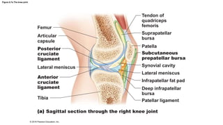
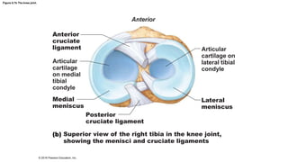
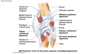
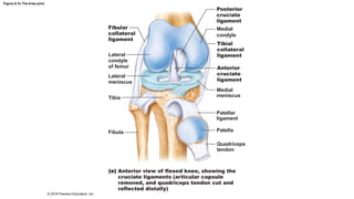
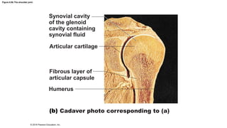
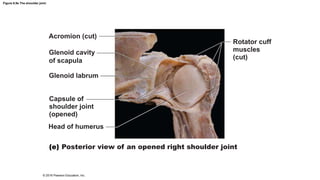
8.5 Selected Synovial Joints
• Synovial joints are diverse
• All have general features, but some also have unique structural
features, abilities, and weaknesses
• Five main synovial joints
– Knee
– Shoulder
– Elbow
– Hip
– Jaw
© 2016 Pearson Education, Inc.
Knee Joint
• Largest, most complex joint of body
• Consists of three joints surrounded by single cavity
1. Femoropatellar joint
• Plane joint
• Allows gliding motion during knee flexion
2. Lateral joint and 3. Medial joint
• Lateral and medial joints together are called tibiofemoral joint
• Joint between femoral condyles and lateral and medial menisci of tibia
• Hinge joint that allows flexion, extension, and some rotation when knee
partly flexed
© 2016 Pearson Education, Inc.
Figure 8.7a The knee joint.
Femur
Articular
capsule
Posterior
cruciate
ligament
Lateral meniscus
Anterior
cruciate
ligament
Tibia
Tendon of
quadriceps
femoris
Suprapatellar
bursa
Patella
Subcutaneous
prepatellar bursa
Synovial cavity
Lateral meniscus
Infrapatellar fat pad
Deep infrapatellar
bursa
Patellar ligament
Sagittal section through the right knee joint
© 2016 Pearson Education, Inc.
Figure 8.7b The knee joint.
Anterior
Anterior
cruciate
ligament
Articular
cartilage
on medial
tibial
condyle
Medial
meniscus
Posterior
cruciate ligament
Superior view of the right tibia in the knee joint,
showing the menisci and cruciate ligaments
Articular
cartilage on
lateral tibial
condyle
Lateral
meniscus
© 2016 Pearson Education, Inc.
Knee Joint (cont.)
• Joint capsule is thin and absent anteriorly
• Anteriorly, quadriceps tendon gives rise to three broad ligaments
that run from patella to tibia
– Medial and lateral patellar retinacula that flank the patellar
ligament
• Doctors tap patellar ligament to test knee-jerk reflex
• At least 12 bursae associated with knee joint
© 2016 Pearson Education, Inc.
Figure 8.7c The knee joint.
Quadriceps
femoris muscle
Tendon of
quadriceps
femoris muscle
Patella
Lateral
patellar
retinaculum
Fibular
collateral
ligament
Fibula
Medial patellar
retinaculum
Tibial collateral
ligament
Patellar
ligament
Tibia
Anterior view of right knee
© 2016 Pearson Education, Inc.
Knee Joint (cont.)
• Capsular, extracapsular, or intracapsular ligaments act to
stabilize knee joint
• Capsular and extracapsular ligaments help prevent
hyperextension of knee
– Fibular and tibial collateral ligaments: prevent rotation when
knee is extended
– Oblique popliteal ligament: stabilizes posterior knee joint
– Arcuate popliteal ligament: reinforces joint capsule posteriorly
© 2016 Pearson Education, Inc.
Figure 8.7d The knee joint.
Tendon of
adductor
magnus
Medial head of
gastrocnemius
muscle
Popliteus
muscle (cut)
Tibial
collateral
ligament
Tendon of
semimembranosus
muscle
Femur
Articular capsule
Oblique popliteal
ligament
Lateral head of
gastrocnemius
muscle
Bursa
Fibular collateral
ligament
Arcuate popliteal
ligament
Tibia
Posterior view of the joint capsule, including ligaments
© 2016 Pearson Education, Inc.
Knee Joint (cont.)
• Intracapsular ligaments reside within capsule, but outside
synovial cavity
• Help to prevent anterior-posterior displacement
– Anterior cruciate ligament (ACL)
• Attaches to anterior tibia
• Prevents forward sliding of tibia and stops hyperextension of knee
– Posterior cruciate ligament
• Attaches to posterior tibia
• Prevents backward sliding of tibia and forward sliding of femur
© 2016 Pearson Education, Inc.
Figure 8.7e The knee joint.
Posterior
cruciate
ligament
Fibular
collateral
ligament
Medial
condyle
Tibial
collateral
ligament
Anterior
cruciate
ligament
Medial
meniscus
Patellar
ligament
Patella
Quadriceps
tendon
Lateral
condyle
of femur
Lateral
meniscus
Tibia
Fibula
Anterior view of flexed knee, showing the
cruciate ligaments (articular capsule
removed, and quadriceps tendon cut and
reflected distally)
© 2016 Pearson Education, Inc.
Figure 8.7f The knee joint.
Medial femoral
condyle
Anterior cruciate
ligament
Medial meniscus
on medial tibial
condyle
Patella
Photograph of an opened knee joint;
view similar to (e)
© 2016 Pearson Education, Inc.
Clinical – Homeostatic Imbalance 8.1
• Knee absorbs great amount of vertical force; however, it is
vulnerable to horizontal blows
– Common knee injuries involved the 3 C’s:
• Collateral ligaments
• Cruciate ligaments
• Cartilages (menisci)
– Lateral blows to extended knee can result in tears in tibial collateral
ligament, medial meniscus, and anterior cruciate ligament
– Injuries affecting just ACL are common in runners who change
direction, twisting ACL
– Surgery usually needed for repairs
© 2016 Pearson Education, Inc.
Figure 8.8 The “unhappy triad:” ruptured ACL, ruptured tibial collateral ligament, and torn meniscus.
Lateral
Hockey puck
Patella
(outline)
Medial
Tibial
collateral
ligament
(torn)
Medial
meniscus
(torn)
Anterior
cruciate
ligament
(torn)
© 2016 Pearson Education, Inc.
Shoulder (Glenohumeral) Joint
• Most freely moving joint in body
• Stability is sacrificed for freedom of movement
• Ball-and-socket joint
– Large, hemispherical head of humerus fits in small, shallow glenoid
cavity of scapula
• Like a golf ball on a tee
• Articular capsule enclosing cavity is also thin and loose
– Contributes to freedom of movement
© 2016 Pearson Education, Inc.
Figure 8.9a The shoulder joint.
Acromion of scapula
Coracoacromial ligament
Subacromial bursa
Fibrous layer of
articular capsule
Tendon sheath
Tendon of long head
of biceps brachii muscle
Synovial cavity of
the glenoid cavity
containing synovial
fluid
Articular cartilage
Synovial membrane
Fibrous layer of
articular capsule
Humerus
Frontal section through right shoulder joint
© 2016 Pearson Education, Inc.
Figure 8.9b The shoulder joint.
Synovial cavity
of the glenoid
cavity containing
synovial fluid
Articular cartilage
Fibrous layer of
articular capsule
Humerus
Cadaver photo corresponding to (a)
© 2016 Pearson Education, Inc.
Shoulder (Glenohumeral) Joint (cont.)
• Glenoid labrum: fibrocartilaginous rim around glenoid cavity
– Helps to add depth to shallow cavity
– Cavity still only holds one-third of head of humerus
• Reinforcing ligaments
– Primarily on anterior aspect
– Coracohumeral ligament
• Helps support weight of upper limb
– Three glenohumeral ligaments
• Strengthen anterior capsule, but are weak support
© 2016 Pearson Education, Inc.
Figure 8.9c The shoulder joint.
Acromion
Coracoacromial ligament
Subacromial bursa
Coracohumeral
ligament
Transverse humeral
ligament
Tendon sheath
Tendon of long head
of biceps brachii
muscle
Coracoid process
Articular capsule
reinforced by
glenohumeral
ligaments
Subscapular
bursa
Tendon of the
subscapularis
muscle
Scapula
Anterior view of right shoulder joint capsule
© 2016 Pearson Education, Inc.
Figure 8.9d The shoulder joint.
Acromion
Coracoid process
Articular capsule
Glenoid cavity
Glenoid labrum
Tendon of long head
of biceps brachii muscle
Glenohumeral ligaments
Tendon of the
subscapularis muscle
Scapula
Posterior Anterior
Lateral view of socket of right shoulder joint,
humerus removed
© 2016 Pearson Education, Inc.
Shoulder (Glenohumeral) Joint (cont.)
• Reinforcing muscle tendons contribute most to joint stability
– Tendon of long head of biceps brachii muscle is “superstabilizer”
• Travels through intertubercular sulcus
• Secures humerus to glenoid cavity
– Four rotator cuff tendons encircle the shoulder joint
• Subscapularis
• Supraspinatus
• Infraspinatus
• Teres minor
© 2016 Pearson Education, Inc.
Figure 8.9c The shoulder joint.
Acromion
Coracoacromial ligament
Subacromial bursa
Coracohumeral
ligament
Transverse humeral
ligament
Tendon sheath
Tendon of long head
of biceps brachii
muscle
Coracoid process
Articular capsule
reinforced by
glenohumeral
ligaments
Subscapular
bursa
Tendon of the
subscapularis
muscle
Scapula
Anterior view of right shoulder joint capsule
© 2016 Pearson Education, Inc.
Figure 8.9d The shoulder joint.
Acromion
Coracoid process
Articular capsule
Glenoid cavity
Glenoid labrum
Tendon of long head
of biceps brachii muscle
Glenohumeral ligaments
Tendon of the
subscapularis muscle
Scapula
Posterior Anterior
Lateral view of socket of right shoulder joint,
humerus removed
© 2016 Pearson Education, Inc.
Figure 8.9e The shoulder joint.
Acromion (cut)
Glenoid cavity
of scapula
Glenoid labrum
Rotator cuff
muscles
(cut)
Capsule of
shoulder joint
(opened)
Head of humerus
Posterior view of an opened right shoulder joint
© 2016 Pearson Education, Inc.
Elbow Joint
• Humerus articulates with radius and ulna
• Hinge joint formed primarily from trochlear notch of ulna
articulating with trochlea of humerus
– Allows for flexion and extension only
• Anular ligament surrounds head of radius
• Two capsular ligaments restrict side-to-side movement
– Ulnar collateral ligament
– Radial collateral ligament
© 2016 Pearson Education, Inc.
Figure 8.10a The elbow joint.
Articular
capsule
Synovial
membrane
Humerus
Fat pad
Tendon of
triceps muscle
Bursa
Trochlea
Articular cartilage
of the trochlear
notch
Median sagittal section through right elbow
(lateral view)
Synovial cavity
Articular cartilage
Coronoid process
Tendon of
brachialis muscle
Ulna
© 2016 Pearson Education, Inc.
Figure 8.10b The elbow joint.
Humerus
Anular
ligament
Lateral
epicondyle
Articular
capsule
Radial
collateral
ligament
Olecranon
Ulna
Lateral view of right elbow joint
Radius
© 2016 Pearson Education, Inc.
Figure 8.10c The elbow joint.
Humerus
Anular
ligament
Medial
epicondyle
Radius
Articular
capsule
Coronoid
process
of ulna
Ulna
Ulnar
collateral
ligament
Cadaver photo of medial view of right elbow
© 2016 Pearson Education, Inc.
Figure 8.10d The elbow joint.
Articular
capsule
Anular
ligament
Coronoid
process
Medial
epicondyle
Ulnar
collateral
ligament
Humerus
Radius
Ulna
Medial view of right elbow
© 2016 Pearson Education, Inc.
Hip (Coxal) Joint
• Ball-and-socket joint
• Large, spherical head of the femur articulates with deep cup-
shaped acetabulum
• Good range of motion, but limited by the deep socket
– Acetabular labrum: rim of fibrocartilage that enhances depth of
socket (hip dislocations are rare)
© 2016 Pearson Education, Inc.
Figure 8.11a The hip joint.
Articular
cartilage
Acetabular
labrum
Femur
Hip (coxal) bone
Ligament of the
head of the femur
(ligamentum teres)
Synovial cavity
Articular capsule
Frontal section through the right hip joint
© 2016 Pearson Education, Inc.
Figure 8.11b The hip joint.
Acetabular
labrum
Synovial
membrane
Ligament of the
head of the femur
(ligamentum teres)
Head of femur
Articular capsule
(cut)
Photo of the interior of the hip joint, lateral view
© 2016 Pearson Education, Inc.
Hip (Coxal) Joint (cont.)
• Reinforcing ligaments include:
– Iliofemoral ligament
– Pubofemoral ligament
– Ischiofemoral ligament
– Ligament of head of femur (ligamentum teres)
• Slack during most hip movements, so not important in stabilizing
• Does contain artery that supplies head of femur
• Greatest stability comes from deep ball-and-socket joint
© 2016 Pearson Education, Inc.
Figure 8.11c The hip joint.
Ischium
Iliofemoral
ligament
Ischiofemoral
ligament
Greater
trochanter
of femur
Posterior view of right hip joint,
capsule in place
© 2016 Pearson Education, Inc.
Figure 8.11d The hip joint.
Anterior
inferior iliac
spine
Greater
trochanter
Iliofemoral
ligament
Pubofemoral
ligament
Anterior view of right hip joint, capsule in place
© 2016 Pearson Education, Inc.
Temporomandibular Joint (TMJ)
• Jaw joint is a modified hinge joint
• Mandibular condyle articulates with temporal bone
– Posterior temporal bone forms mandibular fossa, while anterior
portion forms articular tubercle
• Articular capsule thickens into strong lateral ligament
© 2016 Pearson Education, Inc.
Temporomandibular Joint (TMJ) (cont.)
• Two types of movement
– Hinge: depression and elevation of mandible
– Gliding: side-to-side (lateral excursion) grinding of teeth
• Most easily dislocated joint in the body
© 2016 Pearson Education, Inc.
Figure 8.12a The temporomandibular (jaw) joint.
Mandibular fossa
Articular tubercle
Zygomatic process
Infratemporal fossa
External
acoustic
meatus
Articular
capsule
Ramus of
mandible
Lateral
ligament
Location of the joint in the skull
© 2016 Pearson Education, Inc.
Figure 8.12b The temporomandibular (jaw) joint.
Articular disc
Mandibular
fossa
Articular tubercle
Superior joint
cavity
Articular
capsule
Synovial
membranes
Condylar
process of
mandible
Ramus of
mandible
Inferior joint
cavity
Enlargement of a sagittal section through the joint
© 2016 Pearson Education, Inc.
Figure 8.12c The temporomandibular (jaw) joint.
Superior view
Outline of
the mandibular
fossa
Lateral excursion: lateral (side-to-side) movements of the mandible
© 2016 Pearson Education, Inc.
Clinical – Homeostatic Imbalance 8.2
• Dislocation of TMJ is most common because of shallow socket of
joint
• Almost always dislocates anteriorly, causing mouth to remain
open
– To realign, physician must push mandible back into place
© 2016 Pearson Education, Inc.
Clinical – Homeostatic Imbalance 8.2
• Symptoms: ear and face pain, tender muscles, popping sounds
when opening mouth, joint stiffness
• Usually caused by grinding teeth, but can also be due to jaw
trauma or poor occlusion of teeth
– Treatment for grinding teeth includes bite plate
– Relaxing jaw muscles helps
© 2016 Pearson Education, Inc.

8.5 no vids

  • 1.
    8.5 Selected SynovialJoints • Synovial joints are diverse • All have general features, but some also have unique structural features, abilities, and weaknesses • Five main synovial joints – Knee – Shoulder – Elbow – Hip – Jaw © 2016 Pearson Education, Inc.
  • 2.
    Knee Joint • Largest,most complex joint of body • Consists of three joints surrounded by single cavity 1. Femoropatellar joint • Plane joint • Allows gliding motion during knee flexion 2. Lateral joint and 3. Medial joint • Lateral and medial joints together are called tibiofemoral joint • Joint between femoral condyles and lateral and medial menisci of tibia • Hinge joint that allows flexion, extension, and some rotation when knee partly flexed © 2016 Pearson Education, Inc.
  • 3.
    Figure 8.7a Theknee joint. Femur Articular capsule Posterior cruciate ligament Lateral meniscus Anterior cruciate ligament Tibia Tendon of quadriceps femoris Suprapatellar bursa Patella Subcutaneous prepatellar bursa Synovial cavity Lateral meniscus Infrapatellar fat pad Deep infrapatellar bursa Patellar ligament Sagittal section through the right knee joint © 2016 Pearson Education, Inc.
  • 4.
    Figure 8.7b Theknee joint. Anterior Anterior cruciate ligament Articular cartilage on medial tibial condyle Medial meniscus Posterior cruciate ligament Superior view of the right tibia in the knee joint, showing the menisci and cruciate ligaments Articular cartilage on lateral tibial condyle Lateral meniscus © 2016 Pearson Education, Inc.
  • 5.
    Knee Joint (cont.) •Joint capsule is thin and absent anteriorly • Anteriorly, quadriceps tendon gives rise to three broad ligaments that run from patella to tibia – Medial and lateral patellar retinacula that flank the patellar ligament • Doctors tap patellar ligament to test knee-jerk reflex • At least 12 bursae associated with knee joint © 2016 Pearson Education, Inc.
  • 6.
    Figure 8.7c Theknee joint. Quadriceps femoris muscle Tendon of quadriceps femoris muscle Patella Lateral patellar retinaculum Fibular collateral ligament Fibula Medial patellar retinaculum Tibial collateral ligament Patellar ligament Tibia Anterior view of right knee © 2016 Pearson Education, Inc.
  • 7.
    Knee Joint (cont.) •Capsular, extracapsular, or intracapsular ligaments act to stabilize knee joint • Capsular and extracapsular ligaments help prevent hyperextension of knee – Fibular and tibial collateral ligaments: prevent rotation when knee is extended – Oblique popliteal ligament: stabilizes posterior knee joint – Arcuate popliteal ligament: reinforces joint capsule posteriorly © 2016 Pearson Education, Inc.
  • 8.
    Figure 8.7d Theknee joint. Tendon of adductor magnus Medial head of gastrocnemius muscle Popliteus muscle (cut) Tibial collateral ligament Tendon of semimembranosus muscle Femur Articular capsule Oblique popliteal ligament Lateral head of gastrocnemius muscle Bursa Fibular collateral ligament Arcuate popliteal ligament Tibia Posterior view of the joint capsule, including ligaments © 2016 Pearson Education, Inc.
  • 9.
    Knee Joint (cont.) •Intracapsular ligaments reside within capsule, but outside synovial cavity • Help to prevent anterior-posterior displacement – Anterior cruciate ligament (ACL) • Attaches to anterior tibia • Prevents forward sliding of tibia and stops hyperextension of knee – Posterior cruciate ligament • Attaches to posterior tibia • Prevents backward sliding of tibia and forward sliding of femur © 2016 Pearson Education, Inc.
  • 10.
    Figure 8.7e Theknee joint. Posterior cruciate ligament Fibular collateral ligament Medial condyle Tibial collateral ligament Anterior cruciate ligament Medial meniscus Patellar ligament Patella Quadriceps tendon Lateral condyle of femur Lateral meniscus Tibia Fibula Anterior view of flexed knee, showing the cruciate ligaments (articular capsule removed, and quadriceps tendon cut and reflected distally) © 2016 Pearson Education, Inc.
  • 11.
    Figure 8.7f Theknee joint. Medial femoral condyle Anterior cruciate ligament Medial meniscus on medial tibial condyle Patella Photograph of an opened knee joint; view similar to (e) © 2016 Pearson Education, Inc.
  • 12.
    Clinical – HomeostaticImbalance 8.1 • Knee absorbs great amount of vertical force; however, it is vulnerable to horizontal blows – Common knee injuries involved the 3 C’s: • Collateral ligaments • Cruciate ligaments • Cartilages (menisci) – Lateral blows to extended knee can result in tears in tibial collateral ligament, medial meniscus, and anterior cruciate ligament – Injuries affecting just ACL are common in runners who change direction, twisting ACL – Surgery usually needed for repairs © 2016 Pearson Education, Inc.
  • 13.
    Figure 8.8 The“unhappy triad:” ruptured ACL, ruptured tibial collateral ligament, and torn meniscus. Lateral Hockey puck Patella (outline) Medial Tibial collateral ligament (torn) Medial meniscus (torn) Anterior cruciate ligament (torn) © 2016 Pearson Education, Inc.
  • 14.
    Shoulder (Glenohumeral) Joint •Most freely moving joint in body • Stability is sacrificed for freedom of movement • Ball-and-socket joint – Large, hemispherical head of humerus fits in small, shallow glenoid cavity of scapula • Like a golf ball on a tee • Articular capsule enclosing cavity is also thin and loose – Contributes to freedom of movement © 2016 Pearson Education, Inc.
  • 15.
    Figure 8.9a Theshoulder joint. Acromion of scapula Coracoacromial ligament Subacromial bursa Fibrous layer of articular capsule Tendon sheath Tendon of long head of biceps brachii muscle Synovial cavity of the glenoid cavity containing synovial fluid Articular cartilage Synovial membrane Fibrous layer of articular capsule Humerus Frontal section through right shoulder joint © 2016 Pearson Education, Inc.
  • 16.
    Figure 8.9b Theshoulder joint. Synovial cavity of the glenoid cavity containing synovial fluid Articular cartilage Fibrous layer of articular capsule Humerus Cadaver photo corresponding to (a) © 2016 Pearson Education, Inc.
  • 17.
    Shoulder (Glenohumeral) Joint(cont.) • Glenoid labrum: fibrocartilaginous rim around glenoid cavity – Helps to add depth to shallow cavity – Cavity still only holds one-third of head of humerus • Reinforcing ligaments – Primarily on anterior aspect – Coracohumeral ligament • Helps support weight of upper limb – Three glenohumeral ligaments • Strengthen anterior capsule, but are weak support © 2016 Pearson Education, Inc.
  • 18.
    Figure 8.9c Theshoulder joint. Acromion Coracoacromial ligament Subacromial bursa Coracohumeral ligament Transverse humeral ligament Tendon sheath Tendon of long head of biceps brachii muscle Coracoid process Articular capsule reinforced by glenohumeral ligaments Subscapular bursa Tendon of the subscapularis muscle Scapula Anterior view of right shoulder joint capsule © 2016 Pearson Education, Inc.
  • 19.
    Figure 8.9d Theshoulder joint. Acromion Coracoid process Articular capsule Glenoid cavity Glenoid labrum Tendon of long head of biceps brachii muscle Glenohumeral ligaments Tendon of the subscapularis muscle Scapula Posterior Anterior Lateral view of socket of right shoulder joint, humerus removed © 2016 Pearson Education, Inc.
  • 20.
    Shoulder (Glenohumeral) Joint(cont.) • Reinforcing muscle tendons contribute most to joint stability – Tendon of long head of biceps brachii muscle is “superstabilizer” • Travels through intertubercular sulcus • Secures humerus to glenoid cavity – Four rotator cuff tendons encircle the shoulder joint • Subscapularis • Supraspinatus • Infraspinatus • Teres minor © 2016 Pearson Education, Inc.
  • 21.
    Figure 8.9c Theshoulder joint. Acromion Coracoacromial ligament Subacromial bursa Coracohumeral ligament Transverse humeral ligament Tendon sheath Tendon of long head of biceps brachii muscle Coracoid process Articular capsule reinforced by glenohumeral ligaments Subscapular bursa Tendon of the subscapularis muscle Scapula Anterior view of right shoulder joint capsule © 2016 Pearson Education, Inc.
  • 22.
    Figure 8.9d Theshoulder joint. Acromion Coracoid process Articular capsule Glenoid cavity Glenoid labrum Tendon of long head of biceps brachii muscle Glenohumeral ligaments Tendon of the subscapularis muscle Scapula Posterior Anterior Lateral view of socket of right shoulder joint, humerus removed © 2016 Pearson Education, Inc.
  • 23.
    Figure 8.9e Theshoulder joint. Acromion (cut) Glenoid cavity of scapula Glenoid labrum Rotator cuff muscles (cut) Capsule of shoulder joint (opened) Head of humerus Posterior view of an opened right shoulder joint © 2016 Pearson Education, Inc.
  • 24.
    Elbow Joint • Humerusarticulates with radius and ulna • Hinge joint formed primarily from trochlear notch of ulna articulating with trochlea of humerus – Allows for flexion and extension only • Anular ligament surrounds head of radius • Two capsular ligaments restrict side-to-side movement – Ulnar collateral ligament – Radial collateral ligament © 2016 Pearson Education, Inc.
  • 25.
    Figure 8.10a Theelbow joint. Articular capsule Synovial membrane Humerus Fat pad Tendon of triceps muscle Bursa Trochlea Articular cartilage of the trochlear notch Median sagittal section through right elbow (lateral view) Synovial cavity Articular cartilage Coronoid process Tendon of brachialis muscle Ulna © 2016 Pearson Education, Inc.
  • 26.
    Figure 8.10b Theelbow joint. Humerus Anular ligament Lateral epicondyle Articular capsule Radial collateral ligament Olecranon Ulna Lateral view of right elbow joint Radius © 2016 Pearson Education, Inc.
  • 27.
    Figure 8.10c Theelbow joint. Humerus Anular ligament Medial epicondyle Radius Articular capsule Coronoid process of ulna Ulna Ulnar collateral ligament Cadaver photo of medial view of right elbow © 2016 Pearson Education, Inc.
  • 28.
    Figure 8.10d Theelbow joint. Articular capsule Anular ligament Coronoid process Medial epicondyle Ulnar collateral ligament Humerus Radius Ulna Medial view of right elbow © 2016 Pearson Education, Inc.
  • 29.
    Hip (Coxal) Joint •Ball-and-socket joint • Large, spherical head of the femur articulates with deep cup- shaped acetabulum • Good range of motion, but limited by the deep socket – Acetabular labrum: rim of fibrocartilage that enhances depth of socket (hip dislocations are rare) © 2016 Pearson Education, Inc.
  • 30.
    Figure 8.11a Thehip joint. Articular cartilage Acetabular labrum Femur Hip (coxal) bone Ligament of the head of the femur (ligamentum teres) Synovial cavity Articular capsule Frontal section through the right hip joint © 2016 Pearson Education, Inc.
  • 31.
    Figure 8.11b Thehip joint. Acetabular labrum Synovial membrane Ligament of the head of the femur (ligamentum teres) Head of femur Articular capsule (cut) Photo of the interior of the hip joint, lateral view © 2016 Pearson Education, Inc.
  • 32.
    Hip (Coxal) Joint(cont.) • Reinforcing ligaments include: – Iliofemoral ligament – Pubofemoral ligament – Ischiofemoral ligament – Ligament of head of femur (ligamentum teres) • Slack during most hip movements, so not important in stabilizing • Does contain artery that supplies head of femur • Greatest stability comes from deep ball-and-socket joint © 2016 Pearson Education, Inc.
  • 33.
    Figure 8.11c Thehip joint. Ischium Iliofemoral ligament Ischiofemoral ligament Greater trochanter of femur Posterior view of right hip joint, capsule in place © 2016 Pearson Education, Inc.
  • 34.
    Figure 8.11d Thehip joint. Anterior inferior iliac spine Greater trochanter Iliofemoral ligament Pubofemoral ligament Anterior view of right hip joint, capsule in place © 2016 Pearson Education, Inc.
  • 35.
    Temporomandibular Joint (TMJ) •Jaw joint is a modified hinge joint • Mandibular condyle articulates with temporal bone – Posterior temporal bone forms mandibular fossa, while anterior portion forms articular tubercle • Articular capsule thickens into strong lateral ligament © 2016 Pearson Education, Inc.
  • 36.
    Temporomandibular Joint (TMJ)(cont.) • Two types of movement – Hinge: depression and elevation of mandible – Gliding: side-to-side (lateral excursion) grinding of teeth • Most easily dislocated joint in the body © 2016 Pearson Education, Inc.
  • 37.
    Figure 8.12a Thetemporomandibular (jaw) joint. Mandibular fossa Articular tubercle Zygomatic process Infratemporal fossa External acoustic meatus Articular capsule Ramus of mandible Lateral ligament Location of the joint in the skull © 2016 Pearson Education, Inc.
  • 38.
    Figure 8.12b Thetemporomandibular (jaw) joint. Articular disc Mandibular fossa Articular tubercle Superior joint cavity Articular capsule Synovial membranes Condylar process of mandible Ramus of mandible Inferior joint cavity Enlargement of a sagittal section through the joint © 2016 Pearson Education, Inc.
  • 39.
    Figure 8.12c Thetemporomandibular (jaw) joint. Superior view Outline of the mandibular fossa Lateral excursion: lateral (side-to-side) movements of the mandible © 2016 Pearson Education, Inc.
  • 40.
    Clinical – HomeostaticImbalance 8.2 • Dislocation of TMJ is most common because of shallow socket of joint • Almost always dislocates anteriorly, causing mouth to remain open – To realign, physician must push mandible back into place © 2016 Pearson Education, Inc.
  • 41.
    Clinical – HomeostaticImbalance 8.2 • Symptoms: ear and face pain, tender muscles, popping sounds when opening mouth, joint stiffness • Usually caused by grinding teeth, but can also be due to jaw trauma or poor occlusion of teeth – Treatment for grinding teeth includes bite plate – Relaxing jaw muscles helps © 2016 Pearson Education, Inc.