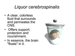
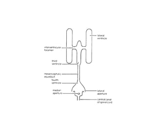
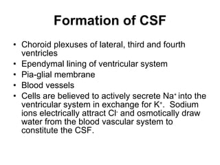
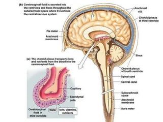
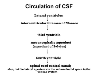
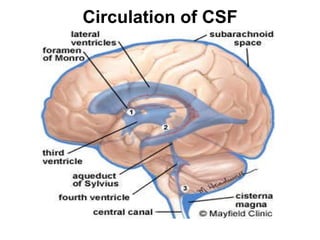
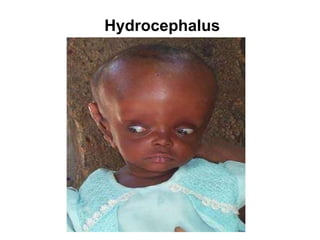
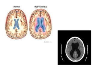
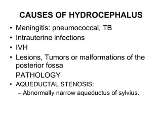
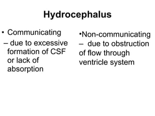
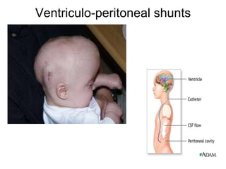
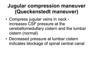
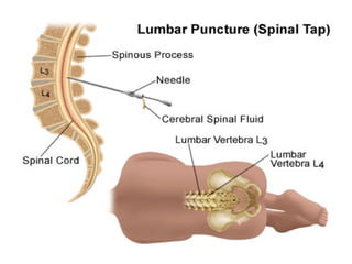
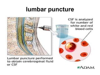
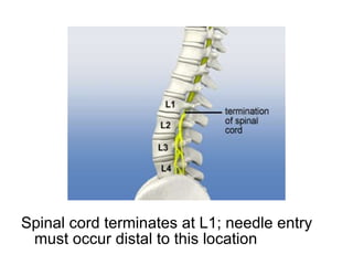
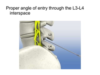
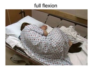
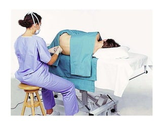
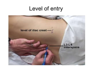
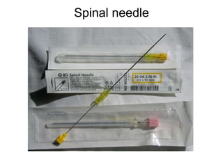
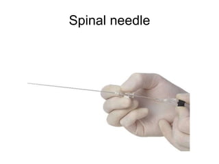
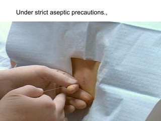
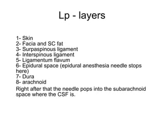
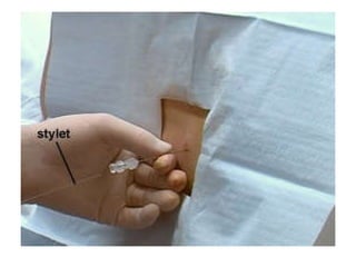
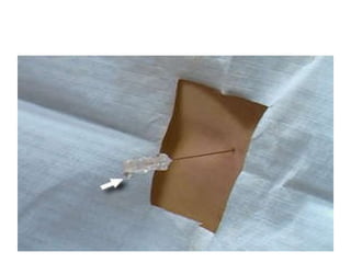
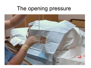
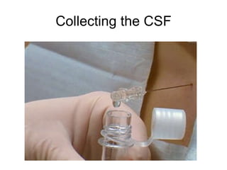
CEREBROSPINAL FLUID CSF DR. U. RATHNA KUMARI MD – Post Graduate IPEM
Liquor cerebrospinalis   A clear, colorless fluid that surrounds and permeates the CNS.  Offers support, protection and nourishment.  In essence, the brain "floats" in it.
Cerebrospinal fluid (CSF) is a clear fluid present in the ventricles of the brain, the central canal of the spinal cord, and the subarachnoid space. CSF is produced in the brain by modified  ependymal  cells  in the  choroid  plexus  (approx. 50-70%), and the remainder is formed around blood vessels and along ventricular walls. 
Functions of CSF Protects, lubricates the brain Provides nutrients, removes waste 90-150 ml adult 10-60 ml in newborn Modulates pressure changes ( Buoyancy)  Serves as a chemical buffer to maintain constant ionic environment Serves as a transport medium for nutrients and metabolites, endocrine substances and even neurotransmitters
 
Location of CSF Two lateral ventricles Third ventricle Fourth ventricle Spinal cord central canal Subarachnoid space Continuous with extracellular fluid of brain parenchyma
CSF flows through the subarachnoid space between the arachnoid and pia mater 20 ml of fluid produced every hr in choroids plexus and reabsorbed by arachnoid villi
 
Formation of CSF Choroid plexuses of lateral, third and fourth ventricles Ependymal lining of ventricular system Pia-glial membrane Blood vessels Cells are believed to actively secrete Na +  into the ventricular system in exchange for K + .  Sodium ions electrically attract Cl -  and osmotically draw water from the blood vascular system to constitute the CSF.
 
Circulation of CSF Lateral ventricles  interventricular foramen of Monroe third ventricle  mesencephalic aqueduct (aqueduct of Sylvius) fourth ventricle  spinal cord central canal;  also, out the lateral apertures to the subarachnoid space to the venous system
foramina of  Magendie  (Median aperture) and  foramina of  Luschka (Lateral apertures);  Circulates, Due to pulsation of blood in choroid plexus Due to pulsation of ependymal cells
Circulation of CSF
Absorption of CSF Through the arachnoid villi, a protrusion of arachnoid membrane into the central venous sinus and other sinuses A valve opens when CSF pressure exceeds venous pressure Absorption by veins and capillaries of CNS
arachnoid granulation
It is suggested that CSF flow along the  cranial nerves  and spinal nerve roots allow it into the lymphatic channels. plays a substantial role in CSF reabsorbtion, in the  neonate , where arachnoid granulations are sparsely distributed.  Absorption of CSF
Composition of CSF
Normal CSF Thin, colourless, clear fluid Pressure 90-180mm WATER (10-100 neonates) 0-5 WBC’s /mm 3  (neonates 0-30/ mm 3  ) (Lymphocytes & monocytes) Occasional ependymal or choroid plexus cells Protein 15-45mg/dl Glucose 50-80mg/dl Chloride 113-130 mEq/L Sterile
CSF GLUCOSE Glucose enters the CSF by selective transport across the blood-brain barrier. normal value 60 – 70 percent that of the plasma glucose.  The diagnostic significance.  low CSF glucose  can be considerable diagnostic value in determining the causative agents in meningitis.
Clinical significance of elevated protein values Elevated total protein values are most frequently seen in pathologic conditions.  elevated CSF protein due to damage to the blood brain barrier.
CSF GLUTAMINE Glutamine is produced in the CNS by the brain cells from ammonia and alpha-ketoglutarate. removes the toxic metabolic waste product ammonia from the CNS.  Normal concentration of ammonia is 8-18 mg/dl.   Elevated levels associated with liver disorders.
CSF LACTATE Aid in the diagnosis and management of meningitis cases Destruction of tissue within the CNS owing to oxygen deprivation (hypoxia) causes the production of increased CSF lactic acid levels.
Blood-Brain Barrier (BBB) physiological barrier to flow of substances from blood to brain tissue The BBB functions to preserve a stable environment for neurons of the CNS
Blood-Brain Barrier (BBB)
Brain capillary Tight junctions Surrounded by astrocytes Carrier mediated transport of glucose and amino acids
Blood-Brain Barrier Lipid soluble molecules will cross easily. High CO 2 /low O 2  produce vasodilation and decrease resistance of BBB Injury or inflammation  decreases the resistance of BBB (allows some antibiotics to be used for treatment)
Circumventricular organs (CVO) Selected brain areas are not protected by the BBB – the circumventricular organs (CVO)  bordering on the 3 rd  and 4 th  ventricles parts of the hypothalamus - median eminence neurohypophysis pineal gland area postrema subfornical organ subcommissural organ
Hydrocephalus An abnormal increase in the volume of CSF Symptoms:  sleep changes, spastic paresis, papilledema, bulging of skull in young, seizures, cranial nerve deficits, depression.
Hydrocephalus
 
CAUSES OF HYDROCEPHALUS Meningitis: pneumococcal, TB Intrauterine infections  IVH  Lesions, Tumors or malformations of the posterior fossa PATHOLOGY AQUEDUCTAL STENOSIS:  Abnormally narrow aqueductus of sylvius.
Hydrocephalus Communicating  –  due to excessive formation of CSF or lack of absorption Non-communicating –  due to obstruction of flow through ventricle system
Ventriculo-peritoneal shunts
Jugular compression maneuver (Queckenstedt maneuver) Compress jugular veins in neck - increases CSF pressure at the cerebellomedullary cistern and the lumbar cistern (normal) Decreased pressure at lumbar cistern indicates blockage of spinal central canal
Lumbar puncture a  lumbar puncture  (or  LP , and colloquially known as a  spinal tap ) is a  diagnostic   procedure that is performed in order to collect a sample of  cerebrospinal fluid  (CSF) for  biochemical ,  microbiological , and  cytological  analysis
 
lumbar puncture
Spinal cord terminates at L1; needle entry must occur distal to this location
Proper angle of entry through the L3-L4 interspace
 
full flexion
 
Level of entry
Spinal needle
Spinal needle
Under strict aseptic precautions.,
Lp - layers 1- Skin 2- Facia and SC fat 3- Surpaspinous ligament 4- Interspinous ligament 5- Ligamentum flavum 6- Epidural space (epidural anesthesia needle stops here) 7- Dura 8- arachnoid Right after that the needle pops into the subarachnoid space where the CSF is.
 
 
The opening pressure 
Collecting the CSF
Appearance of CSF clear and colourless (normal);  bright red - This indicates the presence of blood. The appearance of fresh blood in all three tubes supports the diagnosis of a subarachnoid hemorrhage.  xanthochromic (yellow) - jaundice  xanthochromic (yellow) - suggests  a subarachnoid hemorrhage has recently occurred  turbid - This indicates the presence of white cells and is suggestive of a CNS infection.
Cell counts  increased neutrophils - This indicates a bacterial meningitis, cerebral abscess,  increased lymphocytes - This indicates a viral meningitis, tuberculosis, syphilis, fungal and parasitic infections.  Degenerative diseases of the CNS, such as multiple sclerosis, will also generate elevated lymphocyte counts.
 

Cerebrospinal fluid

  • 1.
    CEREBROSPINAL FLUID CSFDR. U. RATHNA KUMARI MD – Post Graduate IPEM
  • 2.
    Liquor cerebrospinalis A clear, colorless fluid that surrounds and permeates the CNS. Offers support, protection and nourishment. In essence, the brain "floats" in it.
  • 3.
    Cerebrospinal fluid (CSF)is a clear fluid present in the ventricles of the brain, the central canal of the spinal cord, and the subarachnoid space. CSF is produced in the brain by modified  ependymal cells in the  choroid plexus  (approx. 50-70%), and the remainder is formed around blood vessels and along ventricular walls. 
  • 4.
    Functions of CSFProtects, lubricates the brain Provides nutrients, removes waste 90-150 ml adult 10-60 ml in newborn Modulates pressure changes ( Buoyancy) Serves as a chemical buffer to maintain constant ionic environment Serves as a transport medium for nutrients and metabolites, endocrine substances and even neurotransmitters
  • 5.
  • 6.
    Location of CSFTwo lateral ventricles Third ventricle Fourth ventricle Spinal cord central canal Subarachnoid space Continuous with extracellular fluid of brain parenchyma
  • 7.
    CSF flows throughthe subarachnoid space between the arachnoid and pia mater 20 ml of fluid produced every hr in choroids plexus and reabsorbed by arachnoid villi
  • 8.
  • 9.
    Formation of CSFChoroid plexuses of lateral, third and fourth ventricles Ependymal lining of ventricular system Pia-glial membrane Blood vessels Cells are believed to actively secrete Na + into the ventricular system in exchange for K + . Sodium ions electrically attract Cl - and osmotically draw water from the blood vascular system to constitute the CSF.
  • 10.
  • 11.
    Circulation of CSFLateral ventricles interventricular foramen of Monroe third ventricle mesencephalic aqueduct (aqueduct of Sylvius) fourth ventricle spinal cord central canal; also, out the lateral apertures to the subarachnoid space to the venous system
  • 12.
    foramina of Magendie  (Median aperture) and  foramina of Luschka (Lateral apertures); Circulates, Due to pulsation of blood in choroid plexus Due to pulsation of ependymal cells
  • 13.
  • 14.
    Absorption of CSFThrough the arachnoid villi, a protrusion of arachnoid membrane into the central venous sinus and other sinuses A valve opens when CSF pressure exceeds venous pressure Absorption by veins and capillaries of CNS
  • 15.
  • 16.
    It is suggestedthat CSF flow along the  cranial nerves  and spinal nerve roots allow it into the lymphatic channels. plays a substantial role in CSF reabsorbtion, in the  neonate , where arachnoid granulations are sparsely distributed. Absorption of CSF
  • 17.
  • 18.
    Normal CSF Thin,colourless, clear fluid Pressure 90-180mm WATER (10-100 neonates) 0-5 WBC’s /mm 3 (neonates 0-30/ mm 3 ) (Lymphocytes & monocytes) Occasional ependymal or choroid plexus cells Protein 15-45mg/dl Glucose 50-80mg/dl Chloride 113-130 mEq/L Sterile
  • 19.
    CSF GLUCOSE Glucoseenters the CSF by selective transport across the blood-brain barrier. normal value 60 – 70 percent that of the plasma glucose. The diagnostic significance. low CSF glucose can be considerable diagnostic value in determining the causative agents in meningitis.
  • 20.
    Clinical significance ofelevated protein values Elevated total protein values are most frequently seen in pathologic conditions. elevated CSF protein due to damage to the blood brain barrier.
  • 21.
    CSF GLUTAMINE Glutamineis produced in the CNS by the brain cells from ammonia and alpha-ketoglutarate. removes the toxic metabolic waste product ammonia from the CNS. Normal concentration of ammonia is 8-18 mg/dl. Elevated levels associated with liver disorders.
  • 22.
    CSF LACTATE Aidin the diagnosis and management of meningitis cases Destruction of tissue within the CNS owing to oxygen deprivation (hypoxia) causes the production of increased CSF lactic acid levels.
  • 23.
    Blood-Brain Barrier (BBB)physiological barrier to flow of substances from blood to brain tissue The BBB functions to preserve a stable environment for neurons of the CNS
  • 24.
  • 25.
    Brain capillary Tightjunctions Surrounded by astrocytes Carrier mediated transport of glucose and amino acids
  • 26.
    Blood-Brain Barrier Lipidsoluble molecules will cross easily. High CO 2 /low O 2 produce vasodilation and decrease resistance of BBB Injury or inflammation decreases the resistance of BBB (allows some antibiotics to be used for treatment)
  • 27.
    Circumventricular organs (CVO)Selected brain areas are not protected by the BBB – the circumventricular organs (CVO) bordering on the 3 rd and 4 th ventricles parts of the hypothalamus - median eminence neurohypophysis pineal gland area postrema subfornical organ subcommissural organ
  • 28.
    Hydrocephalus An abnormalincrease in the volume of CSF Symptoms: sleep changes, spastic paresis, papilledema, bulging of skull in young, seizures, cranial nerve deficits, depression.
  • 29.
  • 30.
  • 31.
    CAUSES OF HYDROCEPHALUSMeningitis: pneumococcal, TB Intrauterine infections IVH Lesions, Tumors or malformations of the posterior fossa PATHOLOGY AQUEDUCTAL STENOSIS: Abnormally narrow aqueductus of sylvius.
  • 32.
    Hydrocephalus Communicating – due to excessive formation of CSF or lack of absorption Non-communicating – due to obstruction of flow through ventricle system
  • 33.
  • 34.
    Jugular compression maneuver(Queckenstedt maneuver) Compress jugular veins in neck - increases CSF pressure at the cerebellomedullary cistern and the lumbar cistern (normal) Decreased pressure at lumbar cistern indicates blockage of spinal central canal
  • 35.
    Lumbar puncture a lumbar puncture  (or  LP , and colloquially known as a  spinal tap ) is a  diagnostic   procedure that is performed in order to collect a sample of  cerebrospinal fluid  (CSF) for  biochemical , microbiological , and  cytological  analysis
  • 36.
  • 37.
  • 38.
    Spinal cord terminatesat L1; needle entry must occur distal to this location
  • 39.
    Proper angle ofentry through the L3-L4 interspace
  • 40.
  • 41.
  • 42.
  • 43.
  • 44.
  • 45.
  • 46.
    Under strict asepticprecautions.,
  • 47.
    Lp - layers1- Skin 2- Facia and SC fat 3- Surpaspinous ligament 4- Interspinous ligament 5- Ligamentum flavum 6- Epidural space (epidural anesthesia needle stops here) 7- Dura 8- arachnoid Right after that the needle pops into the subarachnoid space where the CSF is.
  • 48.
  • 49.
  • 50.
  • 51.
  • 52.
    Appearance of CSFclear and colourless (normal); bright red - This indicates the presence of blood. The appearance of fresh blood in all three tubes supports the diagnosis of a subarachnoid hemorrhage. xanthochromic (yellow) - jaundice xanthochromic (yellow) - suggests a subarachnoid hemorrhage has recently occurred turbid - This indicates the presence of white cells and is suggestive of a CNS infection.
  • 53.
    Cell counts increased neutrophils - This indicates a bacterial meningitis, cerebral abscess, increased lymphocytes - This indicates a viral meningitis, tuberculosis, syphilis, fungal and parasitic infections. Degenerative diseases of the CNS, such as multiple sclerosis, will also generate elevated lymphocyte counts.
  • 54.