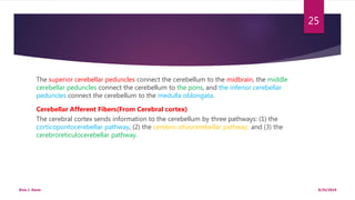
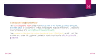
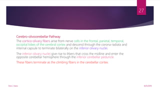
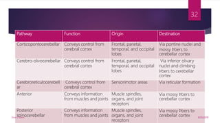
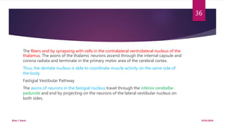
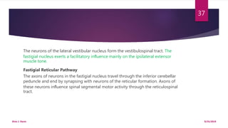
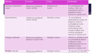
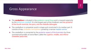
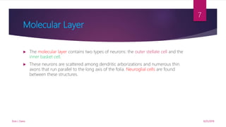
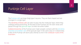
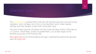
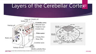
SlideShare a Scribd company logo
1 of 38
NEUROANATOMY
THE CEREBELLUM
Year II Sem. II
RESEARCHED AND PREPARED BY: STUDENT DR. ELVIS J. DAVIS
MEDICAL STUDENT
A. M. DOGLIOTTI COLLEGE OF MEDICINE
Gross Appearance
 The cerebellum is situated in the posterior cranial fossa and is covered superiorly
by the tentorium cerebelli. It is the largest part of the hindbrain and lies posterior
to the fourth ventricle, the pons, and the medulla oblongata.
 The cerebellum is somewhat ovoid in shape and constricted in its median part. It
consists of two cerebellar hemispheres joined by a narrow median vermis.
 The cerebellum is connected to the posterior aspect of the brainstem by three
symmetrical bundles of nerve fibers called the superior, middle, and inferior
cerebellar peduncles.
9/25/2019Elvis J. Davis
2
Gross Appearance of Cerebellum
9/25/2019Elvis J. Davis
3
 The cerebellum is divided into three main lobes: the anterior lobe, the middle
lobe, and the flocculonodular lobe.
 The anterior lobe may be seen on the superior surface of the cerebellum and is
separated from the middle lobe by a wide V-shaped fissure called the primary
fissure.
 The middle lobe (sometimes called the posterior lobe),which is the largest part of
the cerebellum, is situated between the primary and uvulonodular fissures.
 The flocculonodular lobe is situated posterior to the uvulonodular fissure.
9/25/2019Elvis J. Davis
4
Structure of the Cerebellar Cortex
 The cerebellar cortex can be regarded as a large sheet with folds(Folia) lying in the coronal or
transverse plane.
 Each fold or folium contains a core of white matter covered superficially by gray matter.
 At right angles, the cut surface of the folium has a branched appearance, called the arbor vitae.
 The gray matter of the cerebellar cortex has a uniform structure. It may be divided into three
layers:
 the molecular layer(external layer)
 the Purkinje cell layer(middle layer)
 the granular layer(internal layer)
9/25/2019Elvis J. Davis
5
Internal Structure of Cerebellum
9/25/2019Elvis J. Davis
6
Molecular Layer
 The molecular layer contains two types of neurons: the outer stellate cell and the
inner basket cell.
 These neurons are scattered among dendritic arborizations and numerous thin
axons that run parallel to the long axis of the folia. Neuroglial cells are found
between these structures.
9/25/2019Elvis J. Davis
7
Purkinje Cell Layer
The Purkinje cells are large Golgi type I neurons. They are flask shaped and are
arranged in a single layer.
The dendrites of these cells are seen to pass into the molecular layer, where they
undergo profuse branching. At the base of the Purkinje cell, the axon arises and
passes through the granular layer to enter the white matter.
Collateral branches of the Purkinje axon make synaptic contacts with the dendrites
of basket and stellate cells of the granular layer in the same area or in distant folia.
A few of the Purkinje cell axons pass directly to end in the vestibular nuclei of the
brainstem.
9/25/2019Elvis J. Davis
8
Granular Layer
The granular layer is packed with small cells with densely staining nuclei and scanty
cytoplasm. Each cell gives rise to four or five dendrites, which make clawlike
endings and have synaptic contact with mossy fiber input.
The axon of each granule cell passes into the molecular layer, where it bifurcates at
a T junction. These fibers, known as parallel fibers, run at right angles to the
dendritic processes of the Purkinje cells.
Neuroglial cells are found throughout this layer. Scattered throughout the granular
layer are Golgi cells.
9/25/2019Elvis J. Davis
9
Layers of the Cerebellar Cortex
9/25/2019Elvis J. Davis
10
Functions of the Cerebellum
The cerebellum receives afferent information concerning voluntary movement from
the cerebral cortex and from the muscles, tendons, and joints. It also receives
information concerning balance from the vestibular nerve and possibly concerning
sight through the tectocerebellar tract.
All this information is fed into the cerebellar cortical circuitry by the mossy fibers
and the climbing fibers and converges on the Purkinje cells. The axons of the
Purkinje cells project with few exceptions on the deep cerebellar nuclei. The output
of the vermis projects to the fastigial nucleus, the intermediate regions of the cortex
project to the globose and emboliform nuclei, and the output of the lateral part of
the cerebellar hemisphere projects to the dentate nucleus.
9/25/2019Elvis J. Davis
11
A few Purkinje cell axons pass directly out of the cerebellum and end on the lateral
vestibular nucleus in the brainstem. The Purkinje axons exert an inhibitory influence
on the neurons of the cerebellar nuclei and the lateral vestibular nuclei.
The cerebellar output is conducted to the sites of origin of the descending pathways
that influence motor activity at the segmental spinal level. In this respect, the
cerebellum has no direct neuronal connections with the lower motor neurons but
exerts its influence indirectly through the cerebral cortex and brainstem.
9/25/2019Elvis J. Davis
12
The cerebellum functions as a coordinator of precise movements by continually
comparing the output of the motor area of the cerebral cortex with the
proprioceptive information received from the site of muscle action; it is then able to
bring about the necessary adjustments by influencing the activity of the lower motor
neurons (Snell’s Clinical Neuroanatomy, 7th ed, pg.243).
This is accomplished by controlling the timing and sequence of firing of the alpha and
gamma motor neurons. The cerebellum can send back information to the motor
cerebral cortex to inhibit the agonist muscles and stimulate the antagonist muscles,
thus limiting the extent of voluntary movement.
9/25/2019Elvis J. Davis
13
Functional Areas of the Cerebellar Cortex
The cerebellar cortex can be divided into three functional areas.
The cortex of the vermis influences the movements of the long axis of the body,
namely, the neck, the shoulders, the thorax, the abdomen, and the hips.
The intermediate zone which lies immediately lateral to the vermis, has been shown
to control the muscles of the distal parts of the limbs, especially the hands and feet.
The lateral zone of each cerebellar hemisphere is concerned with the planning of
sequential movements of the entire body and is involved with the conscious
assessment of movement errors.
9/25/2019Elvis J. Davis
14
Intracerebellar Nuclei
Four masses of gray matter are embedded in the white matter of the cerebellum on
each side of the midline. From lateral to medial, these nuclei are the dentate, the
emboliform, the globose, and the fastigial.
The dentate nucleus is the largest of the cerebellar nuclei. It has the shape of a
crumpled bag with the opening facing medially.
The interior of the bag is filled with white matter made up of efferent fibers that
leave the nucleus through the opening to form a large part of the superior
cerebellar peduncle.
9/25/2019Elvis J. Davis
15
The emboliform nucleus is ovoid and is situated medial to the dentate nucleus, partially
covering its hilus.
The globose nucleus consists of one or more rounded cell groups that lie medial to the
emboliform nucleus.
The fastigial nucleus lies near the midline in the vermis and close to the roof of the fourth
ventricle; it is larger than the globose nucleus.
The intracerebellar nuclei are composed of large, multipolar neurons with simple
branching dendrites. The axons form the cerebellar outflow in the superior and inferior
cerebellar peduncles.
9/25/2019Elvis J. Davis
16
Intracerebellar Nuclei
9/25/2019Elvis J. Davis
17
White Matter
There is a small amount of white matter in the vermis; it closely resembles the trunk
and branches of a tree and thus is termed the arbor vitae. There is a large amount
of white matter in each cerebellar hemisphere.
The white matter is made up of three groups of fibers:
 Intrinsic fibers
 Afferent fibers
 Efferent fibers
9/25/2019Elvis J. Davis
18
 The intrinsic fibers do not leave the cerebellum but connect different regions of
the organ. Some interconnect folia of the cerebellar cortex and vermis on the
same side; others connect the two cerebellar hemispheres together.
 The afferent fibers form the greater part of the white matter and proceed to the
cerebellar cortex. They enter the cerebellum mainly through the inferior and
middle cerebellar peduncles.
9/25/2019Elvis J. Davis
19
 The efferent fibers constitute the output of the cerebellum and commence as the
axons of the Purkinje cells of the cerebellar cortex. The great majority of the
Purkinje cell axons pass to and synapse with the neurons of the cerebellar nuclei
(fastigial, globose, emboliform, and dentate).
 Fibers from the dentate, emboliform, and globose nuclei leave the cerebellum
through the superior cerebellar peduncle. Fibers from the fastigial nucleus leave
through the inferior cerebellar peduncle.
9/25/2019Elvis J. Davis
20
Cerebellar Cortical Mechanism
The climbing and the mossy fibers constitute the two main lines of input to the
cerebellar cortex and are excitatory to the Purkinje cells.
The climbing fibers are so named because they ascend through the layers of the
cortex like a vine on a tree.
The climbing fibers are the terminal fibers of the olivocerebellar tracts.
A single Purkinje neuron makes synaptic contact with only one climbing fiber.
However, one climbing fiber makes contact with 1 to 10 Purkinje neurons.
9/25/2019Elvis J. Davis
21
The mossy fibers are the terminal fibers of all other cerebellar afferent tracts.
They have multiple branches and exert a much more diffuse excitatory effect. A single
mossy fiber may stimulate thousands of Purkinje cells through the granule cells.
The stellate, basket, and Golgi cells are seen to serve as inhibitory interneurons.
Inhibitory impulses are transmitted by the Purkinje cells to the intracerebellar nuclei,
which, in turn, modify muscular activity through the motor control areas of the
brainstem and cerebral cortex.
9/25/2019Elvis J. Davis
22
Intracerebellar Nuclear Mechanisms
The deep cerebellar nuclei receive afferent nervous information from two sources:
(1) the inhibitory axons from the Purkinje cells of the overlying cortex
(2) the excitatory axons that are branches of the afferent climbing and mossy fibers.
Cerebellar Cortical Neurotransmitters
Pharmacologic research has suggested that the excitatory climbing and mossy
afferent fibers use glutamate (gamma aminobutyric acid [GABA]) as the excitatory
transmitter on the dendrites of the Purkinje cells.
9/25/2019Elvis J. Davis
23
Further research has indicated that other afferent fibers entering the cortex liberate
norepinephrine and serotonin at their endings that possibly modify the action of
glutamate on the Purkinje cells.
Cerebellar Peduncles
The cerebellum is linked to other parts of the central nervous system by numerous
efferent and afferent fibers that are grouped together on each side into three large
bundles, or peduncles.
9/25/2019Elvis J. Davis
24
The superior cerebellar peduncles connect the cerebellum to the midbrain, the middle
cerebellar peduncles connect the cerebellum to the pons, and the inferior cerebellar
peduncles connect the cerebellum to the medulla oblongata.
Cerebellar Afferent Fibers(From Cerebral cortex)
The cerebral cortex sends information to the cerebellum by three pathways: (1) the
corticopontocerebellar pathway, (2) the cerebro-olivocerebellar pathway, and (3) the
cerebroreticulocerebellar pathway.
9/25/2019Elvis J. Davis
25
Corticopontocerebellar Pathway
The corticopontine fibers arise from nerve cells in the frontal, parietal, temporal,
occipital lobes of the cerebral cortex and descend through the corona radiata and
internal capsule and terminate on the pontine nuclei.
The pontine nuclei give rise to the transverse fibers of the pons, which cross the
midline and enter the opposite cerebellar hemisphere as the middle cerebellar
peduncle.
9/25/2019Elvis J. Davis
26
Cerebro-olivocerebellar Pathway
The cortico-olivary fibers arise from nerve cells in the frontal, parietal, temporal,
occipital lobes of the cerebral cortex and descend through the corona radiata and
internal capsule to terminate bilaterally on the inferior olivary nuclei.
The inferior olivary nuclei give rise to fibers that cross the midline and enter the
opposite cerebellar hemisphere through the inferior cerebellar peduncle.
These fibers terminate as the climbing fibers in the cerebellar cortex.
9/25/2019Elvis J. Davis
27
Cerebroreticulocerebellar Pathway
The corticoreticular fibers arise from nerve cells from many areas of the cerebral
cortex, particularly the sensorimotor areas.
They descend to terminate in the reticular formation on the same side and on the
opposite side in the pons and medulla.
The cells in the reticular formation give rise to the reticulocerebellar fibers that enter
the cerebellar hemisphere on the same side through the inferior and middle
cerebellar peduncles.
9/25/2019Elvis J. Davis
28
Cerebellar Afferent Fibers From the Spinal
Cord
The spinal cord sends information to the cerebellum from somatosensory receptors
by three pathways:(1) the anterior spinocerebellar tract,(2) the posterior
spinocerebellar tract, and (3) the cuneocerebellar tract.
Anterior Spinocerebellar Tract
The axons entering the spinal cord from the posterior root ganglion terminate by
synapsing with the neurons in the nucleus dorsalis (Clarke’s column) at the base of
the posterior gray column.
9/25/2019Elvis J. Davis
29
Most of the axons of these neurons cross to the opposite side and ascend as the
anterior spinocerebellar tract in the contralateral white column; some of the axons
ascend as the anterior spinocerebellar tract in the lateral white column of the same
side.
The fibers enter the cerebellum through the superior cerebellar peduncle and
terminate as mossy fibers in the cerebellar cortex.
The anterior spinocerebellar tract is found at all segments of the spinal cord, and its
fibers convey muscle joint information from the muscle spindles, tendon organs, and
joint receptors of the upper and lower limbs.
9/25/2019Elvis J. Davis
30
Posterior Spinocerebellar Tract
The axons entering the spinal cord from the posterior root ganglion enter the
posterior gray column and terminate by synapsing on the neurons at the base of
posterior gray column. These neurons are known collectively as the nucleus dorsalis
(Clarke’s column).
The axons of these neurons enter the posterolateral part of the lateral white column
on the same side and ascend as the posterior spinocerebellar tract to the medulla
oblongata.
The tract then enters the cerebellum through the inferior cerebellar peduncle and
terminates as mossy fibers in the cerebellar cortex.
9/25/2019Elvis J. Davis
31
Pathway Function Origin Destination
Corticopontocerebellar Conveys control from
cerebral cortex
Frontal, parietal,
temporal, and occipital
lobes
Via pontine nuclei and
mossy fibers to
cerebellar cortex
Cerebro-olivocerebellar Conveys control from
cerebral cortex
Frontal, parietal,
temporal, and occipital
lobes
Via inferior olivary
nuclei and climbing
fibers to cerebellar
cortex
Cerebroreticulocerebell
ar
Conveys control from
cerebral cortex
Sensorimotor areas Via reticular formation
Anterior Conveys information
from muscles and joints
Muscle spindles,
organs, and joint
receptors
Via mossy fibers to
cerebellar cortex
Posterior
spinocerebellar
Conveys information
from muscles and joints
Muscle spindles,
organs, and joint
receptors
Via mossy fibers to
cerebellar cortex 9/25/2019Elvis J. Davis
32
Cuneocerebellar Tract
These fibers originate in the nucleus cuneatus of the medulla oblongata and enter the cerebellar
hemisphere on the same side through the inferior cerebellar peduncle. The fibers terminate as
fibers in the cerebellar cortex.
The cuneocerebellar tract receives muscle joint information from the muscle spindles, tendon
and joint receptors of the upper limb and upper part of the thorax.
Fibers From the Vestibular Nerve
The vestibular nerve receives information from the inner ear concerning motion from the
canals and position relative to gravity from the utricle and saccule. All the afferent fibers from the
ear terminate as mossy fibers in the flocculonodular lobe of the cerebellum.
9/25/2019Elvis J. Davis
33
Cerebellar Efferent Fibers
The entire output of the cerebellar cortex is through the axons of the Purkinje cells.
Most of the axons of the Purkinje cells end by synapsing on the neurons of the
deep cerebellar nuclei.
The efferent fibers from the cerebellum connect with the red nucleus, thalamus,
vestibular complex, and reticular formation.
Globose-Emboliform-Rubral Pathway
Axons of neurons in the globose and emboliform nuclei travel through the superior
cerebellar peduncle and cross the midline to the opposite side in the decussation of
the superior cerebellar peduncles.
9/25/2019Elvis J. Davis
34
The fibers end by synapsing with cells of the contralateral red nucleus, which give rise
to axons of the rubrospinal tract. Thus, it is seen that this pathway crosses twice, once
in the decussation of the superior cerebellar peduncle and again in the rubrospinal
tract close to its origin.
By this means, the globose and emboliform nuclei influence motor activity on the
same side of the body.
Dentothalamic Pathway
Axons of neurons in the dentate nucleus travel through the superior cerebellar
peduncle and cross the midline to the opposite side in the decussation of the
superior cerebellar peduncle.
9/25/2019Elvis J. Davis
35
The fibers end by synapsing with cells in the contralateral ventrolateral nucleus of the
thalamus. The axons of the thalamic neurons ascend through the internal capsule and
corona radiata and terminate in the primary motor area of the cerebral cortex.
Thus, the dentate nucleus is able to coordinate muscle activity on the same side of
the body.
Fastigial Vestibular Pathway
The axons of neurons in the fastigial nucleus travel through the inferior cerebellar
peduncle and end by projecting on the neurons of the lateral vestibular nucleus on
both sides.
9/25/2019Elvis J. Davis
36
The neurons of the lateral vestibular nucleus form the vestibulospinal tract. The
fastigial nucleus exerts a facilitatory influence mainly on the ipsilateral extensor
muscle tone.
Fastigial Reticular Pathway
The axons of neurons in the fastigial nucleus travel through the inferior cerebellar
peduncle and end by synapsing with neurons of the reticular formation. Axons of
these neurons influence spinal segmental motor activity through the reticulospinal
tract.
9/25/2019Elvis J. Davis
37
Pathway Function Origin Destination
Globose-emboliform
rubral
Influences ipsilateral
motor activity
Globose and
nuclei
To contralateral red
nucleus, then via
rubrospinal tract to
ipsilateral motor
neurons in spinal cord
Dentothalamic Influences ipsilateral
motor activity
Dentate nucleus To contralateral
ventrolateral nucleus of
thalamus, then to
contralateral motor
cerebral cortex;
corticospinal tract
crosses midline and
controls ipsilateral
neurons in spinal cord
Fastigial vestibular Influences ipsilateral
extensor muscle tone
Fastigial nucleus Mainly to ipsilateral and
to contralateral lateral
vestibular nuclei;
vestibulospinal tract to
ipsilateral motor
neurons in spinal cord
Fastigial reticular Influences ipsilateral Fastigial nucleus To neurons of reticular9/25/2019Elvis J. Davis
38

More Related Content

What's hot

What's hot (20)

connections of cerebellum
connections of cerebellumconnections of cerebellum
connections of cerebellum
 
The cerebellum demo
The cerebellum demoThe cerebellum demo
The cerebellum demo
 
Anatomy of Cerebellum professor dr saeed abuel makareem _ Relevant Connection...
Anatomy of Cerebellum professor dr saeed abuel makareem _ Relevant Connection...Anatomy of Cerebellum professor dr saeed abuel makareem _ Relevant Connection...
Anatomy of Cerebellum professor dr saeed abuel makareem _ Relevant Connection...
 
Cerebellum
CerebellumCerebellum
Cerebellum
 
Cerebellum 2010
Cerebellum 2010Cerebellum 2010
Cerebellum 2010
 
Cerebellum
CerebellumCerebellum
Cerebellum
 
Cerebellum Anatomy and Physiology
Cerebellum Anatomy and PhysiologyCerebellum Anatomy and Physiology
Cerebellum Anatomy and Physiology
 
Cerebellum ppt
Cerebellum pptCerebellum ppt
Cerebellum ppt
 
Cerebellum
CerebellumCerebellum
Cerebellum
 
Cerebellum surgical anatomy
Cerebellum surgical anatomyCerebellum surgical anatomy
Cerebellum surgical anatomy
 
Anatomy of Cerebellum(Part- 2)
Anatomy of Cerebellum(Part- 2)Anatomy of Cerebellum(Part- 2)
Anatomy of Cerebellum(Part- 2)
 
Cerebellum by dr.gourav thakre 20 03-2012
Cerebellum by dr.gourav thakre 20 03-2012Cerebellum by dr.gourav thakre 20 03-2012
Cerebellum by dr.gourav thakre 20 03-2012
 
The cerebellum
The cerebellumThe cerebellum
The cerebellum
 
Physiology of Cerebellum
Physiology of Cerebellum Physiology of Cerebellum
Physiology of Cerebellum
 
Anatomy of cerebellum
Anatomy of cerebellumAnatomy of cerebellum
Anatomy of cerebellum
 
Cerebellum
CerebellumCerebellum
Cerebellum
 
cerebellum
cerebellum cerebellum
cerebellum
 
Cerebellum
CerebellumCerebellum
Cerebellum
 
Cerebellum by DR.ARSHAD
Cerebellum by DR.ARSHADCerebellum by DR.ARSHAD
Cerebellum by DR.ARSHAD
 
Neural circuit of cerebellum,
Neural circuit of cerebellum,Neural circuit of cerebellum,
Neural circuit of cerebellum,
 

Similar to The cerebellum

Neuroanatomy Cerebellum-5 (Muhadharaty).pdf
Neuroanatomy Cerebellum-5 (Muhadharaty).pdfNeuroanatomy Cerebellum-5 (Muhadharaty).pdf
Neuroanatomy Cerebellum-5 (Muhadharaty).pdfmaktabaalarbaeen
 
Embryology development of central nervous system
Embryology   development of central nervous systemEmbryology   development of central nervous system
Embryology development of central nervous systemMBBS IMS MSU
 
The basal nuclei
The basal nuclei The basal nuclei
The basal nuclei Ed1991davis
 
Nervous System- Comparative account of Brain.pdf
Nervous System- Comparative account of Brain.pdfNervous System- Comparative account of Brain.pdf
Nervous System- Comparative account of Brain.pdfPRIMEPRIME4
 
embryology_developmentofcentralnervoussystem_100708132545_phpapp02.pdf
embryology_developmentofcentralnervoussystem_100708132545_phpapp02.pdfembryology_developmentofcentralnervoussystem_100708132545_phpapp02.pdf
embryology_developmentofcentralnervoussystem_100708132545_phpapp02.pdfJoharseid1
 
Embryology developmentofcentralnervoussystem-100708132545-phpapp02
Embryology developmentofcentralnervoussystem-100708132545-phpapp02Embryology developmentofcentralnervoussystem-100708132545-phpapp02
Embryology developmentofcentralnervoussystem-100708132545-phpapp02PTCHANDAN221
 
Cerebellum parts and functions
Cerebellum parts and functionsCerebellum parts and functions
Cerebellum parts and functionsKokachi KP
 
anatomy of cerebellum.pptx
anatomy of cerebellum.pptxanatomy of cerebellum.pptx
anatomy of cerebellum.pptxDipaliTalaviya1
 
anatomyofcerebellum-181202085620.pdf
anatomyofcerebellum-181202085620.pdfanatomyofcerebellum-181202085620.pdf
anatomyofcerebellum-181202085620.pdfSamarAbdElmonaem
 
10-cerebellum.ppt
10-cerebellum.ppt10-cerebellum.ppt
10-cerebellum.pptAyanshSarki
 
Blood supply to the cns
Blood supply to the cnsBlood supply to the cns
Blood supply to the cnsEd1991davis
 
Cerebellar anatomy as applied to cerebellar microsurgical resections
Cerebellar  anatomy as applied to cerebellar microsurgical resectionsCerebellar  anatomy as applied to cerebellar microsurgical resections
Cerebellar anatomy as applied to cerebellar microsurgical resectionsINUB
 
1. Match the decription with the corresponding cranial dural septum.pdf
1. Match the decription with the corresponding cranial dural septum.pdf1. Match the decription with the corresponding cranial dural septum.pdf
1. Match the decription with the corresponding cranial dural septum.pdfarrowit1
 
The_embryonic_(Organogenetic)_Period(3th_to_8th_weeks_).pptx
The_embryonic_(Organogenetic)_Period(3th_to_8th_weeks_).pptxThe_embryonic_(Organogenetic)_Period(3th_to_8th_weeks_).pptx
The_embryonic_(Organogenetic)_Period(3th_to_8th_weeks_).pptxAhmedYousseryBatan
 

Similar to The cerebellum (20)

Neuroanatomy Cerebellum-5 (Muhadharaty).pdf
Neuroanatomy Cerebellum-5 (Muhadharaty).pdfNeuroanatomy Cerebellum-5 (Muhadharaty).pdf
Neuroanatomy Cerebellum-5 (Muhadharaty).pdf
 
Embryology development of central nervous system
Embryology   development of central nervous systemEmbryology   development of central nervous system
Embryology development of central nervous system
 
The basal nuclei
The basal nuclei The basal nuclei
The basal nuclei
 
Nervous System- Comparative account of Brain.pdf
Nervous System- Comparative account of Brain.pdfNervous System- Comparative account of Brain.pdf
Nervous System- Comparative account of Brain.pdf
 
embryology_developmentofcentralnervoussystem_100708132545_phpapp02.pdf
embryology_developmentofcentralnervoussystem_100708132545_phpapp02.pdfembryology_developmentofcentralnervoussystem_100708132545_phpapp02.pdf
embryology_developmentofcentralnervoussystem_100708132545_phpapp02.pdf
 
Embryology developmentofcentralnervoussystem-100708132545-phpapp02
Embryology developmentofcentralnervoussystem-100708132545-phpapp02Embryology developmentofcentralnervoussystem-100708132545-phpapp02
Embryology developmentofcentralnervoussystem-100708132545-phpapp02
 
Cerebellum
 Cerebellum  Cerebellum
Cerebellum
 
Cerebellum ibms
Cerebellum ibmsCerebellum ibms
Cerebellum ibms
 
Cerebellum parts and functions
Cerebellum parts and functionsCerebellum parts and functions
Cerebellum parts and functions
 
anatomy of cerebellum.pptx
anatomy of cerebellum.pptxanatomy of cerebellum.pptx
anatomy of cerebellum.pptx
 
anatomyofcerebellum-181202085620.pdf
anatomyofcerebellum-181202085620.pdfanatomyofcerebellum-181202085620.pdf
anatomyofcerebellum-181202085620.pdf
 
10-cerebellum.ppt
10-cerebellum.ppt10-cerebellum.ppt
10-cerebellum.ppt
 
Blood supply to the cns
Blood supply to the cnsBlood supply to the cns
Blood supply to the cns
 
Brain development
Brain developmentBrain development
Brain development
 
Cerebellar anatomy as applied to cerebellar microsurgical resections
Cerebellar  anatomy as applied to cerebellar microsurgical resectionsCerebellar  anatomy as applied to cerebellar microsurgical resections
Cerebellar anatomy as applied to cerebellar microsurgical resections
 
NS1 EMBRYOLOGY.pptx
NS1 EMBRYOLOGY.pptxNS1 EMBRYOLOGY.pptx
NS1 EMBRYOLOGY.pptx
 
Nervous system
Nervous systemNervous system
Nervous system
 
Embryology of nervous system
Embryology of nervous systemEmbryology of nervous system
Embryology of nervous system
 
1. Match the decription with the corresponding cranial dural septum.pdf
1. Match the decription with the corresponding cranial dural septum.pdf1. Match the decription with the corresponding cranial dural septum.pdf
1. Match the decription with the corresponding cranial dural septum.pdf
 
The_embryonic_(Organogenetic)_Period(3th_to_8th_weeks_).pptx
The_embryonic_(Organogenetic)_Period(3th_to_8th_weeks_).pptxThe_embryonic_(Organogenetic)_Period(3th_to_8th_weeks_).pptx
The_embryonic_(Organogenetic)_Period(3th_to_8th_weeks_).pptx
 

Recently uploaded

Call Girls Colaba Mumbai ❤️ 9920874524 👈 Cash on Delivery
Call Girls Colaba Mumbai ❤️ 9920874524 👈 Cash on DeliveryCall Girls Colaba Mumbai ❤️ 9920874524 👈 Cash on Delivery
Call Girls Colaba Mumbai ❤️ 9920874524 👈 Cash on Deliverynehamumbai
 
VIP Service Call Girls Sindhi Colony 📳 7877925207 For 18+ VIP Call Girl At Th...
VIP Service Call Girls Sindhi Colony 📳 7877925207 For 18+ VIP Call Girl At Th...VIP Service Call Girls Sindhi Colony 📳 7877925207 For 18+ VIP Call Girl At Th...
VIP Service Call Girls Sindhi Colony 📳 7877925207 For 18+ VIP Call Girl At Th...jageshsingh5554
 
Bangalore Call Girl Whatsapp Number 100% Complete Your Sexual Needs
Bangalore Call Girl Whatsapp Number 100% Complete Your Sexual NeedsBangalore Call Girl Whatsapp Number 100% Complete Your Sexual Needs
Bangalore Call Girl Whatsapp Number 100% Complete Your Sexual NeedsGfnyt
 
Best Rate (Hyderabad) Call Girls Jahanuma ⟟ 8250192130 ⟟ High Class Call Girl...
Best Rate (Hyderabad) Call Girls Jahanuma ⟟ 8250192130 ⟟ High Class Call Girl...Best Rate (Hyderabad) Call Girls Jahanuma ⟟ 8250192130 ⟟ High Class Call Girl...
Best Rate (Hyderabad) Call Girls Jahanuma ⟟ 8250192130 ⟟ High Class Call Girl...astropune
 
Russian Escorts Girls Nehru Place ZINATHI 🔝9711199012 ☪ 24/7 Call Girls Delhi
Russian Escorts Girls  Nehru Place ZINATHI 🔝9711199012 ☪ 24/7 Call Girls DelhiRussian Escorts Girls  Nehru Place ZINATHI 🔝9711199012 ☪ 24/7 Call Girls Delhi
Russian Escorts Girls Nehru Place ZINATHI 🔝9711199012 ☪ 24/7 Call Girls DelhiAlinaDevecerski
 
Call Girls Aurangabad Just Call 9907093804 Top Class Call Girl Service Available
Call Girls Aurangabad Just Call 9907093804 Top Class Call Girl Service AvailableCall Girls Aurangabad Just Call 9907093804 Top Class Call Girl Service Available
Call Girls Aurangabad Just Call 9907093804 Top Class Call Girl Service AvailableDipal Arora
 
Call Girls Siliguri Just Call 9907093804 Top Class Call Girl Service Available
Call Girls Siliguri Just Call 9907093804 Top Class Call Girl Service AvailableCall Girls Siliguri Just Call 9907093804 Top Class Call Girl Service Available
Call Girls Siliguri Just Call 9907093804 Top Class Call Girl Service AvailableDipal Arora
 
Book Paid Powai Call Girls Mumbai 𖠋 9930245274 𖠋Low Budget Full Independent H...
Book Paid Powai Call Girls Mumbai 𖠋 9930245274 𖠋Low Budget Full Independent H...Book Paid Powai Call Girls Mumbai 𖠋 9930245274 𖠋Low Budget Full Independent H...
Book Paid Powai Call Girls Mumbai 𖠋 9930245274 𖠋Low Budget Full Independent H...Call Girls in Nagpur High Profile
 
♛VVIP Hyderabad Call Girls Chintalkunta🖕7001035870🖕Riya Kappor Top Call Girl ...
♛VVIP Hyderabad Call Girls Chintalkunta🖕7001035870🖕Riya Kappor Top Call Girl ...♛VVIP Hyderabad Call Girls Chintalkunta🖕7001035870🖕Riya Kappor Top Call Girl ...
♛VVIP Hyderabad Call Girls Chintalkunta🖕7001035870🖕Riya Kappor Top Call Girl ...astropune
 
Night 7k to 12k Navi Mumbai Call Girl Photo 👉 BOOK NOW 9833363713 👈 ♀️ night ...
Night 7k to 12k Navi Mumbai Call Girl Photo 👉 BOOK NOW 9833363713 👈 ♀️ night ...Night 7k to 12k Navi Mumbai Call Girl Photo 👉 BOOK NOW 9833363713 👈 ♀️ night ...
Night 7k to 12k Navi Mumbai Call Girl Photo 👉 BOOK NOW 9833363713 👈 ♀️ night ...aartirawatdelhi
 
Call Girls Service Navi Mumbai Samaira 8617697112 Independent Escort Service ...
Call Girls Service Navi Mumbai Samaira 8617697112 Independent Escort Service ...Call Girls Service Navi Mumbai Samaira 8617697112 Independent Escort Service ...
Call Girls Service Navi Mumbai Samaira 8617697112 Independent Escort Service ...Call girls in Ahmedabad High profile
 
Bangalore Call Girls Hebbal Kempapura Number 7001035870 Meetin With Bangalor...
Bangalore Call Girls Hebbal Kempapura Number 7001035870  Meetin With Bangalor...Bangalore Call Girls Hebbal Kempapura Number 7001035870  Meetin With Bangalor...
Bangalore Call Girls Hebbal Kempapura Number 7001035870 Meetin With Bangalor...narwatsonia7
 
Call Girls Kochi Just Call 9907093804 Top Class Call Girl Service Available
Call Girls Kochi Just Call 9907093804 Top Class Call Girl Service AvailableCall Girls Kochi Just Call 9907093804 Top Class Call Girl Service Available
Call Girls Kochi Just Call 9907093804 Top Class Call Girl Service AvailableDipal Arora
 
Call Girls Darjeeling Just Call 9907093804 Top Class Call Girl Service Available
Call Girls Darjeeling Just Call 9907093804 Top Class Call Girl Service AvailableCall Girls Darjeeling Just Call 9907093804 Top Class Call Girl Service Available
Call Girls Darjeeling Just Call 9907093804 Top Class Call Girl Service AvailableDipal Arora
 
(👑VVIP ISHAAN ) Russian Call Girls Service Navi Mumbai🖕9920874524🖕Independent...
(👑VVIP ISHAAN ) Russian Call Girls Service Navi Mumbai🖕9920874524🖕Independent...(👑VVIP ISHAAN ) Russian Call Girls Service Navi Mumbai🖕9920874524🖕Independent...
(👑VVIP ISHAAN ) Russian Call Girls Service Navi Mumbai🖕9920874524🖕Independent...Taniya Sharma
 
VIP Call Girls Indore Kirti 💚😋 9256729539 🚀 Indore Escorts
VIP Call Girls Indore Kirti 💚😋  9256729539 🚀 Indore EscortsVIP Call Girls Indore Kirti 💚😋  9256729539 🚀 Indore Escorts
VIP Call Girls Indore Kirti 💚😋 9256729539 🚀 Indore Escortsaditipandeya
 
Chandrapur Call girls 8617370543 Provides all area service COD available
Chandrapur Call girls 8617370543 Provides all area service COD availableChandrapur Call girls 8617370543 Provides all area service COD available
Chandrapur Call girls 8617370543 Provides all area service COD availableDipal Arora
 
Call Girls Service Surat Samaira ❤️🍑 8250192130 👄 Independent Escort Service ...
Call Girls Service Surat Samaira ❤️🍑 8250192130 👄 Independent Escort Service ...Call Girls Service Surat Samaira ❤️🍑 8250192130 👄 Independent Escort Service ...
Call Girls Service Surat Samaira ❤️🍑 8250192130 👄 Independent Escort Service ...CALL GIRLS
 
Bangalore Call Girls Nelamangala Number 7001035870 Meetin With Bangalore Esc...
Bangalore Call Girls Nelamangala Number 7001035870  Meetin With Bangalore Esc...Bangalore Call Girls Nelamangala Number 7001035870  Meetin With Bangalore Esc...
Bangalore Call Girls Nelamangala Number 7001035870 Meetin With Bangalore Esc...narwatsonia7
 
Call Girls Cuttack Just Call 9907093804 Top Class Call Girl Service Available
Call Girls Cuttack Just Call 9907093804 Top Class Call Girl Service AvailableCall Girls Cuttack Just Call 9907093804 Top Class Call Girl Service Available
Call Girls Cuttack Just Call 9907093804 Top Class Call Girl Service AvailableDipal Arora
 

Recently uploaded (20)

Call Girls Colaba Mumbai ❤️ 9920874524 👈 Cash on Delivery
Call Girls Colaba Mumbai ❤️ 9920874524 👈 Cash on DeliveryCall Girls Colaba Mumbai ❤️ 9920874524 👈 Cash on Delivery
Call Girls Colaba Mumbai ❤️ 9920874524 👈 Cash on Delivery
 
VIP Service Call Girls Sindhi Colony 📳 7877925207 For 18+ VIP Call Girl At Th...
VIP Service Call Girls Sindhi Colony 📳 7877925207 For 18+ VIP Call Girl At Th...VIP Service Call Girls Sindhi Colony 📳 7877925207 For 18+ VIP Call Girl At Th...
VIP Service Call Girls Sindhi Colony 📳 7877925207 For 18+ VIP Call Girl At Th...
 
Bangalore Call Girl Whatsapp Number 100% Complete Your Sexual Needs
Bangalore Call Girl Whatsapp Number 100% Complete Your Sexual NeedsBangalore Call Girl Whatsapp Number 100% Complete Your Sexual Needs
Bangalore Call Girl Whatsapp Number 100% Complete Your Sexual Needs
 
Best Rate (Hyderabad) Call Girls Jahanuma ⟟ 8250192130 ⟟ High Class Call Girl...
Best Rate (Hyderabad) Call Girls Jahanuma ⟟ 8250192130 ⟟ High Class Call Girl...Best Rate (Hyderabad) Call Girls Jahanuma ⟟ 8250192130 ⟟ High Class Call Girl...
Best Rate (Hyderabad) Call Girls Jahanuma ⟟ 8250192130 ⟟ High Class Call Girl...
 
Russian Escorts Girls Nehru Place ZINATHI 🔝9711199012 ☪ 24/7 Call Girls Delhi
Russian Escorts Girls  Nehru Place ZINATHI 🔝9711199012 ☪ 24/7 Call Girls DelhiRussian Escorts Girls  Nehru Place ZINATHI 🔝9711199012 ☪ 24/7 Call Girls Delhi
Russian Escorts Girls Nehru Place ZINATHI 🔝9711199012 ☪ 24/7 Call Girls Delhi
 
Call Girls Aurangabad Just Call 9907093804 Top Class Call Girl Service Available
Call Girls Aurangabad Just Call 9907093804 Top Class Call Girl Service AvailableCall Girls Aurangabad Just Call 9907093804 Top Class Call Girl Service Available
Call Girls Aurangabad Just Call 9907093804 Top Class Call Girl Service Available
 
Call Girls Siliguri Just Call 9907093804 Top Class Call Girl Service Available
Call Girls Siliguri Just Call 9907093804 Top Class Call Girl Service AvailableCall Girls Siliguri Just Call 9907093804 Top Class Call Girl Service Available
Call Girls Siliguri Just Call 9907093804 Top Class Call Girl Service Available
 
Book Paid Powai Call Girls Mumbai 𖠋 9930245274 𖠋Low Budget Full Independent H...
Book Paid Powai Call Girls Mumbai 𖠋 9930245274 𖠋Low Budget Full Independent H...Book Paid Powai Call Girls Mumbai 𖠋 9930245274 𖠋Low Budget Full Independent H...
Book Paid Powai Call Girls Mumbai 𖠋 9930245274 𖠋Low Budget Full Independent H...
 
♛VVIP Hyderabad Call Girls Chintalkunta🖕7001035870🖕Riya Kappor Top Call Girl ...
♛VVIP Hyderabad Call Girls Chintalkunta🖕7001035870🖕Riya Kappor Top Call Girl ...♛VVIP Hyderabad Call Girls Chintalkunta🖕7001035870🖕Riya Kappor Top Call Girl ...
♛VVIP Hyderabad Call Girls Chintalkunta🖕7001035870🖕Riya Kappor Top Call Girl ...
 
Night 7k to 12k Navi Mumbai Call Girl Photo 👉 BOOK NOW 9833363713 👈 ♀️ night ...
Night 7k to 12k Navi Mumbai Call Girl Photo 👉 BOOK NOW 9833363713 👈 ♀️ night ...Night 7k to 12k Navi Mumbai Call Girl Photo 👉 BOOK NOW 9833363713 👈 ♀️ night ...
Night 7k to 12k Navi Mumbai Call Girl Photo 👉 BOOK NOW 9833363713 👈 ♀️ night ...
 
Call Girls Service Navi Mumbai Samaira 8617697112 Independent Escort Service ...
Call Girls Service Navi Mumbai Samaira 8617697112 Independent Escort Service ...Call Girls Service Navi Mumbai Samaira 8617697112 Independent Escort Service ...
Call Girls Service Navi Mumbai Samaira 8617697112 Independent Escort Service ...
 
Bangalore Call Girls Hebbal Kempapura Number 7001035870 Meetin With Bangalor...
Bangalore Call Girls Hebbal Kempapura Number 7001035870  Meetin With Bangalor...Bangalore Call Girls Hebbal Kempapura Number 7001035870  Meetin With Bangalor...
Bangalore Call Girls Hebbal Kempapura Number 7001035870 Meetin With Bangalor...
 
Call Girls Kochi Just Call 9907093804 Top Class Call Girl Service Available
Call Girls Kochi Just Call 9907093804 Top Class Call Girl Service AvailableCall Girls Kochi Just Call 9907093804 Top Class Call Girl Service Available
Call Girls Kochi Just Call 9907093804 Top Class Call Girl Service Available
 
Call Girls Darjeeling Just Call 9907093804 Top Class Call Girl Service Available
Call Girls Darjeeling Just Call 9907093804 Top Class Call Girl Service AvailableCall Girls Darjeeling Just Call 9907093804 Top Class Call Girl Service Available
Call Girls Darjeeling Just Call 9907093804 Top Class Call Girl Service Available
 
(👑VVIP ISHAAN ) Russian Call Girls Service Navi Mumbai🖕9920874524🖕Independent...
(👑VVIP ISHAAN ) Russian Call Girls Service Navi Mumbai🖕9920874524🖕Independent...(👑VVIP ISHAAN ) Russian Call Girls Service Navi Mumbai🖕9920874524🖕Independent...
(👑VVIP ISHAAN ) Russian Call Girls Service Navi Mumbai🖕9920874524🖕Independent...
 
VIP Call Girls Indore Kirti 💚😋 9256729539 🚀 Indore Escorts
VIP Call Girls Indore Kirti 💚😋  9256729539 🚀 Indore EscortsVIP Call Girls Indore Kirti 💚😋  9256729539 🚀 Indore Escorts
VIP Call Girls Indore Kirti 💚😋 9256729539 🚀 Indore Escorts
 
Chandrapur Call girls 8617370543 Provides all area service COD available
Chandrapur Call girls 8617370543 Provides all area service COD availableChandrapur Call girls 8617370543 Provides all area service COD available
Chandrapur Call girls 8617370543 Provides all area service COD available
 
Call Girls Service Surat Samaira ❤️🍑 8250192130 👄 Independent Escort Service ...
Call Girls Service Surat Samaira ❤️🍑 8250192130 👄 Independent Escort Service ...Call Girls Service Surat Samaira ❤️🍑 8250192130 👄 Independent Escort Service ...
Call Girls Service Surat Samaira ❤️🍑 8250192130 👄 Independent Escort Service ...
 
Bangalore Call Girls Nelamangala Number 7001035870 Meetin With Bangalore Esc...
Bangalore Call Girls Nelamangala Number 7001035870  Meetin With Bangalore Esc...Bangalore Call Girls Nelamangala Number 7001035870  Meetin With Bangalore Esc...
Bangalore Call Girls Nelamangala Number 7001035870 Meetin With Bangalore Esc...
 
Call Girls Cuttack Just Call 9907093804 Top Class Call Girl Service Available
Call Girls Cuttack Just Call 9907093804 Top Class Call Girl Service AvailableCall Girls Cuttack Just Call 9907093804 Top Class Call Girl Service Available
Call Girls Cuttack Just Call 9907093804 Top Class Call Girl Service Available
 

The cerebellum

  • 1. NEUROANATOMY THE CEREBELLUM Year II Sem. II RESEARCHED AND PREPARED BY: STUDENT DR. ELVIS J. DAVIS MEDICAL STUDENT A. M. DOGLIOTTI COLLEGE OF MEDICINE
  • 2. Gross Appearance  The cerebellum is situated in the posterior cranial fossa and is covered superiorly by the tentorium cerebelli. It is the largest part of the hindbrain and lies posterior to the fourth ventricle, the pons, and the medulla oblongata.  The cerebellum is somewhat ovoid in shape and constricted in its median part. It consists of two cerebellar hemispheres joined by a narrow median vermis.  The cerebellum is connected to the posterior aspect of the brainstem by three symmetrical bundles of nerve fibers called the superior, middle, and inferior cerebellar peduncles. 9/25/2019Elvis J. Davis 2
  • 3. Gross Appearance of Cerebellum 9/25/2019Elvis J. Davis 3
  • 4.  The cerebellum is divided into three main lobes: the anterior lobe, the middle lobe, and the flocculonodular lobe.  The anterior lobe may be seen on the superior surface of the cerebellum and is separated from the middle lobe by a wide V-shaped fissure called the primary fissure.  The middle lobe (sometimes called the posterior lobe),which is the largest part of the cerebellum, is situated between the primary and uvulonodular fissures.  The flocculonodular lobe is situated posterior to the uvulonodular fissure. 9/25/2019Elvis J. Davis 4
  • 5. Structure of the Cerebellar Cortex  The cerebellar cortex can be regarded as a large sheet with folds(Folia) lying in the coronal or transverse plane.  Each fold or folium contains a core of white matter covered superficially by gray matter.  At right angles, the cut surface of the folium has a branched appearance, called the arbor vitae.  The gray matter of the cerebellar cortex has a uniform structure. It may be divided into three layers:  the molecular layer(external layer)  the Purkinje cell layer(middle layer)  the granular layer(internal layer) 9/25/2019Elvis J. Davis 5
  • 6. Internal Structure of Cerebellum 9/25/2019Elvis J. Davis 6
  • 7. Molecular Layer  The molecular layer contains two types of neurons: the outer stellate cell and the inner basket cell.  These neurons are scattered among dendritic arborizations and numerous thin axons that run parallel to the long axis of the folia. Neuroglial cells are found between these structures. 9/25/2019Elvis J. Davis 7
  • 8. Purkinje Cell Layer The Purkinje cells are large Golgi type I neurons. They are flask shaped and are arranged in a single layer. The dendrites of these cells are seen to pass into the molecular layer, where they undergo profuse branching. At the base of the Purkinje cell, the axon arises and passes through the granular layer to enter the white matter. Collateral branches of the Purkinje axon make synaptic contacts with the dendrites of basket and stellate cells of the granular layer in the same area or in distant folia. A few of the Purkinje cell axons pass directly to end in the vestibular nuclei of the brainstem. 9/25/2019Elvis J. Davis 8
  • 9. Granular Layer The granular layer is packed with small cells with densely staining nuclei and scanty cytoplasm. Each cell gives rise to four or five dendrites, which make clawlike endings and have synaptic contact with mossy fiber input. The axon of each granule cell passes into the molecular layer, where it bifurcates at a T junction. These fibers, known as parallel fibers, run at right angles to the dendritic processes of the Purkinje cells. Neuroglial cells are found throughout this layer. Scattered throughout the granular layer are Golgi cells. 9/25/2019Elvis J. Davis 9
  • 10. Layers of the Cerebellar Cortex 9/25/2019Elvis J. Davis 10
  • 11. Functions of the Cerebellum The cerebellum receives afferent information concerning voluntary movement from the cerebral cortex and from the muscles, tendons, and joints. It also receives information concerning balance from the vestibular nerve and possibly concerning sight through the tectocerebellar tract. All this information is fed into the cerebellar cortical circuitry by the mossy fibers and the climbing fibers and converges on the Purkinje cells. The axons of the Purkinje cells project with few exceptions on the deep cerebellar nuclei. The output of the vermis projects to the fastigial nucleus, the intermediate regions of the cortex project to the globose and emboliform nuclei, and the output of the lateral part of the cerebellar hemisphere projects to the dentate nucleus. 9/25/2019Elvis J. Davis 11
  • 12. A few Purkinje cell axons pass directly out of the cerebellum and end on the lateral vestibular nucleus in the brainstem. The Purkinje axons exert an inhibitory influence on the neurons of the cerebellar nuclei and the lateral vestibular nuclei. The cerebellar output is conducted to the sites of origin of the descending pathways that influence motor activity at the segmental spinal level. In this respect, the cerebellum has no direct neuronal connections with the lower motor neurons but exerts its influence indirectly through the cerebral cortex and brainstem. 9/25/2019Elvis J. Davis 12
  • 13. The cerebellum functions as a coordinator of precise movements by continually comparing the output of the motor area of the cerebral cortex with the proprioceptive information received from the site of muscle action; it is then able to bring about the necessary adjustments by influencing the activity of the lower motor neurons (Snell’s Clinical Neuroanatomy, 7th ed, pg.243). This is accomplished by controlling the timing and sequence of firing of the alpha and gamma motor neurons. The cerebellum can send back information to the motor cerebral cortex to inhibit the agonist muscles and stimulate the antagonist muscles, thus limiting the extent of voluntary movement. 9/25/2019Elvis J. Davis 13
  • 14. Functional Areas of the Cerebellar Cortex The cerebellar cortex can be divided into three functional areas. The cortex of the vermis influences the movements of the long axis of the body, namely, the neck, the shoulders, the thorax, the abdomen, and the hips. The intermediate zone which lies immediately lateral to the vermis, has been shown to control the muscles of the distal parts of the limbs, especially the hands and feet. The lateral zone of each cerebellar hemisphere is concerned with the planning of sequential movements of the entire body and is involved with the conscious assessment of movement errors. 9/25/2019Elvis J. Davis 14
  • 15. Intracerebellar Nuclei Four masses of gray matter are embedded in the white matter of the cerebellum on each side of the midline. From lateral to medial, these nuclei are the dentate, the emboliform, the globose, and the fastigial. The dentate nucleus is the largest of the cerebellar nuclei. It has the shape of a crumpled bag with the opening facing medially. The interior of the bag is filled with white matter made up of efferent fibers that leave the nucleus through the opening to form a large part of the superior cerebellar peduncle. 9/25/2019Elvis J. Davis 15
  • 16. The emboliform nucleus is ovoid and is situated medial to the dentate nucleus, partially covering its hilus. The globose nucleus consists of one or more rounded cell groups that lie medial to the emboliform nucleus. The fastigial nucleus lies near the midline in the vermis and close to the roof of the fourth ventricle; it is larger than the globose nucleus. The intracerebellar nuclei are composed of large, multipolar neurons with simple branching dendrites. The axons form the cerebellar outflow in the superior and inferior cerebellar peduncles. 9/25/2019Elvis J. Davis 16
  • 18. White Matter There is a small amount of white matter in the vermis; it closely resembles the trunk and branches of a tree and thus is termed the arbor vitae. There is a large amount of white matter in each cerebellar hemisphere. The white matter is made up of three groups of fibers:  Intrinsic fibers  Afferent fibers  Efferent fibers 9/25/2019Elvis J. Davis 18
  • 19.  The intrinsic fibers do not leave the cerebellum but connect different regions of the organ. Some interconnect folia of the cerebellar cortex and vermis on the same side; others connect the two cerebellar hemispheres together.  The afferent fibers form the greater part of the white matter and proceed to the cerebellar cortex. They enter the cerebellum mainly through the inferior and middle cerebellar peduncles. 9/25/2019Elvis J. Davis 19
  • 20.  The efferent fibers constitute the output of the cerebellum and commence as the axons of the Purkinje cells of the cerebellar cortex. The great majority of the Purkinje cell axons pass to and synapse with the neurons of the cerebellar nuclei (fastigial, globose, emboliform, and dentate).  Fibers from the dentate, emboliform, and globose nuclei leave the cerebellum through the superior cerebellar peduncle. Fibers from the fastigial nucleus leave through the inferior cerebellar peduncle. 9/25/2019Elvis J. Davis 20
  • 21. Cerebellar Cortical Mechanism The climbing and the mossy fibers constitute the two main lines of input to the cerebellar cortex and are excitatory to the Purkinje cells. The climbing fibers are so named because they ascend through the layers of the cortex like a vine on a tree. The climbing fibers are the terminal fibers of the olivocerebellar tracts. A single Purkinje neuron makes synaptic contact with only one climbing fiber. However, one climbing fiber makes contact with 1 to 10 Purkinje neurons. 9/25/2019Elvis J. Davis 21
  • 22. The mossy fibers are the terminal fibers of all other cerebellar afferent tracts. They have multiple branches and exert a much more diffuse excitatory effect. A single mossy fiber may stimulate thousands of Purkinje cells through the granule cells. The stellate, basket, and Golgi cells are seen to serve as inhibitory interneurons. Inhibitory impulses are transmitted by the Purkinje cells to the intracerebellar nuclei, which, in turn, modify muscular activity through the motor control areas of the brainstem and cerebral cortex. 9/25/2019Elvis J. Davis 22
  • 23. Intracerebellar Nuclear Mechanisms The deep cerebellar nuclei receive afferent nervous information from two sources: (1) the inhibitory axons from the Purkinje cells of the overlying cortex (2) the excitatory axons that are branches of the afferent climbing and mossy fibers. Cerebellar Cortical Neurotransmitters Pharmacologic research has suggested that the excitatory climbing and mossy afferent fibers use glutamate (gamma aminobutyric acid [GABA]) as the excitatory transmitter on the dendrites of the Purkinje cells. 9/25/2019Elvis J. Davis 23
  • 24. Further research has indicated that other afferent fibers entering the cortex liberate norepinephrine and serotonin at their endings that possibly modify the action of glutamate on the Purkinje cells. Cerebellar Peduncles The cerebellum is linked to other parts of the central nervous system by numerous efferent and afferent fibers that are grouped together on each side into three large bundles, or peduncles. 9/25/2019Elvis J. Davis 24
  • 25. The superior cerebellar peduncles connect the cerebellum to the midbrain, the middle cerebellar peduncles connect the cerebellum to the pons, and the inferior cerebellar peduncles connect the cerebellum to the medulla oblongata. Cerebellar Afferent Fibers(From Cerebral cortex) The cerebral cortex sends information to the cerebellum by three pathways: (1) the corticopontocerebellar pathway, (2) the cerebro-olivocerebellar pathway, and (3) the cerebroreticulocerebellar pathway. 9/25/2019Elvis J. Davis 25
  • 26. Corticopontocerebellar Pathway The corticopontine fibers arise from nerve cells in the frontal, parietal, temporal, occipital lobes of the cerebral cortex and descend through the corona radiata and internal capsule and terminate on the pontine nuclei. The pontine nuclei give rise to the transverse fibers of the pons, which cross the midline and enter the opposite cerebellar hemisphere as the middle cerebellar peduncle. 9/25/2019Elvis J. Davis 26
  • 27. Cerebro-olivocerebellar Pathway The cortico-olivary fibers arise from nerve cells in the frontal, parietal, temporal, occipital lobes of the cerebral cortex and descend through the corona radiata and internal capsule to terminate bilaterally on the inferior olivary nuclei. The inferior olivary nuclei give rise to fibers that cross the midline and enter the opposite cerebellar hemisphere through the inferior cerebellar peduncle. These fibers terminate as the climbing fibers in the cerebellar cortex. 9/25/2019Elvis J. Davis 27
  • 28. Cerebroreticulocerebellar Pathway The corticoreticular fibers arise from nerve cells from many areas of the cerebral cortex, particularly the sensorimotor areas. They descend to terminate in the reticular formation on the same side and on the opposite side in the pons and medulla. The cells in the reticular formation give rise to the reticulocerebellar fibers that enter the cerebellar hemisphere on the same side through the inferior and middle cerebellar peduncles. 9/25/2019Elvis J. Davis 28
  • 29. Cerebellar Afferent Fibers From the Spinal Cord The spinal cord sends information to the cerebellum from somatosensory receptors by three pathways:(1) the anterior spinocerebellar tract,(2) the posterior spinocerebellar tract, and (3) the cuneocerebellar tract. Anterior Spinocerebellar Tract The axons entering the spinal cord from the posterior root ganglion terminate by synapsing with the neurons in the nucleus dorsalis (Clarke’s column) at the base of the posterior gray column. 9/25/2019Elvis J. Davis 29
  • 30. Most of the axons of these neurons cross to the opposite side and ascend as the anterior spinocerebellar tract in the contralateral white column; some of the axons ascend as the anterior spinocerebellar tract in the lateral white column of the same side. The fibers enter the cerebellum through the superior cerebellar peduncle and terminate as mossy fibers in the cerebellar cortex. The anterior spinocerebellar tract is found at all segments of the spinal cord, and its fibers convey muscle joint information from the muscle spindles, tendon organs, and joint receptors of the upper and lower limbs. 9/25/2019Elvis J. Davis 30
  • 31. Posterior Spinocerebellar Tract The axons entering the spinal cord from the posterior root ganglion enter the posterior gray column and terminate by synapsing on the neurons at the base of posterior gray column. These neurons are known collectively as the nucleus dorsalis (Clarke’s column). The axons of these neurons enter the posterolateral part of the lateral white column on the same side and ascend as the posterior spinocerebellar tract to the medulla oblongata. The tract then enters the cerebellum through the inferior cerebellar peduncle and terminates as mossy fibers in the cerebellar cortex. 9/25/2019Elvis J. Davis 31
  • 32. Pathway Function Origin Destination Corticopontocerebellar Conveys control from cerebral cortex Frontal, parietal, temporal, and occipital lobes Via pontine nuclei and mossy fibers to cerebellar cortex Cerebro-olivocerebellar Conveys control from cerebral cortex Frontal, parietal, temporal, and occipital lobes Via inferior olivary nuclei and climbing fibers to cerebellar cortex Cerebroreticulocerebell ar Conveys control from cerebral cortex Sensorimotor areas Via reticular formation Anterior Conveys information from muscles and joints Muscle spindles, organs, and joint receptors Via mossy fibers to cerebellar cortex Posterior spinocerebellar Conveys information from muscles and joints Muscle spindles, organs, and joint receptors Via mossy fibers to cerebellar cortex 9/25/2019Elvis J. Davis 32
  • 33. Cuneocerebellar Tract These fibers originate in the nucleus cuneatus of the medulla oblongata and enter the cerebellar hemisphere on the same side through the inferior cerebellar peduncle. The fibers terminate as fibers in the cerebellar cortex. The cuneocerebellar tract receives muscle joint information from the muscle spindles, tendon and joint receptors of the upper limb and upper part of the thorax. Fibers From the Vestibular Nerve The vestibular nerve receives information from the inner ear concerning motion from the canals and position relative to gravity from the utricle and saccule. All the afferent fibers from the ear terminate as mossy fibers in the flocculonodular lobe of the cerebellum. 9/25/2019Elvis J. Davis 33
  • 34. Cerebellar Efferent Fibers The entire output of the cerebellar cortex is through the axons of the Purkinje cells. Most of the axons of the Purkinje cells end by synapsing on the neurons of the deep cerebellar nuclei. The efferent fibers from the cerebellum connect with the red nucleus, thalamus, vestibular complex, and reticular formation. Globose-Emboliform-Rubral Pathway Axons of neurons in the globose and emboliform nuclei travel through the superior cerebellar peduncle and cross the midline to the opposite side in the decussation of the superior cerebellar peduncles. 9/25/2019Elvis J. Davis 34
  • 35. The fibers end by synapsing with cells of the contralateral red nucleus, which give rise to axons of the rubrospinal tract. Thus, it is seen that this pathway crosses twice, once in the decussation of the superior cerebellar peduncle and again in the rubrospinal tract close to its origin. By this means, the globose and emboliform nuclei influence motor activity on the same side of the body. Dentothalamic Pathway Axons of neurons in the dentate nucleus travel through the superior cerebellar peduncle and cross the midline to the opposite side in the decussation of the superior cerebellar peduncle. 9/25/2019Elvis J. Davis 35
  • 36. The fibers end by synapsing with cells in the contralateral ventrolateral nucleus of the thalamus. The axons of the thalamic neurons ascend through the internal capsule and corona radiata and terminate in the primary motor area of the cerebral cortex. Thus, the dentate nucleus is able to coordinate muscle activity on the same side of the body. Fastigial Vestibular Pathway The axons of neurons in the fastigial nucleus travel through the inferior cerebellar peduncle and end by projecting on the neurons of the lateral vestibular nucleus on both sides. 9/25/2019Elvis J. Davis 36
  • 37. The neurons of the lateral vestibular nucleus form the vestibulospinal tract. The fastigial nucleus exerts a facilitatory influence mainly on the ipsilateral extensor muscle tone. Fastigial Reticular Pathway The axons of neurons in the fastigial nucleus travel through the inferior cerebellar peduncle and end by synapsing with neurons of the reticular formation. Axons of these neurons influence spinal segmental motor activity through the reticulospinal tract. 9/25/2019Elvis J. Davis 37
  • 38. Pathway Function Origin Destination Globose-emboliform rubral Influences ipsilateral motor activity Globose and nuclei To contralateral red nucleus, then via rubrospinal tract to ipsilateral motor neurons in spinal cord Dentothalamic Influences ipsilateral motor activity Dentate nucleus To contralateral ventrolateral nucleus of thalamus, then to contralateral motor cerebral cortex; corticospinal tract crosses midline and controls ipsilateral neurons in spinal cord Fastigial vestibular Influences ipsilateral extensor muscle tone Fastigial nucleus Mainly to ipsilateral and to contralateral lateral vestibular nuclei; vestibulospinal tract to ipsilateral motor neurons in spinal cord Fastigial reticular Influences ipsilateral Fastigial nucleus To neurons of reticular9/25/2019Elvis J. Davis 38