Downloaded 585 times


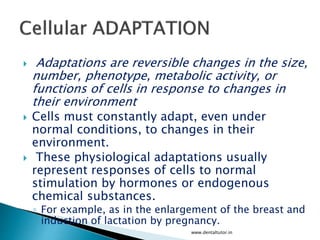
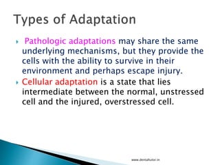
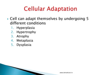
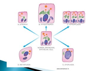
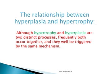
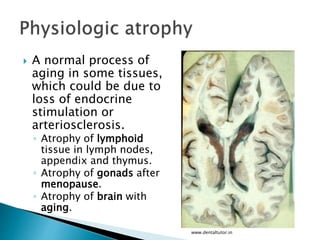
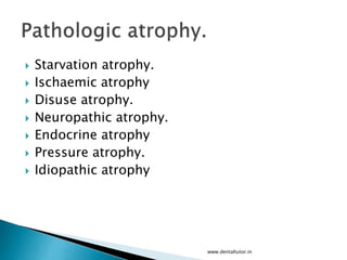
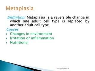
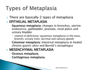
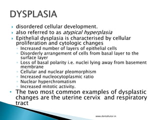
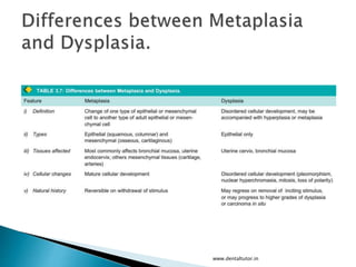


Cells can adapt to changes in their environment through various processes including hyperplasia, hypertrophy, atrophy, metaplasia, and dysplasia. Hyperplasia is an increase in cell number, hypertrophy is an increase in cell size, and atrophy is a decrease in cell size or number. Metaplasia is a reversible change where one adult cell type replaces another. Dysplasia involves disordered cellular development and proliferation with cytological abnormalities. These adaptations allow cells to survive stresses and ensure tissue homeostasis.


![PERI-PROSTHETIC FRACTURE NAIL-PLATE CONSTRUCT [NPC].pptx](https://cdn.slidesharecdn.com/ss_thumbnails/drarunkumardrmohamedashrafperiprostheticfrasturenail-plateconstructnpc-260209164459-7e9d15a1-thumbnail.jpg?width=640&height=640&fit=bounds)


