Download as PDF, PPTX

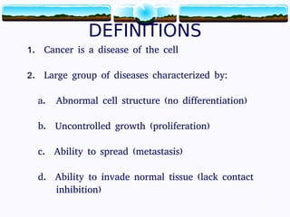
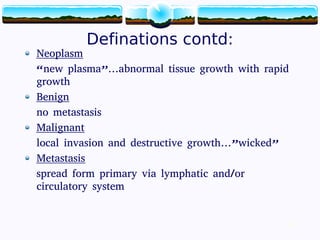
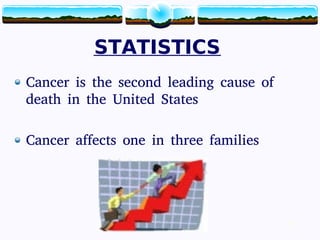
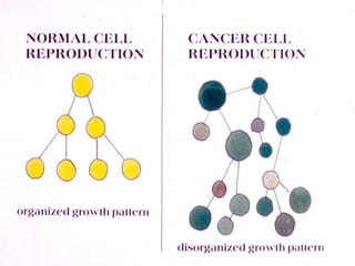

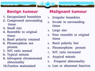
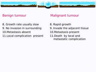

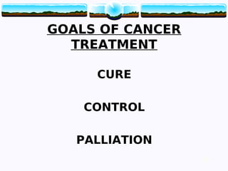
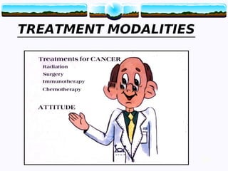
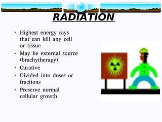
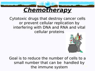


This document provides information about cancer (malignant tumors) including: - Definitions of key terms like neoplasm, benign vs malignant tumors, and metastasis. - Cancer is abnormal cell growth that is uncontrolled, able to invade other tissues, and spread to other parts of the body. - Cancers are classified by site of origin, cell type, grade, and stage. The stage considers tumor size, node involvement, and metastasis. - Cancers are caused by genetic, environmental, and lifestyle factors and can be treated through surgery, radiation, chemotherapy, biotherapy, and hormonal therapy depending on the cancer type and stage.