CELLULAR ADAPTATIONS
Whenthere is increased functional demand, the cell may
adapt to the changes which are expressed morphologically,
which then revert back to normal after the stress is removed.
Reversible changes in the size , shape, phenotype, metabolic
activity or function of cells in response to changes in
environment.
Two types :
Physiologic adaptation
Pathologic adaptation
4.
Common form ofcellular adaptive
response
Atrophy
Hypertrophy
Hyperplasia
Metaplasia
Dysplasia
6.
Atrophy
Reduction ofthe number and size of parenchymal
cells of an organ or its parts which was once normal is
called atrophy.
Two types
Physiological
pathological
7.
Physiologic atrophy
Ageingin some tissues, which could be due to loss of
endocrine stimulation or arteriosclerosis.
For example:
Atrophy of lymphoid tissue with age.
Atrophy of thymus in adult life.
Atrophy of gonads after menopause.
Atrophy of brain with ageing.
9.
Pathological atrophy
Starvationatrophy - In starvation, there is first depletion
of carbohydrate and fat stores followed by protein catabolism.
e.g.
i) cancer
ii)severely ill patients.
Ischaemic atrophy - Gradual diminution of blood supply
due to atherosclerosis may result in shrinkage of the affected organ.
e.g.
i) Small atrophic kidney in atherosclerosis of renal artery.
ii) Atrophy of the brain in cerebral atherosclerosis.
10.
Disuse atrophy- Prolonged diminished functional
activity is associated with disuse atrophy of the organ.
e.g.
i) Wasting of muscles of limb immobilised in cast.
ii) Atrophy of the pancreas in obstruction of pancreatic duct.
Neuropathic atrophy - Interruption in nerve
supply leads to wasting of muscles.
e.g.
i) Poliomyelitis
ii) Motor neuron disease
11.
Endocrine atrophy- Loss of endocrine
regulatory mechanism results in reduced metabolic
activity of tissues and hence atrophy .
e.g.
i) Hypopituitarism may lead to atrophy of thyroid,
adrenal and gonads.
ii) Hypothyroidism may cause atrophy of the skin and
its adnexal structures.
12.
Pressure atrophy- Prolonged pressure from benign tumours
or cyst or aneurysm may cause compression and atrophy of the tissues.
e.g.
i) Erosion of the spine by tumour in nerve root.
ii) Erosion of the sternum by aneurysm of arch of aorta.
Idiopathic atrophy - There are some examples of atrophy
where no obvious cause is present.
e.g.
i) Myopathies.
ii) Testicular atrophy.
13.
Morphological features
Theorgan is small, often shrunken. The cells
become smaller in size but are not dead cells.
Shrinkage in cell size is due to reduction in cell
organelles, chiefly mitochondria,myofilaments
and endoplasmic reticulum.
14.
Hypertrophy
Hypertrophy isan increase in the size of
parenchymal cells resulting in enlargement of the
organ or tissue, without any change in the number of
cells.
Two types :
Physiological
Pathological
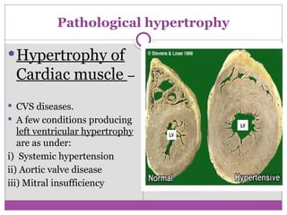
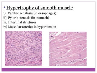
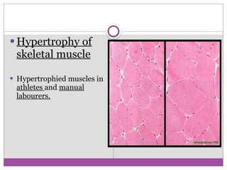
Pathological hypertrophy
Hypertrophy of
Cardiacmuscle –
CVS diseases.
A few conditions producing
left ventricular hypertrophy
are as under:
i) Systemic hypertension
ii) Aortic valve disease
iii) Mitral insufficiency
Compensatory hypertrophymay occur in
an organ when the contralateral organ is removed.
e.g.
Following nephrectomy on one side in a young
patient, there is compensatory hypertrophy as well
as hyperplasia of the nephrons of the other kidney.
22.
Morphological features
Affectedorgan is enlarged and heavy.
Example
Hypertrophied heart of a patient with systemic hypertension
may weigh 700-800 g as compared to average normal adult
weight of 350 g.
There is enlargement of muscle fibres as well as of nuclei.
At Ultra-structural level
Increased synthesis of DNA and RNA,
Increased protein synthesis
Increased number of organelles such as mitochondria,
endoplasmic reticulum and myofibrils.
23.
Hyperplasia
Hyperplasia isan increase in the number
of parenchymal cells resulting in enlargement
of the organ or tissue.
Quite often, both hyperplasia and hypertrophy
occur together.
Two types
Physiological
Pathological
24.
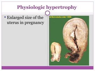
Physiologic hyperplasia
Twotypes : Hormonal and Compensatory
1. Hormonal hyperplasia - Hyperplasia occurring
under the influence of hormonal stimulation.
e.g.
1) Hyperplasia of female breast
at puberty, during pregnancy
and lactation.
2) Hyperplasia of pregnant uterus.
3) Prostatic hyperplasia in old age.
2. Compensatory hyperplasia–
Hyperplasia occurring following removal of part of an organ
or in the contralateral organ in paired organ.
e.g.
1) Regeneration of the liver following partial hepatectomy.
2) Regeneration of epidermis after skin abrasion.
3) Following nephrectomy on one side, there is hyperplasia
of nephrons of the other kidney.
28.
Hyperplasia aredue to excessive stimulation of
hormones or growth factors.
1) Endometrial hyperplasia following estrogen excess.
2) In wound healing, there is formation of granulation tissue
due to proliferation of fibroblasts and endothelial cells.
3) Intraductal epithelial hyperplasia in the breast in
fibrocystic breast disease.
Pathologic hyperplasia
Morphological features
Thereis enlargement of the affected organ or
tissue and increase in the number of cells.
This is due to increased rate of DNA synthesis
and hence increased mitoses of the cells.
31.
Metaplasia
Reversible changeof one type of epithelial or
mesenchymal adult cells to another type of adult
epithelial or mesenchymal cells, usually in response to
abnormal stimuli, and often reverts back to normal on
removal of stimulus.
However, if the stimulus persists for a long time, epithelial
metaplasia may progress to dysplasia and further into cancer.
TWO TYPES
Epithelial
Mesenchymal.
32.
Epithelial metaplasia
Morecommon type.
Metaplastic change may be patchy or diffuse and
usually results in replacement by stronger but less well
specialised epithelium.
Depending upon the type of epithelium transformed,
TWO TYPES of metaplasia.
Squamous metaplasia
Columnar metaplasia
33.
Epithelial metaplasia
1. Squamousmetaplasia.
Variable types of specialised epithelium are capable of
undergoing squamous metaplastic change due to chronic
irritation that may be mechanical, chemical or infective in
origin.
1) In bronchus (normally lined by pseudostratified columnar
ciliated epithelium) in chronic smokers.
2) In uterine endocervix (normally lined by simple columnar
epithelium) in prolapse of the uterus and in old age.
3) In gallbladder(normally lined by simple columnar
epithelium) in chronic cholecystitis with cholelithiasis.
4) In prostate (ducts normally lined by simple columnar
epithelium) in chronic prostatitis and oestrogen therapy.
5) In renal pelvis and urinary bladder (normally lined
by transitional epithelium) in chronic infection and stones.
37.
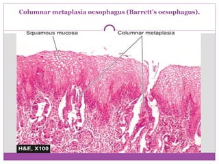
2. Columnar metaplasia
1)Columnar metaplasia in Barrett’s oesophagus,
in which there is change of normal squamous
epithelium to columnar epithelium.
2) Conversion of pseudostratified ciliated columnar
epithelium in chronic bronchitis and
bronchiectasis to columnar type
Mesenchymal metaplasia
Thereis transformation of one adult type of
mesenchymal tissue to another.
TWO TYPES
Osseous metaplasia
Cartilaginous metaplasia
40.
1. Osseous metaplasia- formation of bone in fibrous
tissue, cartilage and myxoid tissue.
1) In arterial wall in old age (Mönckeberg’s sclerosis).
3) In cartilage of larynx and bronchi in elderly people.
4) In scar of chronic inflammation of prolonged duration.
5) In the fibrous stroma of tumour e.g. in leiomyoma.
2. Cartilaginous metaplasia
In healing of fractures, cartilaginous metaplasia may
occur where there is undue mobility.
41.
DYSPLASIA
Dysplasia means‘disordered cellular
development’,
Often preceded or accompanied with
metaplasia and hyperplasia; it is therefore also
referred to as atypical hyperplasia.
Dysplasia occurs most often in epithelial
cells.
42.
Epithelial dysplasiais characterized by cellular
proliferation and cytologic changes as under:
1. Increased number of layers of epithelial cells
2. Disorderly arrangement of cells
3. Loss of basal polarity
4. Cellular and nuclear pleomorphism
5. Increased nucleocytoplasmic ratio
6. Nuclear hyperchromatism
7. Increased mitotic activity.
43.
• Dysplastic changeoccur due to chronic
irritation or prolonged inflammation
• On removal of stimulus changes may disappear.
• In some cases dysplasia may progress into
carcinoma in situ or invasive cancer.
• Example : dysplastic change in uterine cervix.
44.
Uterine cervical dysplasia
-Increased number of layers of squamous epithelium
- Marked cytologic atypia including mitoses
45.
Features METAPLASIA DYSPLASIA
1.Definition Change of one type of
epithelial or mesenchymal
cell to another type of adult
epithelial ormesenchymal cell
Disordered cellular
development, may be
accompanied with
hyperplasia or metaplasia
2. Types Epithelial (squamous,
columnar) and mesenchymal
(osseous,cartilaginous)
Epithelial only
3. Tissues affected Most commonly affects
bronchial mucosa, uterine
endocervix; others
mesenchymal tissues
(cartilage,arteries)
Uterine cervix,
bronchial mucosa
4. Cellular changes Mature cellular
development
Disordered cellular
development
(pleomorphism, nuclear
hyperchromasia, mitosis,
loss of polarity
5. Natural history Reversible on withdrawal of May regress on removal