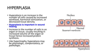
CELL ADAPTATON
Definition
• These are changes a cell goes through in response to an appropriate stimulus and ceases once the need for adaptation has ceased.
• Stimulus can include:
• Physiological
• Hormonal
• Cells adapt by changing their:
• size (atrophy and hypertrophy),
• Number (hyperplasia and atrophy)
• Form (metaplasia).
ATROPHY
• Decrease in size of a cell .
• Decrease in protein synthesis.
• Decreased size results in decreased oxygen consumption and
metabolic needs of the cells and may increase the overall efficiency of
cell function.
• Atrophy is generally a reversible process, except for atrophy caused by
loss of nervous innervation to a tissue.
• Causes of atrophy include:
1. Physiological
2. pathologic
PHYSIOLOGICAL PATHOLOGICAL(LOCAL OR GEENERALISED)
Some embryonic structures during fetal development
e.g notochord, thyroglossal duct
Disuse (paralysis)
Uterus after pregnancy Denervation
Ischemia ( decrease in blood supply)
Aging
Loss of endocrine stimulation
NUTRITION
Kidney: atrophy via renal artery stenosis. NB: decrease in cortex (most metabolically active cells)
MECHANISM
• Reduction in structural componentse.g mitochondria, myofilaments,
ER via proteolysis (lysosomal protease, Increase in number of
autophagic vacuoles, Residual bodies (i.e. lipofuscin  brown
atrophy)
• NB: diminished function but not dead
Pathologic atrophy
• Pathologic atrophy: Shrinkage of our brains as we age
• Disuse atrophy: Essentially when an organ is underused/not used, it
undergoes atrophy.
• Another example of this is an immobilized limb, which undergoes
muscle wasting upon casting.
• Local atrophy: Most often the result of decreased blood flow to that
area
HYPERTROPHY
• Increase in cell size .
• There is increased protein synthesis.
• Occurs when a cell or tissue is exposed to an increased workload.
• Occurs in tissues that cannot undergo mitosis eg cardiac cells,skeletal cells
and nerve cells as an adaptive response.
N.B. THERE IS A LIMIT TO THE AMOUNT OF A TISSUE CAN ENLARGE
• The three basic types of hypertrophy are
1. Physiologic- a response to disease.
2. Compensatory- when cell size increases to take over for non-functioning
cells.
3. Pathologic- is a response to disease.
PATHOLOGICAL PHYSIOLOGICAL COMPENSATORY
Myocardial hypertrophy due to
hypertension
Increased muscle size following
weigh training/ physical labor
e.g. one kidney will undergo
hypertrophy when the other is not
functioning or is removed.
uterus and breast enlarge in
pregnancy
One limb will undergo hypertrophy
when the other is lame
Mechanism
• Increased synthesis of structural proteins via
1. Transcription factors (i. e. c-fos and c-jun)
2. Growth factors (TGF-b, IGF-1, FGF)
3. Vasoactive agents (endothelien-1, AII)
Hypertrophy of uterus
Physiologic Hypertrophy: A: rt = normal uterus, left = gravid uterus; B: left: small, spindle-shaped smooth
muscle cells; rt: larger, more rounded cells of gravid uterus
HYPERPLASIA
• Hyperplasia is an increase in the
number of cells caused by increased
workload, hormonal stimulation, or
decreased tissue density.
• Hyperplasia is important in wound
healing
• Increase in the number of cells in an
organ or tissue, usually resulting in
increased volume of the organ or
tissue hence increased protein
synthesis.
• Like hypertrophy, hyperplasia may
be physiologic, compensatory, or
pathologic.
Physiologic hyperplasia
Physiologic hyperplasia can be divided into:
• Hormonal hyperplasia, which increases the functional capacity of a tissue when
needed,
1. Proliferation of the glandular epithelium of the female breast at puberty and during
pregnancy
2. Occurs in the pregnant uterus.
• Compensatory hyperplasia, which increases tissue mass after damage or partial
resection.
1. Partial hepatectomy --- proliferation of residual liver cells and regeneration of the
liver.
2. After unilateral nephrectomy--- when remaining tissue grows to make up for partial
tissue loss
PATHOLOGIC HYPERPLASIA
• Caused by excessive hormonal stimulation or growth factors acting on target
cells.
• Endometrial hyperplasia: Increase in the amount of oestrogen.
• Benign prostatic hyperplasia: Induced by responses to hormones
(androgens).
• Genital watts.
• Gigantism
METAPLASIA
• Metaplasia is the replacement of one cell type with another cell type.
• A common cause of metaplasia is constant irritation or injury that initiates an
inflammatory response.
• Almost exclusively occurs in epithelial cells.
 May predispose to cancer
 Involves reprogramming of undifferentiated stem cells
 Allows to cells to better survive in a hostile environment
 It is reversible
• Metaplasia may be
1. physiologic
2. pathologic.
PATHOLOGIC METAPLASIA
• Pathologic metaplasia is a response to an extrinsic toxin or stressor
and is generally irreversible.
• For example,
1. after years of exposure to cigarette smoke, stratified squamous
epithelial cells replace the normal ciliated columnar epithelial
cells of the bronchi.
• Although the new cells can better withstand smoke, they don't
secrete mucus nor do they have cilia to protect the airway.
• If exposure to cigarette smoke continues, the squamous cells can
become cancerous.
2. Acid reflux where columnar epithelial cells change into squamous
metaplasia (Barrett esophagus)
3. Connective tissue metaplasia where there is formation of bone,
cartilage, or adipose tissue in tissues that normally don’t contain them
PHYSIOLOGIC METAPLASIA
• Physiologic metaplasia is a normal response to changing conditions
and is generally transient. For example, in the body's normal response
to inflammation, monocytes that migrate to inflamed tissues
transform into macrophages. It is reversible.
Dysplasia
• A derangement of cell growth that leads to tissues with cells of varying size, shape
and appearance.
• Abnormal change in size, shape and organisation of cells within the tissue.
• Generally occurs in response to chronic irritation and inflammation.
• Common examples include dysplasia of epithelial cells of the cervix or the
respiratory tract.
• Dysplasia is considered A STRONG PRECURSOR OF CANCER!!! E.g. Cervical dysplasia
• However, dysplasia is an adaptive process – may or may not lead to cancer
Cont
Dysplasia can be abnormal hyperplasia
Hypertrophy
Atrophy
Metaplasia
ANAPLASIA
• Cells differentiate to a more IMMATURE or embryonic form.
• Malignant tumors are characterized by anaplastic cell growth.

CELL ADAPTATON_091737.pptx

  • 1.
  • 2.
    Definition • These arechanges a cell goes through in response to an appropriate stimulus and ceases once the need for adaptation has ceased. • Stimulus can include: • Physiological • Hormonal • Cells adapt by changing their: • size (atrophy and hypertrophy), • Number (hyperplasia and atrophy) • Form (metaplasia).
  • 4.
    ATROPHY • Decrease insize of a cell . • Decrease in protein synthesis. • Decreased size results in decreased oxygen consumption and metabolic needs of the cells and may increase the overall efficiency of cell function. • Atrophy is generally a reversible process, except for atrophy caused by loss of nervous innervation to a tissue. • Causes of atrophy include: 1. Physiological 2. pathologic
  • 5.
    PHYSIOLOGICAL PATHOLOGICAL(LOCAL ORGEENERALISED) Some embryonic structures during fetal development e.g notochord, thyroglossal duct Disuse (paralysis) Uterus after pregnancy Denervation Ischemia ( decrease in blood supply) Aging Loss of endocrine stimulation NUTRITION
  • 6.
    Kidney: atrophy viarenal artery stenosis. NB: decrease in cortex (most metabolically active cells)
  • 7.
    MECHANISM • Reduction instructural componentse.g mitochondria, myofilaments, ER via proteolysis (lysosomal protease, Increase in number of autophagic vacuoles, Residual bodies (i.e. lipofuscin  brown atrophy) • NB: diminished function but not dead
  • 8.
    Pathologic atrophy • Pathologicatrophy: Shrinkage of our brains as we age • Disuse atrophy: Essentially when an organ is underused/not used, it undergoes atrophy. • Another example of this is an immobilized limb, which undergoes muscle wasting upon casting. • Local atrophy: Most often the result of decreased blood flow to that area
  • 9.
    HYPERTROPHY • Increase incell size . • There is increased protein synthesis. • Occurs when a cell or tissue is exposed to an increased workload. • Occurs in tissues that cannot undergo mitosis eg cardiac cells,skeletal cells and nerve cells as an adaptive response. N.B. THERE IS A LIMIT TO THE AMOUNT OF A TISSUE CAN ENLARGE • The three basic types of hypertrophy are 1. Physiologic- a response to disease. 2. Compensatory- when cell size increases to take over for non-functioning cells. 3. Pathologic- is a response to disease.
  • 10.
    PATHOLOGICAL PHYSIOLOGICAL COMPENSATORY Myocardialhypertrophy due to hypertension Increased muscle size following weigh training/ physical labor e.g. one kidney will undergo hypertrophy when the other is not functioning or is removed. uterus and breast enlarge in pregnancy One limb will undergo hypertrophy when the other is lame
  • 11.
    Mechanism • Increased synthesisof structural proteins via 1. Transcription factors (i. e. c-fos and c-jun) 2. Growth factors (TGF-b, IGF-1, FGF) 3. Vasoactive agents (endothelien-1, AII)
  • 12.
    Hypertrophy of uterus PhysiologicHypertrophy: A: rt = normal uterus, left = gravid uterus; B: left: small, spindle-shaped smooth muscle cells; rt: larger, more rounded cells of gravid uterus
  • 13.
    HYPERPLASIA • Hyperplasia isan increase in the number of cells caused by increased workload, hormonal stimulation, or decreased tissue density. • Hyperplasia is important in wound healing • Increase in the number of cells in an organ or tissue, usually resulting in increased volume of the organ or tissue hence increased protein synthesis. • Like hypertrophy, hyperplasia may be physiologic, compensatory, or pathologic.
  • 14.
    Physiologic hyperplasia Physiologic hyperplasiacan be divided into: • Hormonal hyperplasia, which increases the functional capacity of a tissue when needed, 1. Proliferation of the glandular epithelium of the female breast at puberty and during pregnancy 2. Occurs in the pregnant uterus. • Compensatory hyperplasia, which increases tissue mass after damage or partial resection. 1. Partial hepatectomy --- proliferation of residual liver cells and regeneration of the liver. 2. After unilateral nephrectomy--- when remaining tissue grows to make up for partial tissue loss
  • 15.
    PATHOLOGIC HYPERPLASIA • Causedby excessive hormonal stimulation or growth factors acting on target cells. • Endometrial hyperplasia: Increase in the amount of oestrogen. • Benign prostatic hyperplasia: Induced by responses to hormones (androgens). • Genital watts. • Gigantism
  • 16.
    METAPLASIA • Metaplasia isthe replacement of one cell type with another cell type. • A common cause of metaplasia is constant irritation or injury that initiates an inflammatory response. • Almost exclusively occurs in epithelial cells.  May predispose to cancer  Involves reprogramming of undifferentiated stem cells  Allows to cells to better survive in a hostile environment  It is reversible • Metaplasia may be 1. physiologic 2. pathologic.
  • 18.
    PATHOLOGIC METAPLASIA • Pathologicmetaplasia is a response to an extrinsic toxin or stressor and is generally irreversible. • For example, 1. after years of exposure to cigarette smoke, stratified squamous epithelial cells replace the normal ciliated columnar epithelial cells of the bronchi. • Although the new cells can better withstand smoke, they don't secrete mucus nor do they have cilia to protect the airway. • If exposure to cigarette smoke continues, the squamous cells can become cancerous.
  • 19.
    2. Acid refluxwhere columnar epithelial cells change into squamous metaplasia (Barrett esophagus) 3. Connective tissue metaplasia where there is formation of bone, cartilage, or adipose tissue in tissues that normally don’t contain them
  • 20.
    PHYSIOLOGIC METAPLASIA • Physiologicmetaplasia is a normal response to changing conditions and is generally transient. For example, in the body's normal response to inflammation, monocytes that migrate to inflamed tissues transform into macrophages. It is reversible.
  • 21.
    Dysplasia • A derangementof cell growth that leads to tissues with cells of varying size, shape and appearance. • Abnormal change in size, shape and organisation of cells within the tissue. • Generally occurs in response to chronic irritation and inflammation. • Common examples include dysplasia of epithelial cells of the cervix or the respiratory tract. • Dysplasia is considered A STRONG PRECURSOR OF CANCER!!! E.g. Cervical dysplasia • However, dysplasia is an adaptive process – may or may not lead to cancer
  • 22.
    Cont Dysplasia can beabnormal hyperplasia Hypertrophy Atrophy Metaplasia
  • 24.
    ANAPLASIA • Cells differentiateto a more IMMATURE or embryonic form. • Malignant tumors are characterized by anaplastic cell growth.

Editor's Notes

  • #24 Dysplasia is abnormal hypertrophy,hyperplasia, metaplasia and atrophy