1. Adaptive disorders are changes cells undergo in response to physiological needs or pathological injury, including atrophy, hypertrophy, hyperplasia, metaplasia, and dysplasia.
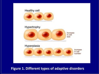
2. Atrophy is a decrease in cell size and number, hypertrophy is an increase in cell size without a change in number, and hyperplasia is an increase in cell number.
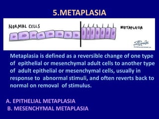
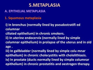
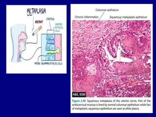
3. Metaplasia and dysplasia involve changes in cell type and abnormal cell development, respectively. Metaplasia is a reversible change from one adult cell type to another, while dysplasia shows disordered cell development and arrangement.
3. 1. INTRODUCTION
Adaptive disorders are the adjustments which the cells
make in response to stresses which may be for
physiologic needs (physiologic adaptation) or a response
to non-lethal pathologic injury (pathologic adaptation).
4. Such physiologic and pathologic adaptations occur by
following processes.
Decreasing or increasing their size i.e. atrophy and
hypertrophy respectively, or by increasing their number
i.e. Hyperplasia (postfi x word -trophy means
nourishment; -plasia means growth of new cells).
Changing the pathway of phenotypic diff eren tia tion
of cells i.e. metaplasia and dysplasia (prefi x word meta-
means transformation; dys- means bad development).
1. INTRODUCTION
6. 2. ATROPHY
Reduction of the number and size of parenchymal cells
of an organ or its parts which was once normal is called
atrophy
CAUSES Atrophy may occur from physiologic or
pathologic causes:
A. Physiologic atrophy
) Atrophy of lymphoid tissue with age.
ii) Atrophy of thymus in adult life.
iii) Atrophy of gonads after menopause.
iv) Atrophy of brain with ageing.
v) Osteoporosis with reduction in size of bony trabeculae
due to ageing.
9. 3.HYPERTROPHY
Hypertrophy is an increase in the size of
parenchymal cells resulting in enlargement of the
organ or tissue, without any change in the number of
cells.
CAUSES
A. Physiologic hypertrophy
Enlarged size of the uterus in Pregnancy is an
example of physiologic hypertrophy as well as
hyperplasia.
10. B. Pathologic hypertrophy
1. Hypertrophy of cardiac muscle may occur in a
number of cardiovasculardiseases. A few conditions
producing left ventricular hypertrophy are as under:
i) Systemic hypertension
ii) Aortic valve disease (stenosis and insuffi ciency)
iii) Mitral insuffi ciency
2.Hypertrophy of skeletal muscle e.g. hypertrophied
mus cles in athletes and manual labourers.
3.HYPERTROPHY
11. 3.Hypertrophy of smooth muscle e.g.
i) Cardiac achalasia (in oesophagus)
ii) Pyloric stenosis (in stomach)
iii) Intestinal strictures
iv) Muscular arteries in hypertension.
4. Compensatory hypertrophy may occur in an organ
when the contralateral organ is removed e.g.
i) Following nephrectomy on one side in a young
patient, there is compensatory hypertrophy as well as
hyperplasia of the nephrons of the other kidney.
ii) Adrenal hyperplasia following removal of one adrenal
gland
3.HYPERTROPHY
12.
13. 4.HYPERPLASIA
Hyperplasia is an increase in the number of paren
chymal cells resulting in enlargement of the organ or
tissue.
. Physiologic hyperplasia
The two most common types are hormonal and
compensatory:
1. Hormonal hyperplasia i.e. hyperplasia occurring under
the influence of hormonal stimulation e.g.
i) Hyperplasia of female breast at puberty, during preg
nancy and lactation.
ii) Hyperplasia of pregnant uterus.
CAUSES
14. iii) Proliferative activity of normal endometrium after
a normal menstrual cycle.
iv) Prostatic hyperplasia in old age
4.HYPERPLASIA
2. Compensatory hyperplasia i.e. hyperplasia occurring
following removal of part of an organ or in the
contralateral organ in paired organ e.g.
i) Regeneration of the liver following partial
hepatectomy.
ii) Regeneration of epidermis after skin abrasion.
iii) Following nephrectomy on one side, there is hyper
plasia of nephrons of the other kidney.
15. 4.HYPERPLASIA
B. Pathologic hyperplasia Most examples of patho logic
hyperplasia are due to excessive stimulation of hormones
or
growth factors e.g.
i) Endometrial hyperplasia following oestrogen excess.
ii) In wound healing, there is formation of granulation
tissue
due to proliferation of fi broblasts and endothelial cells.
iii) Formation of skin warts from hyperplasia of epidermis
due
to human papilloma virus.
iv) Pseudocarcinomatous hyperplasia of the skin occurring
at
the margin of a non-healing ulcer.
16. 5.METAPLASIA
Metaplasia is defined as a reversible change of one type
of epithelial or mesenchymal adult cells to another type
of adult epithelial or mesenchymal cells, usually in
response to abnormal stimuli, and often reverts back to
normal on removal of stimulus.
A. EPITHELIAL METAPLASIA
B. MESENCHYMAL METAPLASIA
17. A. EPITHELIAL METAPLASIA
1. Squamous metaplasia
i) In bronchus (normally lined by pseudostratifi ed
columnar
ciliated epithelium) in chronic smokers.
ii) In uterine endocervix (normally lined by simple
columnar epithelium) in prolapse of the uterus and in old
age .
iii) In gallbladder (normally lined by simple colu mnar
epithelium) in chronic cholecystitis with cholelithiasis.
iv) In prostate (ducts normally lined by simple columnar
epithelium) in chronic prostatitis and oestrogen therapy.
5.METAPLASIA
18.
19. 2. Columnar metaplasia
i) Intestinal metaplasia in healed chronic gastric ulcer.
ii) Columnar metaplasia in Barrett’s oesophagus, in
which
there is change of normal squamous epithelium to
columnar
rpithelium .
iii) Conversion of pseudostratifi ed ciliated columnar
epithelium in chronic bronchitis and bronchiectasis to
columnar
type.
iv) In cervical erosion (congenital and adult type), there
5.METAPLASIA
20. 1. Osseous metaplasia Osseous metaplasia is forma tion
of
bone in fibrous tissue, cartilage and myxoid tissue.
Examples of
osseous metaplasia are as under:
i) In arterial wall in old age (Mönckeberg’s medial calcific
sclerosis)
ii) In soft tissues in myositis ossifi cans
iii) In cartilage of larynx and bronchi in elderly people
iv) In scar of chronic infl ammation of prolonged duration
v) In the fi brous stroma of tumour e.g. in leiomyoma.
2. Cartilaginous metaplasia In healing of fractures,
cartilaginous metaplasia may occur where there is undue
B. MESENCHYMAL METAPLASIA
21. 6. DYSPLASIA
Dysplasia means ‘disordered cellular development’, often
preceded or accompanied with metaplasia and
hyperplasia; it
is therefore also referred to as atypical hyperplasia
22. 1. Increased number of layers of epithelial cells
2. Disorderly arrangement of cells from basal layer to the
surface layer
3. Loss of basal polarity i.e. nuclei lying away from
basement
membrane
4. Cellular and nuclear pleomorphism
5. Increased nucleocytoplasmic ratio
6. Nuclear hyperchromatism
7. Increased mitotic activity.