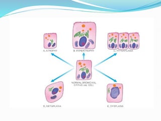
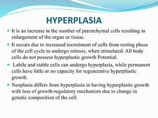
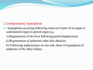
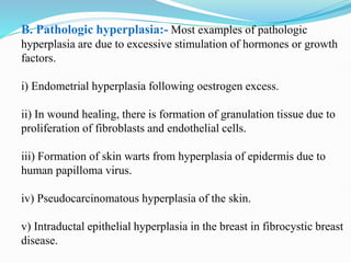
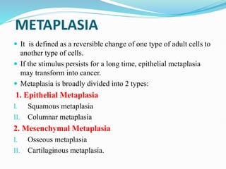
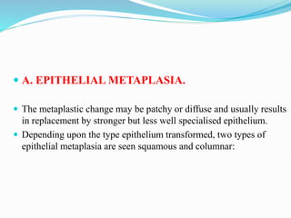
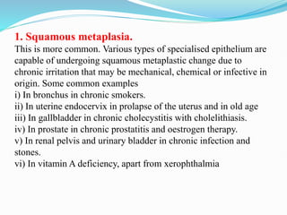
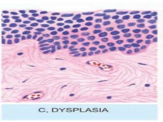
2. CELLULAR ADAPTATIONS
For the sake of survival on exposure to stress, the cells make
adjustments with the changes in their environment to the physiologic
adaptation and to pathologic adaptation.
Decreasing or increasing their size i.e. atrophy and hypertrophy
respectively, or by increasing their number i.e. hyperplasia.
Changing the pathway of phenotypic differentiation of cells i.e.
metaplasia and dysplasia.
Various mechanisms which may be involved in adaptive cellular
responses include the following:
Altered cell surface receptor binding.
Alterations in signal for protein synthesis.
Synthesis of new proteins by the target cell such as heatshock proteins
(HSPs).
3. In general, the adaptive responses are reversible on withdrawal
of stimulus.
However, if the irritant stimulus persists for long time, the cell
may not be able to survive and may either die or progress further
e.g. cell death may occur in sustained atrophy; dysplasia may
progress into carcinoma in situ.
Thus, the concept of evolution ‘survival of the fittest’ holds true
for adaptation as ‘survival of the adaptable’.
4. Common forms of cellular adaptive responses along with
examples of physiologic and pathologic adaptations are briefly
discussed below.
1. ATROPHY
2. HYPERTROPY
3. HYPERPLASIA
4. METAPLASIA
5. DYSPLASIA
5.
6. ATROPHY
Reduction of the number and size of parenchymal cells of an
organ or its parts which was once normal is called atrophy.
CAUSES. Atrophy may occur from physiologic or pathologic.
A. Physiologic atrophy
Atrophy is a normal process of aging in some tissues, which
could be due to loss of endocrine stimulation or
arteriosclerosis. For example:
i) Atrophy of lymphoid tissue in lymph nodes, appendix and
thymus.
ii) Atrophy of gonads after menopause.
iii) Atrophy of brain with aging.
7. B. Pathologic atrophy. The causes are as under:
1. Starvation atrophy:- In starvation, there is first depletion
of carbohydrate and fat stores followed by protein catabolism.
There is general weakness, emaciation and anaemia referred to as
cachexia seen in cancer and severely ill patients.
2. Ischaemic atrophy:- Gradual diminution of blood supply
due to atherosclerosis may result in shrinkage of the affected
organ e.g.
i) Small atrophic kidney in atherosclerosis of renal artery.
ii) Atrophy of brain in cerebral atherosclerosis.
8. 3. Disuse atrophy:- Prolonged diminished functional activity is
associated with disuse atrophy of the organ e.g.
i) Wasting of muscles of limb immobilised in cast.
ii) Atrophy of .the pancreas in obstruction of pancreatic duct.
4. Neuropathic atrophy:- Interruption in nerve supply leads to
wasting of muscles e.g.
i) Poliomyelitis
ii) Motor neuron disease
iii) Nerve section.
5. Endocrine atrophy:- Loss of endocrine regulatory mechanism
results in reduced metabolic activity of tissues and hence atrophy.
i) Hypopituitarism may lead to atrophy of thyroid, adrenal and gonads.
ii) Hypothyroidism may cause atrophy of the skin and its adnexal
structures.
9. 6. Pressure atrophy:- Prolonged pressure from benign tumours or
cyst or aneurysm may cause compression and atrophy of the tissues
e.g.
i) Erosion of spine by tumour in nerve root.
ii) Erosion of skull by meningioma arising from piaarachnoid.
iii) Erosion of sternum by aneurysm of arch of aorta.
7. Idiopathic atrophy:- There are some examples of atrophy where
no obvious cause is present e.g.
i) Myopathies.
ii) Testicular atrophy.
10.
11. HYPERTROPHY
Hypertrophy is an increase in the size of parenchymal cells resulting
in enlargement of the organ or tissue, without any change in the
number of cells.
CAUSES. Hypertrophy may be physiologic or pathologic.
1. Physiologic hypertrophy
Enlarged size of the uterus in pregnancy is an excellent example of
physiologic hypertrophy as well as hyperplasia.
12. 2. Pathologic hypertrophy
1. Hypertrophy of cardiac muscle may occur in a number of
cardiovascular diseases
i) Systemic hypertension
ii) Aortic valve disease
iii) Mitral insufficiency
2. Hypertrophy of smooth muscle e.g. Cardiac achalasia, Pyloric
stenosis, Intestinal strictures, Muscular arteries in hypertension.
3. Hypertrophy of skeletal muscle e.g. hypertrophied muscles in
athletes and manual labourers.
4. Compensatory hypertrophy may occur in an organ when the
contralateral organ is removed e.g. Adrenal hyperplasia.
13.
14. HYPERPLASIA
It is an increase in the number of parenchymal cells resulting in
enlargement of the organ or tissue.
It occurs due to increased recruitment of cells from resting phase
of the cell cycle to undergo mitosis, when stimulated. All body
cells do not possess hyperplastic growth Potential.
Labile and stable cells can undergo hyperplasia, while permanent
cells have little or no capacity for regenerative hyperplastic
growth.
Neoplasia differs from hyperplasia in having hyperplastic growth
with loss of growth-regulatory mechanism due to change in
genetic composition of the cell.
15. CAUSES:- As with other non-neoplastic adaptive disorders of
growth, hyperplasia has also been divided into physiologic and
pathologic.
A. Physiologic hyperplasia:- The two most common types are
as follows:
1. Hormonal hyperplasia i.e. hyperplasia occurring under the
influence of hormonal stimulation e.g.
i) Hyperplasia of female breast at puberty, during pregnancy and
lactation.
ii) Hyperplasia of pregnant uterus.
iii) Proliferative activity of normal endometrium after a normal
menstrual cycle.
iv) Prostatic hyperplasia in old age.
16. 2. Compensatory hyperplasia
hyperplasia occurring following removal of part of an organ or
contralateral organ in paired organ e.g.
i) Regeneration of the liver following partial hepatectomy
ii) Regeneration of epidermis after skin abrasion
iii) Following nephrectomy on one side, there is hyperplasia of
nephrons of the other kidney.
17. B. Pathologic hyperplasia:- Most examples of pathologic
hyperplasia are due to excessive stimulation of hormones or growth
factors.
i) Endometrial hyperplasia following oestrogen excess.
ii) In wound healing, there is formation of granulation tissue due to
proliferation of fibroblasts and endothelial cells.
iii) Formation of skin warts from hyperplasia of epidermis due to
human papilloma virus.
iv) Pseudocarcinomatous hyperplasia of the skin.
v) Intraductal epithelial hyperplasia in the breast in fibrocystic breast
disease.
18.
19. METAPLASIA
It is defined as a reversible change of one type of adult cells to
another type of cells.
If the stimulus persists for a long time, epithelial metaplasia
may transform into cancer.
Metaplasia is broadly divided into 2 types:
1. Epithelial Metaplasia
I. Squamous metaplasia
II. Columnar metaplasia
2. Mesenchymal Metaplasia
I. Osseous metaplasia
II. Cartilaginous metaplasia.
20. A. EPITHELIAL METAPLASIA.
The metaplastic change may be patchy or diffuse and usually results
in replacement by stronger but less well specialised epithelium.
Depending upon the type epithelium transformed, two types of
epithelial metaplasia are seen squamous and columnar:
21. 1. Squamous metaplasia.
This is more common. Various types of specialised epithelium are
capable of undergoing squamous metaplastic change due to
chronic irritation that may be mechanical, chemical or infective in
origin. Some common examples
i) In bronchus in chronic smokers.
ii) In uterine endocervix in prolapse of the uterus and in old age
iii) In gallbladder in chronic cholecystitis with cholelithiasis.
iv) In prostate in chronic prostatitis and oestrogen therapy.
v) In renal pelvis and urinary bladder in chronic infection and
stones.
vi) In vitamin A deficiency, apart from xerophthalmia
22. 2. Columnar metaplasia.
There are some conditions in which there is transformation to
columnar epithelium. For example:
i) Intestinal metaplasia in healed chronic gastric ulcer.
ii) Columnar metaplasia in Barrett’s oesophagus.
iii) Conversion of pseudostratified ciliated columnar epithelium in
chronic bronchitis and bronchiectasis to columnar type.
iv) In cervical erosion there is variable area of endocervical
glandular mucosa everted into the vagina
23. B. MESENCHYMAL METAPLASIA:- Less often, there is
transformation of one adult type of mesenchymal tissue to
another. The examples are as under:
1. Osseous metaplasia:- It is formation of bone in fibrous tissue,
cartilage and myxoid tissue. Examples
i) In arterial wall in old age
ii) In soft tissues in myositis ossificans
iii) In cartilage of larynx and bronchi in elderly people
iv) In scar of chronic inflammation of prolonged duration
v) In the fibrous stroma of tumour
2. Cartilaginous metaplasia:- In healing of fractures,
cartilaginous metaplasia may occur where there is undue mobility.
24.
25. Feature Metaplasia Dysplasia
i)
Definiti
on
Change of one type of
epithelial or mesenchymal cell
to another type of adult
epithelial or mesen-chymal
cell
Disordered cellular development, may be
accompanied with hyperplasia or metaplasia
ii)
Types
Epithelial (squamous,
columnar) and
mesenchymal (osseous,
cartilaginous)
Epithelial only
iv)
Cellular
changes
Mature cellular development Disordered cellular development (pleomorphism,
nuclear hyperchromasia, mitosis, loss of polarity)
v)
Natural
history
Reversible on withdrawal of
stimulus
May regress on removal of inciting stimulus,
or may progress to higher grades of dysplasia
or carcinoma in situ
iii)
Tissues
affected
Most commonly affects
bronchial mucosa, uterine
endocervix others
mesenchymal tissues
(cartilage,
Uterine cervix, bronchial mucosa
26. DYSPLASIA
It means ‘disordered cellular development’, often accompanied with
metaplasia and hyperplasia; it is therefore also referred to as atypical
hyperplasia.
Dysplasia occurs most often in epithelial cells. Epithelial dysplasia is
characterised by cellular proliferation and cytologic changes. These
changes include:
1. Increased number of layers of epithelial cells
2. Disorderly arrangement of cells from basal layer to the surface layer
27. 3. Loss of basal polarity nuclei lying away from basement
membrane.
4. Cellular and nuclear pleomorphism.
5. Increased nucleocytoplasmic ratio.
6. Nuclear hyperchromatism.
7. Increased mitotic activity.
The two most common examples of dysplastic changes are the
uterine cervix and respiratory tract.