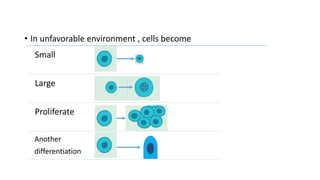
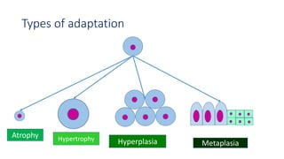
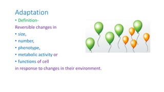
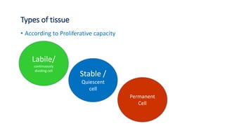
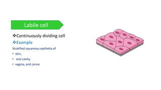
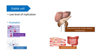
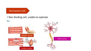
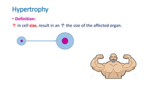
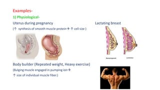
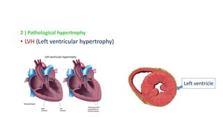
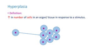
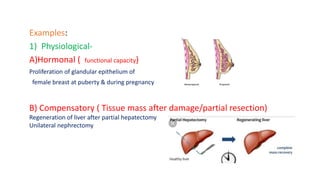
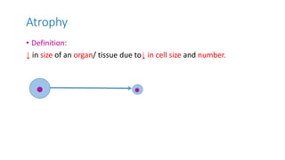
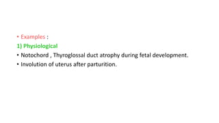
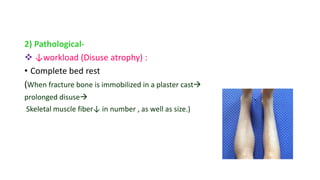
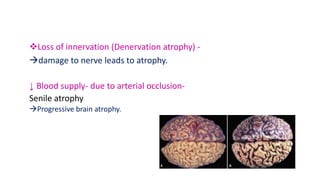
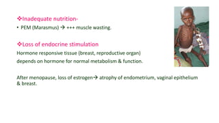
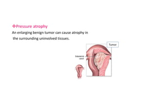
Cellular Adaptation
Dr. Naila Awal
MD (Pathology)
Adaptation in animals
Walrus has thick
skin to protect
against cold
condition
Duck has
webbed feet
Nostrils on the
top of the
snout
The polar bear has small ears &
tail. This adaptation minimizes
heat loss.
Long & bushy
eyelash to
keep out the
sand & sun
Slit like nostrils
 they are able
to open & close
to prevent sand
Hill like humps –
for storing fat, so
that they can go
without eating
long time
So what happens to
our body?
• In unfavorable environment , cells become
Small
Large
Proliferate
Another
differentiation
Types of adaptation
Atrophy Hypertrophy Hyperplasia Metaplasia
Adaptation
• Definition-
Reversible changes in
• size,
• number,
• phenotype,
• metabolic activity or
• functions of cell
in response to changes in their environment.
Types of tissue
• According to Proliferative capacity
Labile/
continuously
dividing cell
Stable /
Quiescent
cell
Permanent
Cell
Continuously dividing cell
Example
Stratified squamous epithelia of
• skin,
• oral cavity,
• vagina, and cervix
Labile cell
• Low level of replication
• Examples
Stable cell
Chondrocyte,
osteocyte Smooth muscle cell
Parenchymal cells of liver, kidneys,
and pancreas
• Non dividing cell, unable to replicate
Ex-
Permanent Cell
Skeletal muscle
Cardiac muscle
Adult neuron
Hypertrophy
• Definition:
↑ in cell size, result in an ↑ the size of the affected organ.
Examples-
1) Physiological-
Uterus during pregnancy Lactating breast
(↑ synthesis of smooth muscle protein ↑ cell size )
Body builder (Repeated weight, Heavy exercise)
(Bulging muscle engaged in pumping ion
↑ size of individual muscle fiber.)
2 ) Pathological hypertrophy
• LVH (Left ventricular hypertrophy)
Left ventricle
Hyperplasia
• Definition:
↑ in number of cells in an organ/ tissue in response to a stimulus.
Examples:
1) Physiological-
A)Hormonal ( functional capacity)
Proliferation of glandular epithelium of
female breast at puberty & during pregnancy
B) Compensatory ( Tissue mass after damage/partial resection)
Regeneration of liver after partial hepatectomy
Unilateral nephrectomy
2) Pathological Hyperplasia
• Endometrial Hyperplasia
• Benign prostatic Hyperplasia
Androgen induced
Ques : Which 1 has more chance of malignancy?
• Skin wart (HPV) CT proliferation in wound
healing
Atrophy
• Definition:
↓ in size of an organ/ tissue due to↓ in cell size and number.
• Examples :
1) Physiological
• Notochord , Thyroglossal duct atrophy during fetal development.
• Involution of uterus after parturition.
2) Pathological-
 ↓workload (Disuse atrophy) :
• Complete bed rest
(When fracture bone is immobilized in a plaster cast
prolonged disuse
Skeletal muscle fiber↓ in number , as well as size.)
Loss of innervation (Denervation atrophy) -
damage to nerve leads to atrophy.
↓ Blood supply- due to arterial occlusion-
Senile atrophy
Progressive brain atrophy.
Inadequate nutrition-
• PEM (Marasmus)  +++ muscle wasting.
Loss of endocrine stimulation
Hormone responsive tissue (breast, reproductive organ)
depends on hormone for normal metabolism & function.
After menopause, loss of estrogen atrophy of endometrium, vaginal epithelium
& breast.
Pressure atrophy
An enlarging benign tumor can cause atrophy in
the surrounding uninvolved tissues.
Tumor
Metaplasia
• Definition
Reversible change in which 1 differentiated cell ( epithelial
/mesenchymal) type is replaced by another cell type.
• Types-
Metaplasia
Epithelial
Squamous Columner
CT
metaplasia
Epithelial Metaplasia
1) Squamous metaplasia-
Respiratory Tract
Ex– Habitual cigarette smoker
Vit A deficiency
• Pathogenesis-
Chronic irritation Normal ciliated columnar epithelium of trachea &
bronchi are replaced by stratified squamous epithelium.
 Stones in the ducts of salivary gland, pancreas and bile duct
• Chronic irritation
Secretory columnar epithelium are
replaced by stratified squamous epithelium.
2) Columnar metaplasia
• Barrett esophagus
Reflux gastric acid squamous epithelium
is replaced by
intestinal like columnar epithelium
• Adenocarcinoma may arise.
Connective Tissue Metaplasia
• Formation of cartilage , bone/ adipose tissue ( mesenchymal ) in
tissues that do not contain these element.
• Example-
Myositis ossificans – Bone formation in muscle, occasionally occurs after
intramuscular hemorrhage.
Any Question?
Cellular adaptation

Cellular adaptation

  • 1.
    Cellular Adaptation Dr. NailaAwal MD (Pathology)
  • 2.
    Adaptation in animals Walrushas thick skin to protect against cold condition Duck has webbed feet Nostrils on the top of the snout
  • 3.
    The polar bearhas small ears & tail. This adaptation minimizes heat loss. Long & bushy eyelash to keep out the sand & sun Slit like nostrils  they are able to open & close to prevent sand Hill like humps – for storing fat, so that they can go without eating long time
  • 4.
    So what happensto our body?
  • 5.
    • In unfavorableenvironment , cells become Small Large Proliferate Another differentiation
  • 6.
    Types of adaptation AtrophyHypertrophy Hyperplasia Metaplasia
  • 7.
    Adaptation • Definition- Reversible changesin • size, • number, • phenotype, • metabolic activity or • functions of cell in response to changes in their environment.
  • 8.
    Types of tissue •According to Proliferative capacity Labile/ continuously dividing cell Stable / Quiescent cell Permanent Cell
  • 9.
    Continuously dividing cell Example Stratifiedsquamous epithelia of • skin, • oral cavity, • vagina, and cervix Labile cell
  • 10.
    • Low levelof replication • Examples Stable cell Chondrocyte, osteocyte Smooth muscle cell Parenchymal cells of liver, kidneys, and pancreas
  • 11.
    • Non dividingcell, unable to replicate Ex- Permanent Cell Skeletal muscle Cardiac muscle Adult neuron
  • 12.
    Hypertrophy • Definition: ↑ incell size, result in an ↑ the size of the affected organ.
  • 13.
    Examples- 1) Physiological- Uterus duringpregnancy Lactating breast (↑ synthesis of smooth muscle protein ↑ cell size ) Body builder (Repeated weight, Heavy exercise) (Bulging muscle engaged in pumping ion ↑ size of individual muscle fiber.)
  • 14.
    2 ) Pathologicalhypertrophy • LVH (Left ventricular hypertrophy) Left ventricle
  • 15.
    Hyperplasia • Definition: ↑ innumber of cells in an organ/ tissue in response to a stimulus.
  • 16.
    Examples: 1) Physiological- A)Hormonal (functional capacity) Proliferation of glandular epithelium of female breast at puberty & during pregnancy B) Compensatory ( Tissue mass after damage/partial resection) Regeneration of liver after partial hepatectomy Unilateral nephrectomy
  • 17.
    2) Pathological Hyperplasia •Endometrial Hyperplasia • Benign prostatic Hyperplasia Androgen induced Ques : Which 1 has more chance of malignancy?
  • 18.
    • Skin wart(HPV) CT proliferation in wound healing
  • 19.
    Atrophy • Definition: ↓ insize of an organ/ tissue due to↓ in cell size and number.
  • 20.
    • Examples : 1)Physiological • Notochord , Thyroglossal duct atrophy during fetal development. • Involution of uterus after parturition.
  • 21.
    2) Pathological-  ↓workload(Disuse atrophy) : • Complete bed rest (When fracture bone is immobilized in a plaster cast prolonged disuse Skeletal muscle fiber↓ in number , as well as size.)
  • 22.
    Loss of innervation(Denervation atrophy) - damage to nerve leads to atrophy. ↓ Blood supply- due to arterial occlusion- Senile atrophy Progressive brain atrophy.
  • 23.
    Inadequate nutrition- • PEM(Marasmus)  +++ muscle wasting. Loss of endocrine stimulation Hormone responsive tissue (breast, reproductive organ) depends on hormone for normal metabolism & function. After menopause, loss of estrogen atrophy of endometrium, vaginal epithelium & breast.
  • 24.
    Pressure atrophy An enlargingbenign tumor can cause atrophy in the surrounding uninvolved tissues. Tumor
  • 25.
    Metaplasia • Definition Reversible changein which 1 differentiated cell ( epithelial /mesenchymal) type is replaced by another cell type.
  • 26.
  • 27.
    Epithelial Metaplasia 1) Squamousmetaplasia- Respiratory Tract Ex– Habitual cigarette smoker Vit A deficiency • Pathogenesis- Chronic irritation Normal ciliated columnar epithelium of trachea & bronchi are replaced by stratified squamous epithelium.
  • 28.
     Stones inthe ducts of salivary gland, pancreas and bile duct • Chronic irritation Secretory columnar epithelium are replaced by stratified squamous epithelium.
  • 29.
    2) Columnar metaplasia •Barrett esophagus Reflux gastric acid squamous epithelium is replaced by intestinal like columnar epithelium • Adenocarcinoma may arise.
  • 30.
    Connective Tissue Metaplasia •Formation of cartilage , bone/ adipose tissue ( mesenchymal ) in tissues that do not contain these element. • Example- Myositis ossificans – Bone formation in muscle, occasionally occurs after intramuscular hemorrhage.
  • 31.