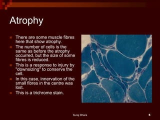
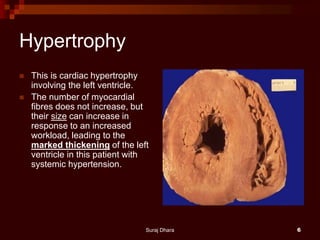
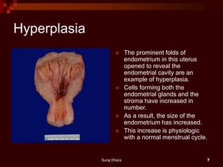
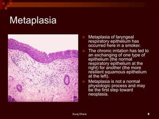
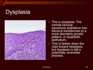
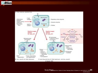
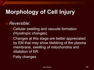
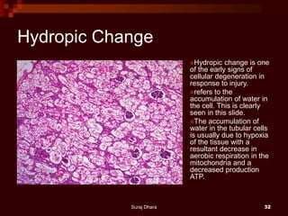
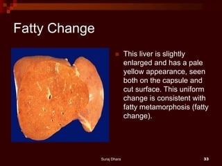
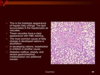
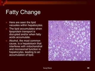
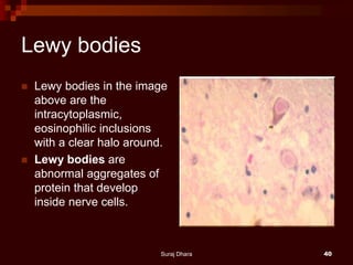
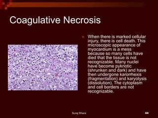
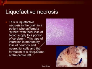
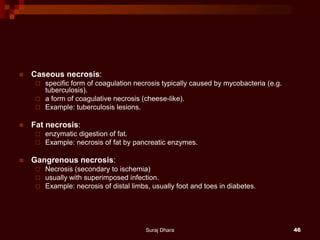
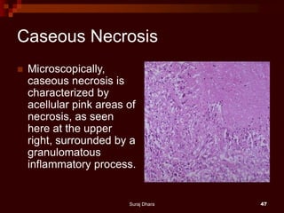
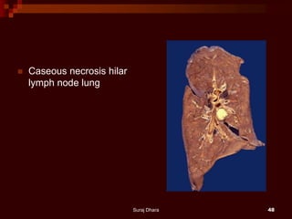
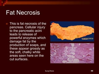
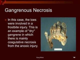
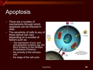
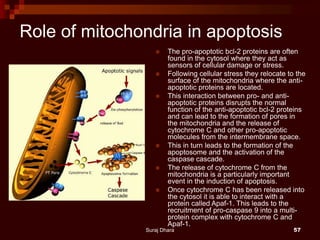
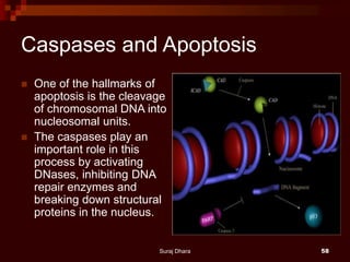
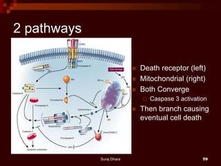
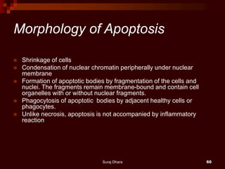
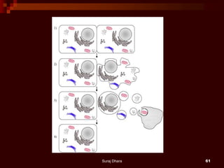
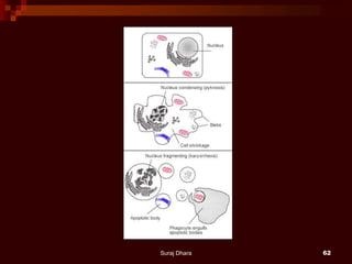
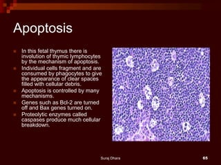
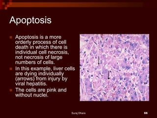
SlideShare a Scribd company logo
PATHOLOGY – PART 1
Objectives
 Upon completion of this lecture you will be able
to:
 Define 4 types of cellular adaptation to physiologic
and pathologic conditions.
 List 7 common causes of cell injury.
 Explain the difference between reversible and
irreversible cell injury.
 Explain the difference between necrosis and
apoptosis.
 Describe patterns of necrosis In tissues or organs.
Suraj Dhara 2
Important Terms
 Cellular Adaptation
 Cell Injury
 Necrosis
 Apoptosis
 Atrophy
 Hypertrophy
 Hyperplasia
 Metaplasia
 For more details see Cotran, R. S., Kumar, V., Collins, T. (1999).
Pathologic Basis of Disease ( 6th ed.). Philadelphia, PA:
Suraj Dhara 3
 Cells are the structural and functional units of tissues and organs.
They are capable of adjusting their structure and functions in
response to various physiological and pathological conditions. This
capability is called cellular adaptation.
 Cellular adaptations include:
 Atrophy--shrinkage of cells
 Hypertrophy--increase in the size of cells which results in enlargement
of the organs
 Hyperplasia--increased number of cells in an organ or tissue
 Metaplasia--transformation or replacement of one adult cell type with
another
Suraj Dhara 4
Atrophy
 There are some muscle fibres
here that show atrophy.
 The number of cells is the
same as before the atrophy
occurred, but the size of some
fibres is reduced.
 This is a response to injury by
"downsizing" to conserve the
cell.
 In this case, innervation of the
small fibres in the centre was
lost.
 This is a trichrome stain.
Suraj Dhara 5
Hypertrophy
 This is cardiac hypertrophy
involving the left ventricle.
 The number of myocardial
fibres does not increase, but
their size can increase in
response to an increased
workload, leading to the
marked thickening of the left
ventricle in this patient with
systemic hypertension.
Suraj Dhara 6
Hyperplasia
 The prominent folds of
endometrium in this uterus
opened to reveal the
endometrial cavity are an
example of hyperplasia.
 Cells forming both the
endometrial glands and the
stroma have increased in
number.
 As a result, the size of the
endometrium has increased.
 This increase is physiologic
with a normal menstrual cycle.
Suraj Dhara 7
Metaplasia
 Metaplasia of laryngeal
respiratory epithelium has
occurred here in a smoker.
 The chronic irritation has led to
an exchanging of one type of
epithelium (the normal
respiratory epithelium at the
right) for another (the more
resilient squamous epithelium
at the left).
 Metaplasia is not a normal
physiologic process and may
be the first step toward
neoplasia.
Suraj Dhara 8
Dysplasia
 This is dysplasia. The
normal cervical
squamous epithelium has
become transformed to a
more disorderly growth
pattern, or dysplastic
epithelium.
 This is farther down the
road toward neoplasia,
but dysplasia is still a
potentially reversible
process.
Suraj Dhara 9
Various Types of Adaptations
 Cells may undergo various adaptations in
physiological and pathological conditions.
These are controlled by complex
molecular mechanisms.
Suraj Dhara 10
Atrophy-
 -shrinkage of cells; classified as:
Physiologic--due to decreased work load
(e.g., decreased size of uterus following child
birth, or disease)
Pathologic--primarily due to denervation of
muscle, diminished blood supply, nutritional
deficiency
Suraj Dhara 11
Hypertrophy-
 Increase in the size of cells which results in
enlargement of the organs.
 It is mostly seen in cells that cannot divide, such
as:
 skeletal muscle (pumping iron),
 cardiac muscle (hypertension).
 These changes usually revert to normal if the
cause is removed.
 Hypertrophy is mediated by different
mechanisms.
Suraj Dhara 12
Hyperplasia-
 Increased number of cells in an organ or tissue.
Hyperplasia may sometimes co-exist with
hypertrophy. Hyperplasia can be classified as:
 physiologic--hormonal (e.g., breast and uterus
during pregnancy)
 compensatory--regeneration of liver following partial
hepatectomy. Various growth factors and interluekins
are important in such hyperplasia.
 pathologic--excessive hormonal stimulation, viral
infection (papilloma viruses); neoplasms
Suraj Dhara 13
Metaplasia-
 Transformation or replacement of one adult cell type to another
adult cell type
 (e.g., the change from columnar to squamous cells in respiratory
tract, from squamous to columnar in Barrett esophagitis).
 Metaplasia also occurs in mesenchymal tissue (e.g., formation of
bone in skeletal muscle).
 Metaplastic changes usually result from chronic irritation.
 Metaplastic changes seem to precede the development of cancer,
in some instances.
 Metaplasia is thought to arise from reprogramming of stem or
undifferentiated cells that are present in adult tissue.
Suraj Dhara 14
Cell Injury
 If the cells fail to adapt under stress, they undergo
certain changes called cell injury. The affected cells
may recover from the injury (reversible) or may die
(irreversible).
 Causes of Cell Injury
 oxygen deprivation (anoxia)
 physical agents
 chemical agents
 infections agents
 immunologic reactions
 genetic defects
 nutritional imbalances
Suraj Dhara 15
IMPORTANT TARGETS OF CELL INJURY
 Aerobic respiration –
 ATP depletion or decreased synthesis.
 Cell membranes - plasma membranes, mitochondrial, lysosomal
and other organelle membranes.
 Protein synthesis.
 Cytoskeleton.
 Genetic apparatus.
Suraj Dhara 16
Downloaded from: Robbins & Cotran Pathologic Basis of Disease (on 4 April 2005 06:11 PM)
© 2005 Elsevier
Suraj Dhara 17
ATP DEPLETION - HYPOXIA/ISCHAEMIA
 Mitochondria - reduced oxidative phosphorylation.
 Cell membrane - reduced sodium pump.
 Sodium and water enter the cell; potassium exits.
 Endoplasmic reticulum dilates, the cell swells, blebs appear.
 Anaerobic glycolysis occurs with loss of glycogen, accumulation of
lactic acid, acid pH which interferes with enzymes.
 Failure of the calcium pump leads to influx of Ca++ into the cell,
activate various enzymes to the detriment of the cell.
 RER loses ribosomes and protein synthesis falls - structural proteins
(membranes,cytoskeleton) and enzymes.
 Misfolded proteins lead to the unfolded protein response which may
further injure the cell.
Suraj Dhara 18
Downloaded from: Robbins & Cotran Pathologic Basis of Disease (on 4 April 2005 06:11 PM)
© 2005 Elsevier
Suraj Dhara 19
THE IMPORTANCE OF CALCIUM
 Influx of calcium to the cytosol comes from the extracellular fluid and
stores in mitochondria and endoplasmic reticulum.
 Ca++ activates phospholipases (damages cell membranes),proteases
(damages cell membranes and cytoskeleton) and endonucleases
(damages DNA).
 This is one of the main mechanisms of cell death, either through severe
damage to membranes of lysosomes and leakage of lysosomal
enzymes or triggering apoptosis.
 Occurs particularly in hypoxia and ischaemia and with certain toxins.
Preventing the rise in Ca++ or restoring to normal levels prevents cell
death.
Suraj Dhara 20
Downloaded from: Robbins & Cotran Pathologic Basis of Disease (on 4 April 2005 06:11 PM)
© 2005 Elsevier
Suraj Dhara 21
THE IMPORTANCE OF FREE RADICALS
 Free radicals have a single unpaired electron in the outer orbit. They
are highly reactive with adjacent molecules.
 Are usually derived from oxygen to produce reactive oxygen species,
superoxide, hydroxyl radicals,H2O2,etc.
 Are normally produced during cellular respiration. Protective molecules
include superoxide dismutase, glutathione peroxidase, vitamin E,
vitamin C, catalase.
 Produced in excess, they react with, and damage proteins, lipids,
carbohydrates, nucleic acids.
 These damaged molecules may themselves be reactive species with a
chain reaction being set up with widespread damage.
Suraj Dhara 22
FREE RADICALS
 In addition to oxygen-derived free radicals, nitric oxide (NO) can act as
a free radical and be converted to an even more reactive anion.
 Iron and copper catalyze free radical formation and are thus important
in the generation of reactive oxygen species.
 Binding to molecules such as transferrin, ferritin and ceruloplasmin is
protective.
 Free radicals cause lipid peroxidation in cell membranes, oxidation of
amino acids and proteins resulting in fragmentation, and protein-protein
cross linkages. Altered proteins are acted on by the proteosomes with
further cell damage.
Suraj Dhara 23
Downloaded from: Robbins & Cotran Pathologic Basis of Disease (on 4 April 2005 06:11 PM)
© 2005 Elsevier
Suraj Dhara 24
FREE RADICALS AND DISEASE
 Free radicals may be a common pathway for most types of cell
damage, particularly oxygen-derived free radicals (oxidative stress).
 Some examples are:-
 oxygen toxicity, ischaemia/reperfusion injury, radiation injury (hydrolyses
H2O to OH & H), metabolism of drugs,toxins,pollutants (eg Paracetamol to
reactive metabolite;CCl4 to CCl3,cigarette smoke);
 leukocyte killing of bacteria or in non-bacterial inflammations,release of iron
in haemorrhages enhances oxidative stress (important in CNS),
 lipid peroxidation of low-density lipoproteins in atherosclerosis,cancer
production (damage to DNA), ageing.
 Therapies for combating oxidative stress are available for prevention or
treatment with antioxidants and/or free-radical scavengers.
Suraj Dhara 25
ISCHAEMIA/REPERFUSION INJURY
 If cells are reversibly injured due to ischaemia, complete recovery
occurs following restoration of blood flow.
 However, reperfusion can result in more damage including cell death.
 This is due to incompletely metabolised products producing reactive
oxygen species on re-introduction of oxygen
 (especially damaging to mitochondria;
 loss of anti-oxidants during ischaemia;
 inflow of calcium with the renewed blood flow;
 recruitment of leukocytes to the injured area.
 Reperfusion injury is especially important in ischaemic damage to the
heart and brain and in organ transplantation.
 Various therapies and preventive measures are in use.
Suraj Dhara 26
MEMBRANE DAMAGE
 Mitochondria –
 mitochondrial permeability transition;
 this non-selective pore may be reversible or become permanent leading to
cell death.
 Leakage of cytochrome c can trigger apoptosis.
 Plasma membrane –
 mechanisms include those occuring with hypoxia/ischaemia and free
radicals, but also
 immune mechanisms as with complement activation and
 perforin from lymphocyte attack on cells infected with a virus.
 All membranes may be damaged and ruptured by
 mechanical force as in trauma, or by
 ice crystals as in extreme cold.
 Damage to lysosomal membranes can lead to cell death by necrosis.
Suraj Dhara 27
Suraj Dhara 28
CHEMICAL & BIOLOGICAL AGENTS
 Chemical agents - direct effects, e.g.
cyanine on cytochrome oxidase, HgCl
on sulphydryl groups of proteins, ricin
on ribosomes, cytotoxic effects of
chemotherapy & antibiotics.
-indirect effects via free radicals.
Suraj Dhara 29
CHEMICAL & BIOLOGICAL AGENTS
 Biological agents - direct effects of
bacterial toxins; cytopathic effects of
viruses and other actions such as:
 interfering with DNA,RNA, proteins, cell
membranes or
inducing apoptosis.
-indirect effects via the host immune reaction.
Suraj Dhara 30
Morphology of Cell Injury
 Reversible:
Cellular swelling and vacuole formation
(Hyodropic changes)
Changes at this stage are better appreciated
by EM that may show blebbing of the plasma
membrane, swelling of mitochondria and
dilatation of ER
Fatty changes
Suraj Dhara 31
Hydropic Change
Hydropic change is one
of the early signs of
cellular degeneration in
response to injury.
refers to the
accumulation of water in
the cell. This is clearly
seen in this slide.
The accumulation of
water in the tubular cells
is usually due to hypoxia
of the tissue with a
resultant decrease in
aerobic respiration in the
mitochondria and a
decreased production
ATP.
Suraj Dhara 32
Fatty Change
 This liver is slightly
enlarged and has a pale
yellow appearance, seen
both on the capsule and
cut surface. This uniform
change is consistent with
fatty metamorphosis (fatty
change).
Suraj Dhara 33
 This is the histologic appearance
of hepatic fatty change. The lipid
accumulates in the hepatocytes as
vacuoles.
 These vacuoles have a clear
appearance with H&E staining.
 The most common cause of fatty
change in developed nations is
alcoholism.
 In developing nations, kwashiorkor
in children is another cause.
 Diabetes mellitus, obesity, and
severe gastrointestinal
malabsorption are additional
causes.
Suraj Dhara 34
Fatty Change
 Here are seen the lipid
vacuoles within hepatocytes.
 The lipid accumulates when
lipoprotein transport is
disrupted and/or when fatty
acids accumulate.
 Alcohol, the most common
cause, is a hepatotoxin that
interferes with mitochondrial
and microsomal function in
hepatocytes, leading to an
accumulation of lipid.
Suraj Dhara 35
Morphology of Cell Injury
 Irreversible/Necrosis
 The changes are produced by enzymatic digestion of
dead cellular elements, denatunation of proteins and
autolysis (by lysosomal enzymes)
 Cytoplasm - increased eosinophilia
 Nucleus - nonspecific breakdown of DNA leading to
 pyknosis (shrinkage),
 karyolysis (fading) and
 karyorrhexis (fragmentation).
Suraj Dhara 36
Chronic Cell Injury
 Non-lethal injury may cause subcellular changes
some of which are characteristically seen in
certain pathologic conditions. The following are
examples of some of these changes:
 Changes in mitochondria seen in various conditions in
some of which there is an increase in the number of
mitochondria with various morphological
abnormalities.
 Cytoskeletal changes with formation of distinctive
intracellular inclusions such as Mallory body
Neurofibrillary tangles, or Lewy body.
Suraj Dhara 37
Mallory Bodies
 Cytoplasmic organelle damage leads
to a variety of injury patterns, most of
which are best seen by electron
microscopy.
 Acute injuries tend to damage an
entire cell, so specific organelle
damage is beside the point.
 However, in some cases the damage
can be cumulative over many years.
 Here are Mallory bodies (the red
globular material) composed of
cytoskeletal filaments in liver cells
chronically damaged from alcoholism.
 These are a type of "intermediate"
filament between the size of actin
(thin) and myosin (thick).
Suraj Dhara 38
Neurofibrillary Tangles
 Here are neurofibrillary
tangles in neurons of a
patient with Alzheimer's
disease.
 The cytoskeletal filaments
are grouped together in
the elongated pink
tangles.
Suraj Dhara 39
Lewy bodies
 Lewy bodies in the image
above are the
intracytoplasmic,
eosinophilic inclusions
with a clear halo around.
 Lewy bodies are
abnormal aggregates of
protein that develop
inside nerve cells.
Suraj Dhara 40
Cell Death
 Death of cells occurs in two ways:
Necrosis--(irreversible injury) changes
produced by enzymatic digestion of dead
cellular elements
Apoptosis--vital process that helps eliminate
unwanted cells--an internally programmed
series of events effected by dedicated gene
products
Suraj Dhara 41
Mechanisms of Cell Death
 Mechanisms of cell death caused by different agents
may vary. However, certain biochemical events are seen
in the process of cell necrosis:
 ATP depletion
 Loss of calcium homeostasis and free cytosolic calcium
 Free radicals: superoxide anions, Hydroxyl radicals, hydrogen
peroxide
 Defective membrane permeability
 Mitochondrial damage
 Cytoskeletal damage
Suraj Dhara 42
Patterns of Necrosis In Tissues
or Organs
 As a result of cell death the tissues or organs display certain
macroscopic changes:
 Coagulative necrosis:
 typically seen in hypoxic environments
 the outline of the dead cells are maintained and the tissue is somewhat firm.
 Example: myocardial infarction
 Liquifactive necrosis: the dead cells undergo disintegration and
affected tissue is liquified.
 Example: cerebral infarction.
 usually associated with cellular destruction and pus formation (e.g.
pneumonia).
 ischemia (restriction of blood supply) in the brain produces liquefactive
rather than coagulative necrosis.
Suraj Dhara 43
Coagulative Necrosis
 When there is marked cellular
injury, there is cell death. This
microscopic appearance of
myocardium is a mess
because so many cells have
died that the tissue is not
recognizable. Many nuclei
have become pyknotic
(shrunken and dark) and have
then undergone karorrhexis
(fragmentation) and karyolysis
(dissolution). The cytoplasm
and cell borders are not
recognizable.
Suraj Dhara 44
Liquefactive necrosis
 This is liquefactive
necrosis in the brain in a
patient who suffered a
"stroke" with focal loss of
blood supply to a portion
of cerebrum. This type of
infarction is marked by
loss of neurons and
neuroglial cells and the
formation of a clear space
at the centre left.
Suraj Dhara 45
 Caseous necrosis:
 specific form of coagulation necrosis typically caused by mycobacteria (e.g.
tuberculosis).
 a form of coagulative necrosis (cheese-like).
 Example: tuberculosis lesions.
 Fat necrosis:
 enzymatic digestion of fat.
 Example: necrosis of fat by pancreatic enzymes.
 Gangrenous necrosis:
 Necrosis (secondary to ischemia)
 usually with superimposed infection.
 Example: necrosis of distal limbs, usually foot and toes in diabetes.
Suraj Dhara 46
Caseous Necrosis
 Microscopically,
caseous necrosis is
characterized by
acellular pink areas of
necrosis, as seen
here at the upper
right, surrounded by a
granulomatous
inflammatory process.
Suraj Dhara 47
 Caseous necrosis hilar
lymph node lung
Suraj Dhara 48
Fat Necrosis
 This is fat necrosis of the
pancreas. Cellular injury
to the pancreatic acini
leads to release of
powerful enzymes which
damage fat by the
production of soaps, and
these appear grossly as
the soft, chalky white
areas seen here on the
cut surfaces.
Suraj Dhara 49
Gangrenous Necrosis
 In this case, the toes
were involved in a
frostbite injury. This is
an example of "dry"
gangrene in which
there is mainly
coagulative necrosis
from the anoxic injury.
Suraj Dhara 50
 Gummatous necrosis is restricted to necrosis
involving spirochaetal infections (e.g. syphilis).
 Haemorrhagic necrosis is due to blockage of the
venous drainage of an organ or tissue (e.g. in
testicular torsion).
 Fibrinoid necrosis is caused by immune-mediated
vascular damage. It is marked by deposition of fibrin-
like proteinaceous material in arterial walls, which
appears smudgy and eosinophilic on light microscopy.
Suraj Dhara 51
Morphological Forms of
Programmed Cell Death
Type I = Apoptosis
Type II = Autophagic Cell Death
Type III = Non-lysosomal
Kerr, Wyllie, and Currie, 1972; Schweicheland Merker, 1973; Clarke
1990Kerr, Wyllie, and Currie, 1972; Schweicheland Merker, 1973
Suraj Dhara 52
Apoptosis
 This process helps to eliminate unwanted cells by an internally
programmed series of events effected by dedicated gene products.
It serves several vital functions and is seen under various settings.
 During development for removal of excess cells during embryogenesis
 To maintain cell population in tissues with high turnover of cells, such as
skin, bowels.
 To eliminate immune cells after cytokine depletion, and autoreactive T-
cells in developing thymus.
 To remove damaged cells by virus
 To eliminate cells with DNA damage by radiation, cytotoxic agents etc.
 Hormone-dependent involution - Endometrium, ovary, breasts etc.
 Cell death in tumours.
Suraj Dhara 53
Apoptosis
 In the human body ~ 100,000 cells are
produced every second by mitosis and a
similar number die by apoptosis.
 Development and morphogenesis
 During limb formation separate digits evolve
 Ablation of cells no longer needed (tadpole)
 Homeostasis
 Immune system
 >95% T and B cells die during maturation
(negative selection)
 Deletion of damaged/ dangerous cells
Suraj Dhara 54
Mechanisms of Apoptosis
 Apoptosis can be induced by various factors
under both physiological and pathological
conditions:
 It is an energy-dependent cascade of molecular
events which include protein cleavage by a
group of enzymes (caspases), protein cross-
linking, DNA breakdown.
 Apoptosis is regulated by a large family of genes
some of which are inhibitory (bcl-2) and some
are stimulatory (bax).
Suraj Dhara 55
Apoptosis
 There are a number of
mechanisms through which
apoptosis can be induced in
cells.
 The sensitivity of cells to any of
these stimuli can vary
depending on a number of
factors such as:
 the expression of pro- and
anti-apoptotic proteins (eg. the
Bcl-2 proteins or the Inhibitor
of Apoptosis Proteins),
 the severity of the stimulus
and
 the stage of the cell cycle.
Suraj Dhara 56
Role of mitochondria in apoptosis
 The pro-apoptotic bcl-2 proteins are often
found in the cytosol where they act as
sensors of cellular damage or stress.
 Following cellular stress they relocate to the
surface of the mitochondria where the anti-
apoptotic proteins are located.
 This interaction between pro- and anti-
apoptotic proteins disrupts the normal
function of the anti-apoptotic bcl-2 proteins
and can lead to the formation of pores in
the mitochondria and the release of
cytochrome C and other pro-apoptotic
molecules from the intermembrane space.
 This in turn leads to the formation of the
apoptosome and the activation of the
caspase cascade.
 The release of cytochrome C from the
mitochondria is a particularly important
event in the induction of apoptosis.
 Once cytochrome C has been released into
the cytosol it is able to interact with a
protein called Apaf-1. This leads to the
recruitment of pro-caspase 9 into a multi-
protein complex with cytochrome C and
Apaf-1.
Suraj Dhara 57
Caspases and Apoptosis
 One of the hallmarks of
apoptosis is the cleavage
of chromosomal DNA into
nucleosomal units.
 The caspases play an
important role in this
process by activating
DNases, inhibiting DNA
repair enzymes and
breaking down structural
proteins in the nucleus.
Suraj Dhara 58
2 pathways
 Death receptor (left)
 Mitochondrial (right)
 Both Converge
 Caspase 3 activation
 Then branch causing
eventual cell death
Suraj Dhara 59
Morphology of Apoptosis
 Shrinkage of cells
 Condensation of nuclear chromatin peripherally under nuclear
membrane
 Formation of apoptotic bodies by fragmentation of the cells and
nuclei. The fragments remain membrane-bound and contain cell
organelles with or without nuclear fragments.
 Phagocytosis of apoptotic bodies by adjacent healthy cells or
phagocytes.
 Unlike necrosis, apoptosis is not accompanied by inflammatory
reaction
Suraj Dhara 60
Suraj Dhara 61
Suraj Dhara 62
Suraj Dhara 63
Suraj Dhara 64
Apoptosis
 In this fetal thymus there is
involution of thymic lymphocytes
by the mechanism of apoptosis.
 Individual cells fragment and are
consumed by phagocytes to give
the appearance of clear spaces
filled with cellular debris.
 Apoptosis is controlled by many
mechanisms.
 Genes such as Bcl-2 are turned
off and Bax genes turned on.
 Proteolytic enzymes called
caspases produce much cellular
breakdown.
Suraj Dhara 65
Apoptosis
 Apoptosis is a more
orderly process of cell
death in which there is
individual cell necrosis,
not necrosis of large
numbers of cells.
 In this example, liver cells
are dying individually
(arrows) from injury by
viral hepatitis.
 The cells are pink and
without nuclei.
Suraj Dhara 66
Autophagy
 When cells are faced with an inadequate
supply of nutrients in their extracellular
fluid (ECF), they may begin to cannibalize
some of their internal organelles (e.g.
mitochondria) for re-use of their
components.
Suraj Dhara 67
 Autophagy refers to a set of diverse
processes whereby intracytoplasmic
material is delivered to lysosomes.
Suraj Dhara 68
Autophagy
Autophagy is a regulated process for the
removal of damaged proteins and
organelles.
Autophagy occurs under basal conditions and is
stimulated by environmental factors such as
starvation.
There is evidence that proteins that are linked to
tumorigenesis can regulate the rate of
autophagy, with oncogenes in general
blocking and tumour suppressors stimulating
the process.
The removal of damaged cellular components,
especially damaged mitochondria, might
decrease the level of reactive oxygen
species (ROS), which in turn might reduce
genomic instability or forestall cellular
senescence.
Such mechanisms might allow moderate
increases in autophagy to reduce the
incidence of cancer and prolong lifespan.
Suraj Dhara 69
Suraj Dhara 70
Autophagy involves:
 formation of a double membrane within the cell which envelops the
materials to be degraded into a vesicle called an autophagosome.
 The autophagosome then fuses with a lysosome forming an
autolysosome whose hydrolytic enzymes degrade the materials.
Suraj Dhara 71
 The confocal microscopy
image shows stable HeLa cells
expressing EGFP-LC3.
 Treatment with an autophagy-
inducing small molecule
increases the formation of
autophagosomes (green
punctate structures) in these
cells.
 Sarkar et al. Nature Chemical
Biology 3(6):331-338 (2007).
Suraj Dhara 72
Apoptosis
Suraj Dhara 73
Necrosis
Suraj Dhara 74

More Related Content

What's hot

Pathology cell injury i
Pathology   cell injury iPathology   cell injury i
Pathology cell injury i
MBBS IMS MSU
 
Acute and chronic inflammation
Acute and chronic inflammationAcute and chronic inflammation
Acute and chronic inflammation
Hamzeh AlBattikhi
 
Acute inflammation
Acute inflammationAcute inflammation
Acute inflammation
Dr. Varughese George
 
Cell injury and Cellular Adaptation: Pathology
Cell injury and Cellular Adaptation: PathologyCell injury and Cellular Adaptation: Pathology
Cell injury and Cellular Adaptation: Pathology
Harshit Jadav
 
Healing and repair
Healing and repair Healing and repair
Healing and repair
dinesh
 
Cell injury
Cell injuryCell injury
Cell injury
Appy Akshay Agarwal
 
Ch 2 adaptations, cell injury, cell death
Ch 2 adaptations, cell injury, cell deathCh 2 adaptations, cell injury, cell death
Ch 2 adaptations, cell injury, cell death
Ashish Jawarkar
 
Neoplasia
NeoplasiaNeoplasia
Neoplasia
Suraj Dhara
 
Tissue repair
Tissue repairTissue repair
Tissue repair
Lubna Abu Alrub,DDS
 
Difference between reversible and irreversible cell injury,Mechanism of cell ...
Difference between reversible and irreversible cell injury,Mechanism of cell ...Difference between reversible and irreversible cell injury,Mechanism of cell ...
Difference between reversible and irreversible cell injury,Mechanism of cell ...
Rukhshanda Ramzaan
 
Chronic inflammation
Chronic inflammationChronic inflammation
Chronic inflammation
Mohammad Muztaba
 
Acute and chronic inflammation 1 robbins
Acute and chronic inflammation 1  robbinsAcute and chronic inflammation 1  robbins
Acute and chronic inflammation 1 robbinssujiiss
 
Healing and repair
Healing and repairHealing and repair
Healing and repair
MAHMOUD IBRAHIM
 
Inflammation
InflammationInflammation
Inflammation
Soujanya Pharm.D
 
Necrosis
NecrosisNecrosis
Necrosis
Ekbalmolla1
 
Acute inflammation handouts 30 9-2016
Acute inflammation handouts 30 9-2016Acute inflammation handouts 30 9-2016
Acute inflammation handouts 30 9-2016
pathologydept
 
Pathologic calcification
Pathologic calcificationPathologic calcification
Pathologic calcificationStudent
 
INFLAMMATION,types,morphological patterns,acute inflammation,chronic inflamma...
INFLAMMATION,types,morphological patterns,acute inflammation,chronic inflamma...INFLAMMATION,types,morphological patterns,acute inflammation,chronic inflamma...
INFLAMMATION,types,morphological patterns,acute inflammation,chronic inflamma...
Bahaudin zakriya university multan pakistan
 
healing and tissue repair
healing and tissue repairhealing and tissue repair
healing and tissue repair
Government Medical College
 

What's hot (20)

Pathology cell injury i
Pathology   cell injury iPathology   cell injury i
Pathology cell injury i
 
Acute and chronic inflammation
Acute and chronic inflammationAcute and chronic inflammation
Acute and chronic inflammation
 
Acute inflammation
Acute inflammationAcute inflammation
Acute inflammation
 
Cell injury and Cellular Adaptation: Pathology
Cell injury and Cellular Adaptation: PathologyCell injury and Cellular Adaptation: Pathology
Cell injury and Cellular Adaptation: Pathology
 
Healing and repair
Healing and repair Healing and repair
Healing and repair
 
Cell injury
Cell injuryCell injury
Cell injury
 
Ch 2 adaptations, cell injury, cell death
Ch 2 adaptations, cell injury, cell deathCh 2 adaptations, cell injury, cell death
Ch 2 adaptations, cell injury, cell death
 
Neoplasia
NeoplasiaNeoplasia
Neoplasia
 
Tissue repair
Tissue repairTissue repair
Tissue repair
 
Neoplasia
NeoplasiaNeoplasia
Neoplasia
 
Difference between reversible and irreversible cell injury,Mechanism of cell ...
Difference between reversible and irreversible cell injury,Mechanism of cell ...Difference between reversible and irreversible cell injury,Mechanism of cell ...
Difference between reversible and irreversible cell injury,Mechanism of cell ...
 
Chronic inflammation
Chronic inflammationChronic inflammation
Chronic inflammation
 
Acute and chronic inflammation 1 robbins
Acute and chronic inflammation 1  robbinsAcute and chronic inflammation 1  robbins
Acute and chronic inflammation 1 robbins
 
Healing and repair
Healing and repairHealing and repair
Healing and repair
 
Inflammation
InflammationInflammation
Inflammation
 
Necrosis
NecrosisNecrosis
Necrosis
 
Acute inflammation handouts 30 9-2016
Acute inflammation handouts 30 9-2016Acute inflammation handouts 30 9-2016
Acute inflammation handouts 30 9-2016
 
Pathologic calcification
Pathologic calcificationPathologic calcification
Pathologic calcification
 
INFLAMMATION,types,morphological patterns,acute inflammation,chronic inflamma...
INFLAMMATION,types,morphological patterns,acute inflammation,chronic inflamma...INFLAMMATION,types,morphological patterns,acute inflammation,chronic inflamma...
INFLAMMATION,types,morphological patterns,acute inflammation,chronic inflamma...
 
healing and tissue repair
healing and tissue repairhealing and tissue repair
healing and tissue repair
 

Similar to Cell injury & Cell death

Cell- injury-and-Cell-Death-1.ppt
Cell- injury-and-Cell-Death-1.pptCell- injury-and-Cell-Death-1.ppt
Cell- injury-and-Cell-Death-1.ppt
ZORAIZ HAIDER
 
cell injury...VAN.ppt
cell injury...VAN.pptcell injury...VAN.ppt
cell injury...VAN.ppt
TalentMulilo
 
cell adaptation and cell injury - Copy-4 copy.pptx
cell adaptation and cell injury - Copy-4 copy.pptxcell adaptation and cell injury - Copy-4 copy.pptx
cell adaptation and cell injury - Copy-4 copy.pptx
NadiirMahamoud
 
2 obj331 cellinjury
2 obj331 cellinjury2 obj331 cellinjury
2 obj331 cellinjurymchibuzor
 
2 obj331 cellinjury
2 obj331 cellinjury2 obj331 cellinjury
2 obj331 cellinjurymchibuzor
 
pathology-cell _injury.pdf
pathology-cell _injury.pdfpathology-cell _injury.pdf
pathology-cell _injury.pdf
Dhanush Karma
 
Cell injury l1 medical sept 2018
Cell injury l1 medical sept 2018Cell injury l1 medical sept 2018
Cell injury l1 medical sept 2018
Sufia Husain
 
Lab 10
Lab  10Lab  10
Biology of ageing
Biology of ageingBiology of ageing
Biology of ageing
Doha Rasheedy
 
Cell injury
Cell injuryCell injury
Cell injury
Hamzeh AlBattikhi
 
Tissue Injury
Tissue InjuryTissue Injury
Tissue Injury
mvraveendrambbs
 
CELL INJURY NEW.pathology of cell injury .pptx
CELL INJURY NEW.pathology of cell injury .pptxCELL INJURY NEW.pathology of cell injury .pptx
CELL INJURY NEW.pathology of cell injury .pptx
ABIDOFFICIALCHANNEL
 
Cell injury lecture #01 &02
Cell injury lecture #01 &02Cell injury lecture #01 &02
Cell injury lecture #01 &02
abdul aziz
 
General Pathology Review
General Pathology ReviewGeneral Pathology Review
General Pathology Review
DJ CrissCross
 
Basic pathophysiology.pptx
Basic pathophysiology.pptxBasic pathophysiology.pptx
Basic pathophysiology.pptx
DrirFaisalHasan
 
Basic pathophysiology.pptx
Basic pathophysiology.pptxBasic pathophysiology.pptx
Basic pathophysiology.pptx
Dr-Faisal Al-Shormany
 
Pathomorhology
PathomorhologyPathomorhology
PathomorhologyRaj Twix
 
cell injury1.pptx
cell injury1.pptxcell injury1.pptx
cell injury1.pptx
SirnaEmana1
 
Introduction to pathology.pptx
Introduction to pathology.pptxIntroduction to pathology.pptx
Introduction to pathology.pptx
Yomif3
 

Similar to Cell injury & Cell death (20)

Cell- injury-and-Cell-Death-1.ppt
Cell- injury-and-Cell-Death-1.pptCell- injury-and-Cell-Death-1.ppt
Cell- injury-and-Cell-Death-1.ppt
 
cell injury...VAN.ppt
cell injury...VAN.pptcell injury...VAN.ppt
cell injury...VAN.ppt
 
cell adaptation and cell injury - Copy-4 copy.pptx
cell adaptation and cell injury - Copy-4 copy.pptxcell adaptation and cell injury - Copy-4 copy.pptx
cell adaptation and cell injury - Copy-4 copy.pptx
 
2 obj331 cellinjury
2 obj331 cellinjury2 obj331 cellinjury
2 obj331 cellinjury
 
2 obj331 cellinjury
2 obj331 cellinjury2 obj331 cellinjury
2 obj331 cellinjury
 
pathology-cell _injury.pdf
pathology-cell _injury.pdfpathology-cell _injury.pdf
pathology-cell _injury.pdf
 
Cell injury l1 medical sept 2018
Cell injury l1 medical sept 2018Cell injury l1 medical sept 2018
Cell injury l1 medical sept 2018
 
Lab 10
Lab  10Lab  10
Lab 10
 
Biology of ageing
Biology of ageingBiology of ageing
Biology of ageing
 
Cell injury
Cell injuryCell injury
Cell injury
 
Tissue Injury
Tissue InjuryTissue Injury
Tissue Injury
 
CELL INJURY NEW.pathology of cell injury .pptx
CELL INJURY NEW.pathology of cell injury .pptxCELL INJURY NEW.pathology of cell injury .pptx
CELL INJURY NEW.pathology of cell injury .pptx
 
Cell injury lecture #01 &02
Cell injury lecture #01 &02Cell injury lecture #01 &02
Cell injury lecture #01 &02
 
cell injury
cell injurycell injury
cell injury
 
General Pathology Review
General Pathology ReviewGeneral Pathology Review
General Pathology Review
 
Basic pathophysiology.pptx
Basic pathophysiology.pptxBasic pathophysiology.pptx
Basic pathophysiology.pptx
 
Basic pathophysiology.pptx
Basic pathophysiology.pptxBasic pathophysiology.pptx
Basic pathophysiology.pptx
 
Pathomorhology
PathomorhologyPathomorhology
Pathomorhology
 
cell injury1.pptx
cell injury1.pptxcell injury1.pptx
cell injury1.pptx
 
Introduction to pathology.pptx
Introduction to pathology.pptxIntroduction to pathology.pptx
Introduction to pathology.pptx
 

More from Suraj Dhara

SHOCK
SHOCKSHOCK
ACUTE INFLAMMATION
ACUTE INFLAMMATIONACUTE INFLAMMATION
ACUTE INFLAMMATION
Suraj Dhara
 
EXAMINATION OF SWELLING
EXAMINATION OF SWELLING EXAMINATION OF SWELLING
EXAMINATION OF SWELLING
Suraj Dhara
 
PAIN : HISTORY TAKING
PAIN : HISTORY TAKINGPAIN : HISTORY TAKING
PAIN : HISTORY TAKING
Suraj Dhara
 
MANAGEMENT OF BREAST CARCINOMA
MANAGEMENT OF BREAST CARCINOMAMANAGEMENT OF BREAST CARCINOMA
MANAGEMENT OF BREAST CARCINOMA
Suraj Dhara
 
DISEASE AND DENGUE
DISEASE AND DENGUEDISEASE AND DENGUE
DISEASE AND DENGUE
Suraj Dhara
 
THE VESTIGIALS
THE VESTIGIALSTHE VESTIGIALS
THE VESTIGIALS
Suraj Dhara
 
STAPHYLOCOCCUS
STAPHYLOCOCCUSSTAPHYLOCOCCUS
STAPHYLOCOCCUS
Suraj Dhara
 
PNEUMOCOCCUS
PNEUMOCOCCUSPNEUMOCOCCUS
PNEUMOCOCCUS
Suraj Dhara
 
SALMONELLA
SALMONELLASALMONELLA
SALMONELLA
Suraj Dhara
 
PAIN : PATHOPHYSIOLOGY
PAIN : PATHOPHYSIOLOGYPAIN : PATHOPHYSIOLOGY
PAIN : PATHOPHYSIOLOGY
Suraj Dhara
 
EPISTAXIS
EPISTAXISEPISTAXIS
EPISTAXIS
Suraj Dhara
 
NASAL POLYP
NASAL POLYPNASAL POLYP
NASAL POLYP
Suraj Dhara
 
FORENSIC MEDICINE MCQS
FORENSIC MEDICINE MCQSFORENSIC MEDICINE MCQS
FORENSIC MEDICINE MCQS
Suraj Dhara
 
AMYLOIDOSIS
AMYLOIDOSISAMYLOIDOSIS
AMYLOIDOSIS
Suraj Dhara
 
RABIES (RHABDOVIRUS)
RABIES (RHABDOVIRUS)RABIES (RHABDOVIRUS)
RABIES (RHABDOVIRUS)
Suraj Dhara
 
ANATOMY OF EXTERNAL EAR
ANATOMY OF EXTERNAL EARANATOMY OF EXTERNAL EAR
ANATOMY OF EXTERNAL EAR
Suraj Dhara
 
ANATOMY OF LARYNX
ANATOMY OF LARYNXANATOMY OF LARYNX
ANATOMY OF LARYNX
Suraj Dhara
 
CONCEPTS OF HEALTH AND DISEASE
CONCEPTS OF HEALTH AND DISEASECONCEPTS OF HEALTH AND DISEASE
CONCEPTS OF HEALTH AND DISEASE
Suraj Dhara
 
LOW BIRTH WEIGHT (ACTIONS TAKEN BY BMOH)
LOW BIRTH WEIGHT (ACTIONS TAKEN BY BMOH)LOW BIRTH WEIGHT (ACTIONS TAKEN BY BMOH)
LOW BIRTH WEIGHT (ACTIONS TAKEN BY BMOH)
Suraj Dhara
 

More from Suraj Dhara (20)

SHOCK
SHOCKSHOCK
SHOCK
 
ACUTE INFLAMMATION
ACUTE INFLAMMATIONACUTE INFLAMMATION
ACUTE INFLAMMATION
 
EXAMINATION OF SWELLING
EXAMINATION OF SWELLING EXAMINATION OF SWELLING
EXAMINATION OF SWELLING
 
PAIN : HISTORY TAKING
PAIN : HISTORY TAKINGPAIN : HISTORY TAKING
PAIN : HISTORY TAKING
 
MANAGEMENT OF BREAST CARCINOMA
MANAGEMENT OF BREAST CARCINOMAMANAGEMENT OF BREAST CARCINOMA
MANAGEMENT OF BREAST CARCINOMA
 
DISEASE AND DENGUE
DISEASE AND DENGUEDISEASE AND DENGUE
DISEASE AND DENGUE
 
THE VESTIGIALS
THE VESTIGIALSTHE VESTIGIALS
THE VESTIGIALS
 
STAPHYLOCOCCUS
STAPHYLOCOCCUSSTAPHYLOCOCCUS
STAPHYLOCOCCUS
 
PNEUMOCOCCUS
PNEUMOCOCCUSPNEUMOCOCCUS
PNEUMOCOCCUS
 
SALMONELLA
SALMONELLASALMONELLA
SALMONELLA
 
PAIN : PATHOPHYSIOLOGY
PAIN : PATHOPHYSIOLOGYPAIN : PATHOPHYSIOLOGY
PAIN : PATHOPHYSIOLOGY
 
EPISTAXIS
EPISTAXISEPISTAXIS
EPISTAXIS
 
NASAL POLYP
NASAL POLYPNASAL POLYP
NASAL POLYP
 
FORENSIC MEDICINE MCQS
FORENSIC MEDICINE MCQSFORENSIC MEDICINE MCQS
FORENSIC MEDICINE MCQS
 
AMYLOIDOSIS
AMYLOIDOSISAMYLOIDOSIS
AMYLOIDOSIS
 
RABIES (RHABDOVIRUS)
RABIES (RHABDOVIRUS)RABIES (RHABDOVIRUS)
RABIES (RHABDOVIRUS)
 
ANATOMY OF EXTERNAL EAR
ANATOMY OF EXTERNAL EARANATOMY OF EXTERNAL EAR
ANATOMY OF EXTERNAL EAR
 
ANATOMY OF LARYNX
ANATOMY OF LARYNXANATOMY OF LARYNX
ANATOMY OF LARYNX
 
CONCEPTS OF HEALTH AND DISEASE
CONCEPTS OF HEALTH AND DISEASECONCEPTS OF HEALTH AND DISEASE
CONCEPTS OF HEALTH AND DISEASE
 
LOW BIRTH WEIGHT (ACTIONS TAKEN BY BMOH)
LOW BIRTH WEIGHT (ACTIONS TAKEN BY BMOH)LOW BIRTH WEIGHT (ACTIONS TAKEN BY BMOH)
LOW BIRTH WEIGHT (ACTIONS TAKEN BY BMOH)
 

Recently uploaded

678020731-Sumas-y-Restas-Para-Colorear.pdf
678020731-Sumas-y-Restas-Para-Colorear.pdf678020731-Sumas-y-Restas-Para-Colorear.pdf
678020731-Sumas-y-Restas-Para-Colorear.pdf
CarlosHernanMontoyab2
 
Synthetic Fiber Construction in lab .pptx
Synthetic Fiber Construction in lab .pptxSynthetic Fiber Construction in lab .pptx
Synthetic Fiber Construction in lab .pptx
Pavel ( NSTU)
 
Sha'Carri Richardson Presentation 202345
Sha'Carri Richardson Presentation 202345Sha'Carri Richardson Presentation 202345
Sha'Carri Richardson Presentation 202345
beazzy04
 
BÀI TẬP BỔ TRỢ TIẾNG ANH GLOBAL SUCCESS LỚP 3 - CẢ NĂM (CÓ FILE NGHE VÀ ĐÁP Á...
BÀI TẬP BỔ TRỢ TIẾNG ANH GLOBAL SUCCESS LỚP 3 - CẢ NĂM (CÓ FILE NGHE VÀ ĐÁP Á...BÀI TẬP BỔ TRỢ TIẾNG ANH GLOBAL SUCCESS LỚP 3 - CẢ NĂM (CÓ FILE NGHE VÀ ĐÁP Á...
BÀI TẬP BỔ TRỢ TIẾNG ANH GLOBAL SUCCESS LỚP 3 - CẢ NĂM (CÓ FILE NGHE VÀ ĐÁP Á...
Nguyen Thanh Tu Collection
 
CLASS 11 CBSE B.St Project AIDS TO TRADE - INSURANCE
CLASS 11 CBSE B.St Project AIDS TO TRADE - INSURANCECLASS 11 CBSE B.St Project AIDS TO TRADE - INSURANCE
CLASS 11 CBSE B.St Project AIDS TO TRADE - INSURANCE
BhavyaRajput3
 
Model Attribute Check Company Auto Property
Model Attribute  Check Company Auto PropertyModel Attribute  Check Company Auto Property
Model Attribute Check Company Auto Property
Celine George
 
How libraries can support authors with open access requirements for UKRI fund...
How libraries can support authors with open access requirements for UKRI fund...How libraries can support authors with open access requirements for UKRI fund...
How libraries can support authors with open access requirements for UKRI fund...
Jisc
 
Digital Tools and AI for Teaching Learning and Research
Digital Tools and AI for Teaching Learning and ResearchDigital Tools and AI for Teaching Learning and Research
Digital Tools and AI for Teaching Learning and Research
Vikramjit Singh
 
Acetabularia Information For Class 9 .docx
Acetabularia Information For Class 9  .docxAcetabularia Information For Class 9  .docx
Acetabularia Information For Class 9 .docx
vaibhavrinwa19
 
Guidance_and_Counselling.pdf B.Ed. 4th Semester
Guidance_and_Counselling.pdf B.Ed. 4th SemesterGuidance_and_Counselling.pdf B.Ed. 4th Semester
Guidance_and_Counselling.pdf B.Ed. 4th Semester
Atul Kumar Singh
 
aaaaaaaaaaaaaaaaaaaaaaaaaaaaaaaaaaaaaaaaa
aaaaaaaaaaaaaaaaaaaaaaaaaaaaaaaaaaaaaaaaaaaaaaaaaaaaaaaaaaaaaaaaaaaaaaaaaaaaaaaaaa
aaaaaaaaaaaaaaaaaaaaaaaaaaaaaaaaaaaaaaaaa
siemaillard
 
Unit 8 - Information and Communication Technology (Paper I).pdf
Unit 8 - Information and Communication Technology (Paper I).pdfUnit 8 - Information and Communication Technology (Paper I).pdf
Unit 8 - Information and Communication Technology (Paper I).pdf
Thiyagu K
 
1.4 modern child centered education - mahatma gandhi-2.pptx
1.4 modern child centered education - mahatma gandhi-2.pptx1.4 modern child centered education - mahatma gandhi-2.pptx
1.4 modern child centered education - mahatma gandhi-2.pptx
JosvitaDsouza2
 
Introduction to AI for Nonprofits with Tapp Network
Introduction to AI for Nonprofits with Tapp NetworkIntroduction to AI for Nonprofits with Tapp Network
Introduction to AI for Nonprofits with Tapp Network
TechSoup
 
The geography of Taylor Swift - some ideas
The geography of Taylor Swift - some ideasThe geography of Taylor Swift - some ideas
The geography of Taylor Swift - some ideas
GeoBlogs
 
Operation Blue Star - Saka Neela Tara
Operation Blue Star   -  Saka Neela TaraOperation Blue Star   -  Saka Neela Tara
Operation Blue Star - Saka Neela Tara
Balvir Singh
 
Instructions for Submissions thorugh G- Classroom.pptx
Instructions for Submissions thorugh G- Classroom.pptxInstructions for Submissions thorugh G- Classroom.pptx
Instructions for Submissions thorugh G- Classroom.pptx
Jheel Barad
 
Biological Screening of Herbal Drugs in detailed.
Biological Screening of Herbal Drugs in detailed.Biological Screening of Herbal Drugs in detailed.
Biological Screening of Herbal Drugs in detailed.
Ashokrao Mane college of Pharmacy Peth-Vadgaon
 
Embracing GenAI - A Strategic Imperative
Embracing GenAI - A Strategic ImperativeEmbracing GenAI - A Strategic Imperative
Embracing GenAI - A Strategic Imperative
Peter Windle
 
Unit 2- Research Aptitude (UGC NET Paper I).pdf
Unit 2- Research Aptitude (UGC NET Paper I).pdfUnit 2- Research Aptitude (UGC NET Paper I).pdf
Unit 2- Research Aptitude (UGC NET Paper I).pdf
Thiyagu K
 

Recently uploaded (20)

678020731-Sumas-y-Restas-Para-Colorear.pdf
678020731-Sumas-y-Restas-Para-Colorear.pdf678020731-Sumas-y-Restas-Para-Colorear.pdf
678020731-Sumas-y-Restas-Para-Colorear.pdf
 
Synthetic Fiber Construction in lab .pptx
Synthetic Fiber Construction in lab .pptxSynthetic Fiber Construction in lab .pptx
Synthetic Fiber Construction in lab .pptx
 
Sha'Carri Richardson Presentation 202345
Sha'Carri Richardson Presentation 202345Sha'Carri Richardson Presentation 202345
Sha'Carri Richardson Presentation 202345
 
BÀI TẬP BỔ TRỢ TIẾNG ANH GLOBAL SUCCESS LỚP 3 - CẢ NĂM (CÓ FILE NGHE VÀ ĐÁP Á...
BÀI TẬP BỔ TRỢ TIẾNG ANH GLOBAL SUCCESS LỚP 3 - CẢ NĂM (CÓ FILE NGHE VÀ ĐÁP Á...BÀI TẬP BỔ TRỢ TIẾNG ANH GLOBAL SUCCESS LỚP 3 - CẢ NĂM (CÓ FILE NGHE VÀ ĐÁP Á...
BÀI TẬP BỔ TRỢ TIẾNG ANH GLOBAL SUCCESS LỚP 3 - CẢ NĂM (CÓ FILE NGHE VÀ ĐÁP Á...
 
CLASS 11 CBSE B.St Project AIDS TO TRADE - INSURANCE
CLASS 11 CBSE B.St Project AIDS TO TRADE - INSURANCECLASS 11 CBSE B.St Project AIDS TO TRADE - INSURANCE
CLASS 11 CBSE B.St Project AIDS TO TRADE - INSURANCE
 
Model Attribute Check Company Auto Property
Model Attribute  Check Company Auto PropertyModel Attribute  Check Company Auto Property
Model Attribute Check Company Auto Property
 
How libraries can support authors with open access requirements for UKRI fund...
How libraries can support authors with open access requirements for UKRI fund...How libraries can support authors with open access requirements for UKRI fund...
How libraries can support authors with open access requirements for UKRI fund...
 
Digital Tools and AI for Teaching Learning and Research
Digital Tools and AI for Teaching Learning and ResearchDigital Tools and AI for Teaching Learning and Research
Digital Tools and AI for Teaching Learning and Research
 
Acetabularia Information For Class 9 .docx
Acetabularia Information For Class 9  .docxAcetabularia Information For Class 9  .docx
Acetabularia Information For Class 9 .docx
 
Guidance_and_Counselling.pdf B.Ed. 4th Semester
Guidance_and_Counselling.pdf B.Ed. 4th SemesterGuidance_and_Counselling.pdf B.Ed. 4th Semester
Guidance_and_Counselling.pdf B.Ed. 4th Semester
 
aaaaaaaaaaaaaaaaaaaaaaaaaaaaaaaaaaaaaaaaa
aaaaaaaaaaaaaaaaaaaaaaaaaaaaaaaaaaaaaaaaaaaaaaaaaaaaaaaaaaaaaaaaaaaaaaaaaaaaaaaaaa
aaaaaaaaaaaaaaaaaaaaaaaaaaaaaaaaaaaaaaaaa
 
Unit 8 - Information and Communication Technology (Paper I).pdf
Unit 8 - Information and Communication Technology (Paper I).pdfUnit 8 - Information and Communication Technology (Paper I).pdf
Unit 8 - Information and Communication Technology (Paper I).pdf
 
1.4 modern child centered education - mahatma gandhi-2.pptx
1.4 modern child centered education - mahatma gandhi-2.pptx1.4 modern child centered education - mahatma gandhi-2.pptx
1.4 modern child centered education - mahatma gandhi-2.pptx
 
Introduction to AI for Nonprofits with Tapp Network
Introduction to AI for Nonprofits with Tapp NetworkIntroduction to AI for Nonprofits with Tapp Network
Introduction to AI for Nonprofits with Tapp Network
 
The geography of Taylor Swift - some ideas
The geography of Taylor Swift - some ideasThe geography of Taylor Swift - some ideas
The geography of Taylor Swift - some ideas
 
Operation Blue Star - Saka Neela Tara
Operation Blue Star   -  Saka Neela TaraOperation Blue Star   -  Saka Neela Tara
Operation Blue Star - Saka Neela Tara
 
Instructions for Submissions thorugh G- Classroom.pptx
Instructions for Submissions thorugh G- Classroom.pptxInstructions for Submissions thorugh G- Classroom.pptx
Instructions for Submissions thorugh G- Classroom.pptx
 
Biological Screening of Herbal Drugs in detailed.
Biological Screening of Herbal Drugs in detailed.Biological Screening of Herbal Drugs in detailed.
Biological Screening of Herbal Drugs in detailed.
 
Embracing GenAI - A Strategic Imperative
Embracing GenAI - A Strategic ImperativeEmbracing GenAI - A Strategic Imperative
Embracing GenAI - A Strategic Imperative
 
Unit 2- Research Aptitude (UGC NET Paper I).pdf
Unit 2- Research Aptitude (UGC NET Paper I).pdfUnit 2- Research Aptitude (UGC NET Paper I).pdf
Unit 2- Research Aptitude (UGC NET Paper I).pdf
 

Cell injury & Cell death

  • 2. Objectives  Upon completion of this lecture you will be able to:  Define 4 types of cellular adaptation to physiologic and pathologic conditions.  List 7 common causes of cell injury.  Explain the difference between reversible and irreversible cell injury.  Explain the difference between necrosis and apoptosis.  Describe patterns of necrosis In tissues or organs. Suraj Dhara 2
  • 3. Important Terms  Cellular Adaptation  Cell Injury  Necrosis  Apoptosis  Atrophy  Hypertrophy  Hyperplasia  Metaplasia  For more details see Cotran, R. S., Kumar, V., Collins, T. (1999). Pathologic Basis of Disease ( 6th ed.). Philadelphia, PA: Suraj Dhara 3
  • 4.  Cells are the structural and functional units of tissues and organs. They are capable of adjusting their structure and functions in response to various physiological and pathological conditions. This capability is called cellular adaptation.  Cellular adaptations include:  Atrophy--shrinkage of cells  Hypertrophy--increase in the size of cells which results in enlargement of the organs  Hyperplasia--increased number of cells in an organ or tissue  Metaplasia--transformation or replacement of one adult cell type with another Suraj Dhara 4
  • 5. Atrophy  There are some muscle fibres here that show atrophy.  The number of cells is the same as before the atrophy occurred, but the size of some fibres is reduced.  This is a response to injury by "downsizing" to conserve the cell.  In this case, innervation of the small fibres in the centre was lost.  This is a trichrome stain. Suraj Dhara 5
  • 6. Hypertrophy  This is cardiac hypertrophy involving the left ventricle.  The number of myocardial fibres does not increase, but their size can increase in response to an increased workload, leading to the marked thickening of the left ventricle in this patient with systemic hypertension. Suraj Dhara 6
  • 7. Hyperplasia  The prominent folds of endometrium in this uterus opened to reveal the endometrial cavity are an example of hyperplasia.  Cells forming both the endometrial glands and the stroma have increased in number.  As a result, the size of the endometrium has increased.  This increase is physiologic with a normal menstrual cycle. Suraj Dhara 7
  • 8. Metaplasia  Metaplasia of laryngeal respiratory epithelium has occurred here in a smoker.  The chronic irritation has led to an exchanging of one type of epithelium (the normal respiratory epithelium at the right) for another (the more resilient squamous epithelium at the left).  Metaplasia is not a normal physiologic process and may be the first step toward neoplasia. Suraj Dhara 8
  • 9. Dysplasia  This is dysplasia. The normal cervical squamous epithelium has become transformed to a more disorderly growth pattern, or dysplastic epithelium.  This is farther down the road toward neoplasia, but dysplasia is still a potentially reversible process. Suraj Dhara 9
  • 10. Various Types of Adaptations  Cells may undergo various adaptations in physiological and pathological conditions. These are controlled by complex molecular mechanisms. Suraj Dhara 10
  • 11. Atrophy-  -shrinkage of cells; classified as: Physiologic--due to decreased work load (e.g., decreased size of uterus following child birth, or disease) Pathologic--primarily due to denervation of muscle, diminished blood supply, nutritional deficiency Suraj Dhara 11
  • 12. Hypertrophy-  Increase in the size of cells which results in enlargement of the organs.  It is mostly seen in cells that cannot divide, such as:  skeletal muscle (pumping iron),  cardiac muscle (hypertension).  These changes usually revert to normal if the cause is removed.  Hypertrophy is mediated by different mechanisms. Suraj Dhara 12
  • 13. Hyperplasia-  Increased number of cells in an organ or tissue. Hyperplasia may sometimes co-exist with hypertrophy. Hyperplasia can be classified as:  physiologic--hormonal (e.g., breast and uterus during pregnancy)  compensatory--regeneration of liver following partial hepatectomy. Various growth factors and interluekins are important in such hyperplasia.  pathologic--excessive hormonal stimulation, viral infection (papilloma viruses); neoplasms Suraj Dhara 13
  • 14. Metaplasia-  Transformation or replacement of one adult cell type to another adult cell type  (e.g., the change from columnar to squamous cells in respiratory tract, from squamous to columnar in Barrett esophagitis).  Metaplasia also occurs in mesenchymal tissue (e.g., formation of bone in skeletal muscle).  Metaplastic changes usually result from chronic irritation.  Metaplastic changes seem to precede the development of cancer, in some instances.  Metaplasia is thought to arise from reprogramming of stem or undifferentiated cells that are present in adult tissue. Suraj Dhara 14
  • 15. Cell Injury  If the cells fail to adapt under stress, they undergo certain changes called cell injury. The affected cells may recover from the injury (reversible) or may die (irreversible).  Causes of Cell Injury  oxygen deprivation (anoxia)  physical agents  chemical agents  infections agents  immunologic reactions  genetic defects  nutritional imbalances Suraj Dhara 15
  • 16. IMPORTANT TARGETS OF CELL INJURY  Aerobic respiration –  ATP depletion or decreased synthesis.  Cell membranes - plasma membranes, mitochondrial, lysosomal and other organelle membranes.  Protein synthesis.  Cytoskeleton.  Genetic apparatus. Suraj Dhara 16
  • 17. Downloaded from: Robbins & Cotran Pathologic Basis of Disease (on 4 April 2005 06:11 PM) © 2005 Elsevier Suraj Dhara 17
  • 18. ATP DEPLETION - HYPOXIA/ISCHAEMIA  Mitochondria - reduced oxidative phosphorylation.  Cell membrane - reduced sodium pump.  Sodium and water enter the cell; potassium exits.  Endoplasmic reticulum dilates, the cell swells, blebs appear.  Anaerobic glycolysis occurs with loss of glycogen, accumulation of lactic acid, acid pH which interferes with enzymes.  Failure of the calcium pump leads to influx of Ca++ into the cell, activate various enzymes to the detriment of the cell.  RER loses ribosomes and protein synthesis falls - structural proteins (membranes,cytoskeleton) and enzymes.  Misfolded proteins lead to the unfolded protein response which may further injure the cell. Suraj Dhara 18
  • 19. Downloaded from: Robbins & Cotran Pathologic Basis of Disease (on 4 April 2005 06:11 PM) © 2005 Elsevier Suraj Dhara 19
  • 20. THE IMPORTANCE OF CALCIUM  Influx of calcium to the cytosol comes from the extracellular fluid and stores in mitochondria and endoplasmic reticulum.  Ca++ activates phospholipases (damages cell membranes),proteases (damages cell membranes and cytoskeleton) and endonucleases (damages DNA).  This is one of the main mechanisms of cell death, either through severe damage to membranes of lysosomes and leakage of lysosomal enzymes or triggering apoptosis.  Occurs particularly in hypoxia and ischaemia and with certain toxins. Preventing the rise in Ca++ or restoring to normal levels prevents cell death. Suraj Dhara 20
  • 21. Downloaded from: Robbins & Cotran Pathologic Basis of Disease (on 4 April 2005 06:11 PM) © 2005 Elsevier Suraj Dhara 21
  • 22. THE IMPORTANCE OF FREE RADICALS  Free radicals have a single unpaired electron in the outer orbit. They are highly reactive with adjacent molecules.  Are usually derived from oxygen to produce reactive oxygen species, superoxide, hydroxyl radicals,H2O2,etc.  Are normally produced during cellular respiration. Protective molecules include superoxide dismutase, glutathione peroxidase, vitamin E, vitamin C, catalase.  Produced in excess, they react with, and damage proteins, lipids, carbohydrates, nucleic acids.  These damaged molecules may themselves be reactive species with a chain reaction being set up with widespread damage. Suraj Dhara 22
  • 23. FREE RADICALS  In addition to oxygen-derived free radicals, nitric oxide (NO) can act as a free radical and be converted to an even more reactive anion.  Iron and copper catalyze free radical formation and are thus important in the generation of reactive oxygen species.  Binding to molecules such as transferrin, ferritin and ceruloplasmin is protective.  Free radicals cause lipid peroxidation in cell membranes, oxidation of amino acids and proteins resulting in fragmentation, and protein-protein cross linkages. Altered proteins are acted on by the proteosomes with further cell damage. Suraj Dhara 23
  • 24. Downloaded from: Robbins & Cotran Pathologic Basis of Disease (on 4 April 2005 06:11 PM) © 2005 Elsevier Suraj Dhara 24
  • 25. FREE RADICALS AND DISEASE  Free radicals may be a common pathway for most types of cell damage, particularly oxygen-derived free radicals (oxidative stress).  Some examples are:-  oxygen toxicity, ischaemia/reperfusion injury, radiation injury (hydrolyses H2O to OH & H), metabolism of drugs,toxins,pollutants (eg Paracetamol to reactive metabolite;CCl4 to CCl3,cigarette smoke);  leukocyte killing of bacteria or in non-bacterial inflammations,release of iron in haemorrhages enhances oxidative stress (important in CNS),  lipid peroxidation of low-density lipoproteins in atherosclerosis,cancer production (damage to DNA), ageing.  Therapies for combating oxidative stress are available for prevention or treatment with antioxidants and/or free-radical scavengers. Suraj Dhara 25
  • 26. ISCHAEMIA/REPERFUSION INJURY  If cells are reversibly injured due to ischaemia, complete recovery occurs following restoration of blood flow.  However, reperfusion can result in more damage including cell death.  This is due to incompletely metabolised products producing reactive oxygen species on re-introduction of oxygen  (especially damaging to mitochondria;  loss of anti-oxidants during ischaemia;  inflow of calcium with the renewed blood flow;  recruitment of leukocytes to the injured area.  Reperfusion injury is especially important in ischaemic damage to the heart and brain and in organ transplantation.  Various therapies and preventive measures are in use. Suraj Dhara 26
  • 27. MEMBRANE DAMAGE  Mitochondria –  mitochondrial permeability transition;  this non-selective pore may be reversible or become permanent leading to cell death.  Leakage of cytochrome c can trigger apoptosis.  Plasma membrane –  mechanisms include those occuring with hypoxia/ischaemia and free radicals, but also  immune mechanisms as with complement activation and  perforin from lymphocyte attack on cells infected with a virus.  All membranes may be damaged and ruptured by  mechanical force as in trauma, or by  ice crystals as in extreme cold.  Damage to lysosomal membranes can lead to cell death by necrosis. Suraj Dhara 27
  • 29. CHEMICAL & BIOLOGICAL AGENTS  Chemical agents - direct effects, e.g. cyanine on cytochrome oxidase, HgCl on sulphydryl groups of proteins, ricin on ribosomes, cytotoxic effects of chemotherapy & antibiotics. -indirect effects via free radicals. Suraj Dhara 29
  • 30. CHEMICAL & BIOLOGICAL AGENTS  Biological agents - direct effects of bacterial toxins; cytopathic effects of viruses and other actions such as:  interfering with DNA,RNA, proteins, cell membranes or inducing apoptosis. -indirect effects via the host immune reaction. Suraj Dhara 30
  • 31. Morphology of Cell Injury  Reversible: Cellular swelling and vacuole formation (Hyodropic changes) Changes at this stage are better appreciated by EM that may show blebbing of the plasma membrane, swelling of mitochondria and dilatation of ER Fatty changes Suraj Dhara 31
  • 32. Hydropic Change Hydropic change is one of the early signs of cellular degeneration in response to injury. refers to the accumulation of water in the cell. This is clearly seen in this slide. The accumulation of water in the tubular cells is usually due to hypoxia of the tissue with a resultant decrease in aerobic respiration in the mitochondria and a decreased production ATP. Suraj Dhara 32
  • 33. Fatty Change  This liver is slightly enlarged and has a pale yellow appearance, seen both on the capsule and cut surface. This uniform change is consistent with fatty metamorphosis (fatty change). Suraj Dhara 33
  • 34.  This is the histologic appearance of hepatic fatty change. The lipid accumulates in the hepatocytes as vacuoles.  These vacuoles have a clear appearance with H&E staining.  The most common cause of fatty change in developed nations is alcoholism.  In developing nations, kwashiorkor in children is another cause.  Diabetes mellitus, obesity, and severe gastrointestinal malabsorption are additional causes. Suraj Dhara 34
  • 35. Fatty Change  Here are seen the lipid vacuoles within hepatocytes.  The lipid accumulates when lipoprotein transport is disrupted and/or when fatty acids accumulate.  Alcohol, the most common cause, is a hepatotoxin that interferes with mitochondrial and microsomal function in hepatocytes, leading to an accumulation of lipid. Suraj Dhara 35
  • 36. Morphology of Cell Injury  Irreversible/Necrosis  The changes are produced by enzymatic digestion of dead cellular elements, denatunation of proteins and autolysis (by lysosomal enzymes)  Cytoplasm - increased eosinophilia  Nucleus - nonspecific breakdown of DNA leading to  pyknosis (shrinkage),  karyolysis (fading) and  karyorrhexis (fragmentation). Suraj Dhara 36
  • 37. Chronic Cell Injury  Non-lethal injury may cause subcellular changes some of which are characteristically seen in certain pathologic conditions. The following are examples of some of these changes:  Changes in mitochondria seen in various conditions in some of which there is an increase in the number of mitochondria with various morphological abnormalities.  Cytoskeletal changes with formation of distinctive intracellular inclusions such as Mallory body Neurofibrillary tangles, or Lewy body. Suraj Dhara 37
  • 38. Mallory Bodies  Cytoplasmic organelle damage leads to a variety of injury patterns, most of which are best seen by electron microscopy.  Acute injuries tend to damage an entire cell, so specific organelle damage is beside the point.  However, in some cases the damage can be cumulative over many years.  Here are Mallory bodies (the red globular material) composed of cytoskeletal filaments in liver cells chronically damaged from alcoholism.  These are a type of "intermediate" filament between the size of actin (thin) and myosin (thick). Suraj Dhara 38
  • 39. Neurofibrillary Tangles  Here are neurofibrillary tangles in neurons of a patient with Alzheimer's disease.  The cytoskeletal filaments are grouped together in the elongated pink tangles. Suraj Dhara 39
  • 40. Lewy bodies  Lewy bodies in the image above are the intracytoplasmic, eosinophilic inclusions with a clear halo around.  Lewy bodies are abnormal aggregates of protein that develop inside nerve cells. Suraj Dhara 40
  • 41. Cell Death  Death of cells occurs in two ways: Necrosis--(irreversible injury) changes produced by enzymatic digestion of dead cellular elements Apoptosis--vital process that helps eliminate unwanted cells--an internally programmed series of events effected by dedicated gene products Suraj Dhara 41
  • 42. Mechanisms of Cell Death  Mechanisms of cell death caused by different agents may vary. However, certain biochemical events are seen in the process of cell necrosis:  ATP depletion  Loss of calcium homeostasis and free cytosolic calcium  Free radicals: superoxide anions, Hydroxyl radicals, hydrogen peroxide  Defective membrane permeability  Mitochondrial damage  Cytoskeletal damage Suraj Dhara 42
  • 43. Patterns of Necrosis In Tissues or Organs  As a result of cell death the tissues or organs display certain macroscopic changes:  Coagulative necrosis:  typically seen in hypoxic environments  the outline of the dead cells are maintained and the tissue is somewhat firm.  Example: myocardial infarction  Liquifactive necrosis: the dead cells undergo disintegration and affected tissue is liquified.  Example: cerebral infarction.  usually associated with cellular destruction and pus formation (e.g. pneumonia).  ischemia (restriction of blood supply) in the brain produces liquefactive rather than coagulative necrosis. Suraj Dhara 43
  • 44. Coagulative Necrosis  When there is marked cellular injury, there is cell death. This microscopic appearance of myocardium is a mess because so many cells have died that the tissue is not recognizable. Many nuclei have become pyknotic (shrunken and dark) and have then undergone karorrhexis (fragmentation) and karyolysis (dissolution). The cytoplasm and cell borders are not recognizable. Suraj Dhara 44
  • 45. Liquefactive necrosis  This is liquefactive necrosis in the brain in a patient who suffered a "stroke" with focal loss of blood supply to a portion of cerebrum. This type of infarction is marked by loss of neurons and neuroglial cells and the formation of a clear space at the centre left. Suraj Dhara 45
  • 46.  Caseous necrosis:  specific form of coagulation necrosis typically caused by mycobacteria (e.g. tuberculosis).  a form of coagulative necrosis (cheese-like).  Example: tuberculosis lesions.  Fat necrosis:  enzymatic digestion of fat.  Example: necrosis of fat by pancreatic enzymes.  Gangrenous necrosis:  Necrosis (secondary to ischemia)  usually with superimposed infection.  Example: necrosis of distal limbs, usually foot and toes in diabetes. Suraj Dhara 46
  • 47. Caseous Necrosis  Microscopically, caseous necrosis is characterized by acellular pink areas of necrosis, as seen here at the upper right, surrounded by a granulomatous inflammatory process. Suraj Dhara 47
  • 48.  Caseous necrosis hilar lymph node lung Suraj Dhara 48
  • 49. Fat Necrosis  This is fat necrosis of the pancreas. Cellular injury to the pancreatic acini leads to release of powerful enzymes which damage fat by the production of soaps, and these appear grossly as the soft, chalky white areas seen here on the cut surfaces. Suraj Dhara 49
  • 50. Gangrenous Necrosis  In this case, the toes were involved in a frostbite injury. This is an example of "dry" gangrene in which there is mainly coagulative necrosis from the anoxic injury. Suraj Dhara 50
  • 51.  Gummatous necrosis is restricted to necrosis involving spirochaetal infections (e.g. syphilis).  Haemorrhagic necrosis is due to blockage of the venous drainage of an organ or tissue (e.g. in testicular torsion).  Fibrinoid necrosis is caused by immune-mediated vascular damage. It is marked by deposition of fibrin- like proteinaceous material in arterial walls, which appears smudgy and eosinophilic on light microscopy. Suraj Dhara 51
  • 52. Morphological Forms of Programmed Cell Death Type I = Apoptosis Type II = Autophagic Cell Death Type III = Non-lysosomal Kerr, Wyllie, and Currie, 1972; Schweicheland Merker, 1973; Clarke 1990Kerr, Wyllie, and Currie, 1972; Schweicheland Merker, 1973 Suraj Dhara 52
  • 53. Apoptosis  This process helps to eliminate unwanted cells by an internally programmed series of events effected by dedicated gene products. It serves several vital functions and is seen under various settings.  During development for removal of excess cells during embryogenesis  To maintain cell population in tissues with high turnover of cells, such as skin, bowels.  To eliminate immune cells after cytokine depletion, and autoreactive T- cells in developing thymus.  To remove damaged cells by virus  To eliminate cells with DNA damage by radiation, cytotoxic agents etc.  Hormone-dependent involution - Endometrium, ovary, breasts etc.  Cell death in tumours. Suraj Dhara 53
  • 54. Apoptosis  In the human body ~ 100,000 cells are produced every second by mitosis and a similar number die by apoptosis.  Development and morphogenesis  During limb formation separate digits evolve  Ablation of cells no longer needed (tadpole)  Homeostasis  Immune system  >95% T and B cells die during maturation (negative selection)  Deletion of damaged/ dangerous cells Suraj Dhara 54
  • 55. Mechanisms of Apoptosis  Apoptosis can be induced by various factors under both physiological and pathological conditions:  It is an energy-dependent cascade of molecular events which include protein cleavage by a group of enzymes (caspases), protein cross- linking, DNA breakdown.  Apoptosis is regulated by a large family of genes some of which are inhibitory (bcl-2) and some are stimulatory (bax). Suraj Dhara 55
  • 56. Apoptosis  There are a number of mechanisms through which apoptosis can be induced in cells.  The sensitivity of cells to any of these stimuli can vary depending on a number of factors such as:  the expression of pro- and anti-apoptotic proteins (eg. the Bcl-2 proteins or the Inhibitor of Apoptosis Proteins),  the severity of the stimulus and  the stage of the cell cycle. Suraj Dhara 56
  • 57. Role of mitochondria in apoptosis  The pro-apoptotic bcl-2 proteins are often found in the cytosol where they act as sensors of cellular damage or stress.  Following cellular stress they relocate to the surface of the mitochondria where the anti- apoptotic proteins are located.  This interaction between pro- and anti- apoptotic proteins disrupts the normal function of the anti-apoptotic bcl-2 proteins and can lead to the formation of pores in the mitochondria and the release of cytochrome C and other pro-apoptotic molecules from the intermembrane space.  This in turn leads to the formation of the apoptosome and the activation of the caspase cascade.  The release of cytochrome C from the mitochondria is a particularly important event in the induction of apoptosis.  Once cytochrome C has been released into the cytosol it is able to interact with a protein called Apaf-1. This leads to the recruitment of pro-caspase 9 into a multi- protein complex with cytochrome C and Apaf-1. Suraj Dhara 57
  • 58. Caspases and Apoptosis  One of the hallmarks of apoptosis is the cleavage of chromosomal DNA into nucleosomal units.  The caspases play an important role in this process by activating DNases, inhibiting DNA repair enzymes and breaking down structural proteins in the nucleus. Suraj Dhara 58
  • 59. 2 pathways  Death receptor (left)  Mitochondrial (right)  Both Converge  Caspase 3 activation  Then branch causing eventual cell death Suraj Dhara 59
  • 60. Morphology of Apoptosis  Shrinkage of cells  Condensation of nuclear chromatin peripherally under nuclear membrane  Formation of apoptotic bodies by fragmentation of the cells and nuclei. The fragments remain membrane-bound and contain cell organelles with or without nuclear fragments.  Phagocytosis of apoptotic bodies by adjacent healthy cells or phagocytes.  Unlike necrosis, apoptosis is not accompanied by inflammatory reaction Suraj Dhara 60
  • 65. Apoptosis  In this fetal thymus there is involution of thymic lymphocytes by the mechanism of apoptosis.  Individual cells fragment and are consumed by phagocytes to give the appearance of clear spaces filled with cellular debris.  Apoptosis is controlled by many mechanisms.  Genes such as Bcl-2 are turned off and Bax genes turned on.  Proteolytic enzymes called caspases produce much cellular breakdown. Suraj Dhara 65
  • 66. Apoptosis  Apoptosis is a more orderly process of cell death in which there is individual cell necrosis, not necrosis of large numbers of cells.  In this example, liver cells are dying individually (arrows) from injury by viral hepatitis.  The cells are pink and without nuclei. Suraj Dhara 66
  • 67. Autophagy  When cells are faced with an inadequate supply of nutrients in their extracellular fluid (ECF), they may begin to cannibalize some of their internal organelles (e.g. mitochondria) for re-use of their components. Suraj Dhara 67
  • 68.  Autophagy refers to a set of diverse processes whereby intracytoplasmic material is delivered to lysosomes. Suraj Dhara 68
  • 69. Autophagy Autophagy is a regulated process for the removal of damaged proteins and organelles. Autophagy occurs under basal conditions and is stimulated by environmental factors such as starvation. There is evidence that proteins that are linked to tumorigenesis can regulate the rate of autophagy, with oncogenes in general blocking and tumour suppressors stimulating the process. The removal of damaged cellular components, especially damaged mitochondria, might decrease the level of reactive oxygen species (ROS), which in turn might reduce genomic instability or forestall cellular senescence. Such mechanisms might allow moderate increases in autophagy to reduce the incidence of cancer and prolong lifespan. Suraj Dhara 69
  • 71. Autophagy involves:  formation of a double membrane within the cell which envelops the materials to be degraded into a vesicle called an autophagosome.  The autophagosome then fuses with a lysosome forming an autolysosome whose hydrolytic enzymes degrade the materials. Suraj Dhara 71
  • 72.  The confocal microscopy image shows stable HeLa cells expressing EGFP-LC3.  Treatment with an autophagy- inducing small molecule increases the formation of autophagosomes (green punctate structures) in these cells.  Sarkar et al. Nature Chemical Biology 3(6):331-338 (2007). Suraj Dhara 72