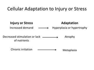
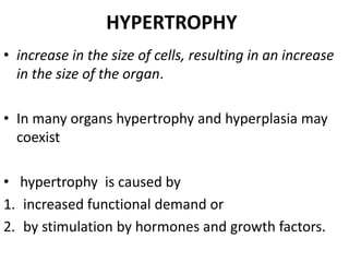
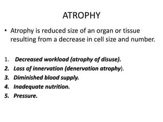
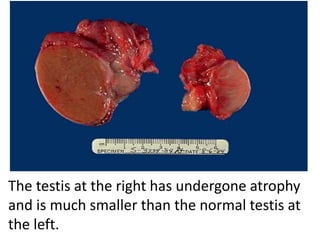
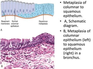
Adaptations of
Cellular Growth
and Differentiation
Cellular Adaptation to Injury or Stress
Injury or Stress
Increased demand
Decreased stimulation or lack
of nutrients
Chronic irritation
Adaptation
Hyperplasia or hypertrophy
Atrophy
Metaplasia
HYPERTROPHY
• increase in the size of cells, resulting in an increase
in the size of the organ.
• In many organs hypertrophy and hyperplasia may
coexist
• hypertrophy is caused by
1. increased functional demand or
2. by stimulation by hormones and growth factors.
• The most common stimulus for hypertrophy of
muscle is increased workload.
• the muscle cells synthesize more proteins and the
number of myofilaments increases.
• growth of the uterus during pregnancy is hormone-
induced hypertrophy .(is stimulated by estrogenic)
• Physiologic hypertrophy of the uterus during pregnancy.
• A, Gross appearance of a normal uterus (right) and a gravid uterus
(removed for postpartum bleeding) (left).
• B, Small spindle-shaped uterine smooth muscle cells from a normal
uterus, compared with
• C, large plump cells from the gravid uterus, at the same magnification.
Normal heart
Cardiac hypertrophy
HYPERPLASIA
• increase in the number of cells in an organ or tissue,
usually resulting in increased mass of the organ or
tissue.
• hyperplasia and hypertrophy frequently occur
together,
• Hyperplasia takes place if the cell population is
capable of dividing,
• .
Examples of Hyperplasia
• Endometrial hyperplasia is caused by increases in
the amount of estrogen
• Benign prostatic hyperplasia is pathologic
hyperplasia induced by responses to, androgens.
• hyperplasia is distinct from cancer, but pathologic
hyperplasia constitutes a fertile soil in which
cancerous proliferation may eventually arise.
ATROPHY
• Atrophy is reduced size of an organ or tissue
resulting from a decrease in cell size and number.
1. Decreased workload (atrophy of disuse).
2. Loss of innervation (denervation atrophy).
3. Diminished blood supply.
4. Inadequate nutrition.
5. Pressure.
• Pressure.
• Tissue compression for any length of time can
cause atrophy.
• An enlarging benign tumor can cause atrophy
in the surrounding uninvolved tissues.
• Atrophy in this setting is probably the result of
ischemic changes
The testis at the right has undergone atrophy
and is much smaller than the normal testis at
the left.
Atrophy.
A, Normal brain of a young adult.
B, Atrophy of the brain in an 82-year-old male with atherosclerotic cerebrovascular disease,
resulting in reduced blood supply.
Note that loss of brain substance narrows the gyri and widens the sulci.
METAPLASIA
• Metaplasia is a reversible change in which one
differentiated cell type is replaced by another
cell type.
• The most common epithelial metaplasia is
columnar to squamous
• as occurs in the respiratory tract in response to
chronic irritation.
• In the habitual cigarette smoker,
• the influences that predispose to metaplasia, if
persistent, may initiate malignant transformation
in metaplastic epithelium.
• Metaplasia of
columnar to
squamous
epithelium.
• A, Schematic
diagram.
• B, Metaplasia of
columnar
epithelium (left)
to squamous
epithelium
(right) in a
bronchus.

2-__cell_injury_-_adaptation.pptx

  • 1.
  • 2.
    Cellular Adaptation toInjury or Stress Injury or Stress Increased demand Decreased stimulation or lack of nutrients Chronic irritation Adaptation Hyperplasia or hypertrophy Atrophy Metaplasia
  • 3.
    HYPERTROPHY • increase inthe size of cells, resulting in an increase in the size of the organ. • In many organs hypertrophy and hyperplasia may coexist • hypertrophy is caused by 1. increased functional demand or 2. by stimulation by hormones and growth factors.
  • 4.
    • The mostcommon stimulus for hypertrophy of muscle is increased workload. • the muscle cells synthesize more proteins and the number of myofilaments increases. • growth of the uterus during pregnancy is hormone- induced hypertrophy .(is stimulated by estrogenic)
  • 5.
    • Physiologic hypertrophyof the uterus during pregnancy. • A, Gross appearance of a normal uterus (right) and a gravid uterus (removed for postpartum bleeding) (left). • B, Small spindle-shaped uterine smooth muscle cells from a normal uterus, compared with • C, large plump cells from the gravid uterus, at the same magnification.
  • 6.
  • 8.
    HYPERPLASIA • increase inthe number of cells in an organ or tissue, usually resulting in increased mass of the organ or tissue. • hyperplasia and hypertrophy frequently occur together, • Hyperplasia takes place if the cell population is capable of dividing, • .
  • 9.
    Examples of Hyperplasia •Endometrial hyperplasia is caused by increases in the amount of estrogen • Benign prostatic hyperplasia is pathologic hyperplasia induced by responses to, androgens. • hyperplasia is distinct from cancer, but pathologic hyperplasia constitutes a fertile soil in which cancerous proliferation may eventually arise.
  • 10.
    ATROPHY • Atrophy isreduced size of an organ or tissue resulting from a decrease in cell size and number. 1. Decreased workload (atrophy of disuse). 2. Loss of innervation (denervation atrophy). 3. Diminished blood supply. 4. Inadequate nutrition. 5. Pressure.
  • 11.
    • Pressure. • Tissuecompression for any length of time can cause atrophy. • An enlarging benign tumor can cause atrophy in the surrounding uninvolved tissues. • Atrophy in this setting is probably the result of ischemic changes
  • 12.
    The testis atthe right has undergone atrophy and is much smaller than the normal testis at the left.
  • 14.
    Atrophy. A, Normal brainof a young adult. B, Atrophy of the brain in an 82-year-old male with atherosclerotic cerebrovascular disease, resulting in reduced blood supply. Note that loss of brain substance narrows the gyri and widens the sulci.
  • 15.
    METAPLASIA • Metaplasia isa reversible change in which one differentiated cell type is replaced by another cell type.
  • 16.
    • The mostcommon epithelial metaplasia is columnar to squamous • as occurs in the respiratory tract in response to chronic irritation. • In the habitual cigarette smoker, • the influences that predispose to metaplasia, if persistent, may initiate malignant transformation in metaplastic epithelium.
  • 17.
    • Metaplasia of columnarto squamous epithelium. • A, Schematic diagram. • B, Metaplasia of columnar epithelium (left) to squamous epithelium (right) in a bronchus.