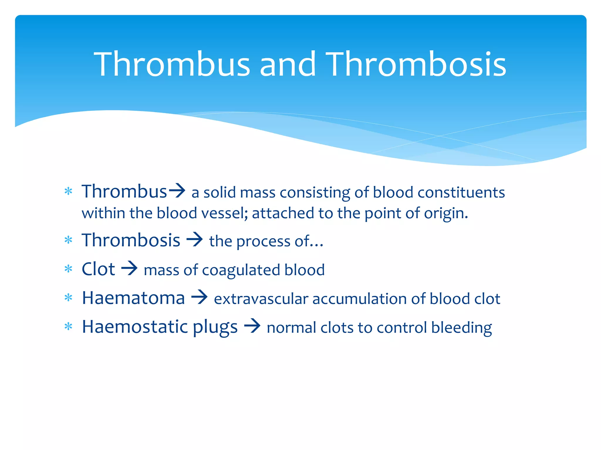
Thrombosis And Embolism
Dr. Saugat Chapagain
Thrombosis
 Thrombus a solid mass consisting of blood constituents
within the blood vessel; attached to the point of origin.
 Thrombosis  the process of…
 Clot  mass of coagulated blood
 Haematoma  extravascular accumulation of blood clot
 Haemostatic plugs  normal clots to control bleeding
Thrombus and Thrombosis
 Virchow’s Triad
 Endothelial injury
 Altered blood flow (stasis/ turbulence)
 Hypercoagulability of blood
Pathophysiology
Coagulation cascade
 Vascular injury exposes subendothelial thrombogenic
connective tissues (collagen, elastin, fibronectin,
laminin and glycosaminoglycans)
 Initial vasoconstriction to reduce blood loss.
 Tissue factors initiate extrinsic pathway
 Collagen initiate intrinsic pathway.
Endothelial injury
 Ulcerated plaque in
advanced atherosclerosis.
 Haemodynamic stress in HTN
 Arterial diseases
 Diabetes mellitus
 Endogenous chemical
agents
(hypercholesterolemia)
 Exogenous chemical agents
(smoking)
 TAO
 Bacterial toxins
 Trauma
 IV cannulation
 Radiation injury
 Turbulent flow of blood in
aneurysm
 Mural thrombus following
cardiac abnormality
Causes of endothelial injury
 Turbulence: unequal flow (arterial); may aggravate
vascular injury as well.
 Stasis: slowing (venous)
 Blood cells and platelets migrate to periphery and
pavement the endothelium
 Normal flow has:
 Central fast moving red and white blood cells.
 Slow moving laminar stream of platelets.
 Peripheral slowest moving cell-free plasma.
Alteration of blood flow
Effects:
 Prevent dilution by fresh flowing blood of activated clotting
factors.
 Disrupt laminar blood flow and bring platelets in contact of
endothelium
 Retard the inflow of clotting factor inhibitors and permit
thrombosis
 Promote endothelial cell activation
Examples:
 DVT of lower limbs
 Mural thrombus
 Thrombosis in aneurism
Favoured by:
 Smoking
 Ageing
 Use of OCPs
 Obesity
Causes:
 Increased coagulation factors
 Increased platelet count and adhesiveness
 Decreased levels of coagulation inhibitors
Hypercoagulability
Primary (genetic)
 Deficiency of anti thrombin
 Deficiency of protein C or S
 Defects in fibrinolysis
 Mutation of factor V
Secondary (acquired)
 Risk factors:
 Advanced age
 Sedentary life style
 Immobilization
 Smoking
 Clinical conditions leading to
thrombosis:
 Heart diseases (MI, CHF, RHD)
 Vascular diseases
(atherosclerosis, aneurysms,
varicosities)
 Hypercoagulibility
(polycythemia, dehydration,
nephrotic syndrome)
 Shock
 Late pregnancy and puerperium
 drugs
Factors favoring thrombosis
 Gross:
 Shape and size depends on origin
 Arterial  white and mural (firm and pale)
 Venous  red and occlusive(soft, red and gelatinous)
 Mixed/ laminated – alt white and red layers separated by
line of ZAHN.
 Microscopic:
 Lines of Zahn seen where visible
 Collection of cells and fibrin
Morphology
Based on colour and components:
 Pale – primarily platelets
 Red – primarily RBCs
 Mixed – visible lines of zahn
Based on site and mode of formation:
 Occlusive
 Mural – occurs in heart chambers/ aorta/ major arteries
Based on infection:
 Bland – non infected
 Septic – infected.
Types
 Resolution – plasmin may dissolve the thrombus
completely
 Organization – eventually covered by endothelial cells
after granulation tissue formation (or calcification/
ossification)
 Propagation – enlargement of size may block
important vessels.
 Thromboembolism – detachment and released into
blood stream
Fate of thrombus
Embolism
 The process of partial or complete obstruction of
some part of the CVS by any mass carried in the
circulation.
 Commonly thromboembolism (90%)
What is it?
 Depending on matter of embolus
 Solid- thrombus, atheroma, malignant cell, parasites, foreign
bodies.
 Liquid- fat globules, amniotic fluid, bone marrow
 Gasseous- air, other gasses
 Depending on infection:
 Bland
 Septic
 Depending on source of emboli:
 Cardiac emboli from left side of the heart – vegetations of
endocarditis, atrial appendages
 Arterial – in systemic arteries of brain, spleen, kidney and
intestine.
 Venous – in pulmonary arteries
 Lymphatic emboli
TYPE
 A detached thrombus or a part of thrombus.
 Site of lodgement:
 Arterial (systemic)
 Venous
 Pulmonary
THROMBOEMBOLISM
 Most common and fatal form.
 Occlusion of the pulmonary arterial tree.
 Pulmonary thrombosis (rare)
ETIOLOGY
 More common in bed ridden patients.
Causes:
 Thrombi originating from large veins of lower legs
(popliteal, femoral and iliac)
 Less common sources ( varicosities of superficial veins,
pelvic veins)
PULMONARY THROMBOEMBOLISM
Detachment of thrombus  thromboembolism 
drains into right atrium.
 Large thrombus is impacted at the bifurcation of main
pulmonary artery (saddle embolus) or on RV or its
outflow tract.
 If large embolus fragments  small embolus impacts
in a number of vessels (esp. on lower lobes of lungs)
 Paradoxical embolism – passage of embolus from rt.
heart to lt. heart via ASD or VSD.
Pathogenesis
 Sudden death – d/t massive pulm. Embolism.
 Acute cor pulmonale – RHF d/t pulmonary HTN
 Pulm. Infarction – obstruction of small pulm. arteries.
 Pulm. Haemorrhage – central pulm. Haemorrhage d/t obstruction of
flow in endarteries.
 Resolution – 60-80 % by fibrinolysis.
 Pulm. HTN, chronic cor-pulmonale and pulm.
Atreriosclerosis.
Consequences
 Arterial embolism
 Esp. originating in LV (MI, cardiomyopathy, RHD,
congenital heart disease, IE, prosthetic valves)
Site of origin:
 Heart (80%)
 Aorta – thrombi over ulcerated atheroma in aortic
aneurysm
 Paradoxical – rt.  lt. in ASD or VSD
 Idiopathic – 10-15%
Systemic thromboembolism
 Infarction – kidney, spleen, mesentery
 Stroke – brain (10%)
 Upper and lower extremeties- peripheral vascular diseases,
gangrene, etc.
Effects
 Embolism of fat globules.
 Obstruction by fragments of adipose tissue  fat
tissue embolism.
Etiology
 Traumatic –
 Trauma to bones, trauma to soft tissue
 Non traumatic –
 Burns, DM, fatty liver, pancreatitis, decompression
sickness.
Fat embolism
 Mechanical obstruction – micro aggregates of fat
causes obstruction of vessels.
 Chemmical injury – FFA causes toxic injury.
 Thrombocytopenia – d/t coating of fat globules with
platelets.
Consequences
 Pulmonary fat embolism
 Systemic fat embolism (in case of ASD/ VSD)
Pathogenesis
 Air, nitrogen and other gases,
 Air embolism – entry of air due to trauma, injury or surgery.
 Decompression sickness/ caisson’s disease, diver’s palsy or
aeroembolism seen in deep sea divers.
Causes:
 During delivery or abortion
 Acquired/ accidental pneumothorax
 Haemodialysis
 Accidental opening of large veins
 Angiography
 Cardiothoracic surgery / trauma
Gas Embolism
 Most serious, unpredictable and unpreventable cause of
maternal mortality.
 Amniotic fluid may contain – epithelial squames, vernix caseosa,
lanugo hair, bile from meconium and mucus
s/s
 Sudden respiratory distress and dyspnoea
 Deep cyanosis
 CV shock
 Convulsions
 Coma
 Unexpected death
Amniotic fluid embolism
 Mechanical blockage of pulmonary circulation in
extensive embolism
 Anaphylactic reaction to components
 DIC d/t liberation of thromboplastin
 Haemorrhagic manifestations d/t thrombocytopenia
and afibrinogenaemia.
Cause of death
Thank you

5. thrombosis and embolism

  • 1.
  • 2.
  • 3.
     Thrombus asolid mass consisting of blood constituents within the blood vessel; attached to the point of origin.  Thrombosis  the process of…  Clot  mass of coagulated blood  Haematoma  extravascular accumulation of blood clot  Haemostatic plugs  normal clots to control bleeding Thrombus and Thrombosis
  • 4.
     Virchow’s Triad Endothelial injury  Altered blood flow (stasis/ turbulence)  Hypercoagulability of blood Pathophysiology
  • 5.
  • 7.
     Vascular injuryexposes subendothelial thrombogenic connective tissues (collagen, elastin, fibronectin, laminin and glycosaminoglycans)  Initial vasoconstriction to reduce blood loss.  Tissue factors initiate extrinsic pathway  Collagen initiate intrinsic pathway. Endothelial injury
  • 8.
     Ulcerated plaquein advanced atherosclerosis.  Haemodynamic stress in HTN  Arterial diseases  Diabetes mellitus  Endogenous chemical agents (hypercholesterolemia)  Exogenous chemical agents (smoking)  TAO  Bacterial toxins  Trauma  IV cannulation  Radiation injury  Turbulent flow of blood in aneurysm  Mural thrombus following cardiac abnormality Causes of endothelial injury
  • 9.
     Turbulence: unequalflow (arterial); may aggravate vascular injury as well.  Stasis: slowing (venous)  Blood cells and platelets migrate to periphery and pavement the endothelium  Normal flow has:  Central fast moving red and white blood cells.  Slow moving laminar stream of platelets.  Peripheral slowest moving cell-free plasma. Alteration of blood flow
  • 10.
    Effects:  Prevent dilutionby fresh flowing blood of activated clotting factors.  Disrupt laminar blood flow and bring platelets in contact of endothelium  Retard the inflow of clotting factor inhibitors and permit thrombosis  Promote endothelial cell activation Examples:  DVT of lower limbs  Mural thrombus  Thrombosis in aneurism
  • 11.
    Favoured by:  Smoking Ageing  Use of OCPs  Obesity Causes:  Increased coagulation factors  Increased platelet count and adhesiveness  Decreased levels of coagulation inhibitors Hypercoagulability
  • 12.
    Primary (genetic)  Deficiencyof anti thrombin  Deficiency of protein C or S  Defects in fibrinolysis  Mutation of factor V Secondary (acquired)  Risk factors:  Advanced age  Sedentary life style  Immobilization  Smoking  Clinical conditions leading to thrombosis:  Heart diseases (MI, CHF, RHD)  Vascular diseases (atherosclerosis, aneurysms, varicosities)  Hypercoagulibility (polycythemia, dehydration, nephrotic syndrome)  Shock  Late pregnancy and puerperium  drugs Factors favoring thrombosis
  • 13.
     Gross:  Shapeand size depends on origin  Arterial  white and mural (firm and pale)  Venous  red and occlusive(soft, red and gelatinous)  Mixed/ laminated – alt white and red layers separated by line of ZAHN.  Microscopic:  Lines of Zahn seen where visible  Collection of cells and fibrin Morphology
  • 14.
    Based on colourand components:  Pale – primarily platelets  Red – primarily RBCs  Mixed – visible lines of zahn Based on site and mode of formation:  Occlusive  Mural – occurs in heart chambers/ aorta/ major arteries Based on infection:  Bland – non infected  Septic – infected. Types
  • 15.
     Resolution –plasmin may dissolve the thrombus completely  Organization – eventually covered by endothelial cells after granulation tissue formation (or calcification/ ossification)  Propagation – enlargement of size may block important vessels.  Thromboembolism – detachment and released into blood stream Fate of thrombus
  • 16.
  • 17.
     The processof partial or complete obstruction of some part of the CVS by any mass carried in the circulation.  Commonly thromboembolism (90%) What is it?
  • 18.
     Depending onmatter of embolus  Solid- thrombus, atheroma, malignant cell, parasites, foreign bodies.  Liquid- fat globules, amniotic fluid, bone marrow  Gasseous- air, other gasses  Depending on infection:  Bland  Septic  Depending on source of emboli:  Cardiac emboli from left side of the heart – vegetations of endocarditis, atrial appendages  Arterial – in systemic arteries of brain, spleen, kidney and intestine.  Venous – in pulmonary arteries  Lymphatic emboli TYPE
  • 19.
     A detachedthrombus or a part of thrombus.  Site of lodgement:  Arterial (systemic)  Venous  Pulmonary THROMBOEMBOLISM
  • 20.
     Most commonand fatal form.  Occlusion of the pulmonary arterial tree.  Pulmonary thrombosis (rare) ETIOLOGY  More common in bed ridden patients. Causes:  Thrombi originating from large veins of lower legs (popliteal, femoral and iliac)  Less common sources ( varicosities of superficial veins, pelvic veins) PULMONARY THROMBOEMBOLISM
  • 21.
    Detachment of thrombus thromboembolism  drains into right atrium.  Large thrombus is impacted at the bifurcation of main pulmonary artery (saddle embolus) or on RV or its outflow tract.  If large embolus fragments  small embolus impacts in a number of vessels (esp. on lower lobes of lungs)  Paradoxical embolism – passage of embolus from rt. heart to lt. heart via ASD or VSD. Pathogenesis
  • 22.
     Sudden death– d/t massive pulm. Embolism.  Acute cor pulmonale – RHF d/t pulmonary HTN  Pulm. Infarction – obstruction of small pulm. arteries.  Pulm. Haemorrhage – central pulm. Haemorrhage d/t obstruction of flow in endarteries.  Resolution – 60-80 % by fibrinolysis.  Pulm. HTN, chronic cor-pulmonale and pulm. Atreriosclerosis. Consequences
  • 23.
     Arterial embolism Esp. originating in LV (MI, cardiomyopathy, RHD, congenital heart disease, IE, prosthetic valves) Site of origin:  Heart (80%)  Aorta – thrombi over ulcerated atheroma in aortic aneurysm  Paradoxical – rt.  lt. in ASD or VSD  Idiopathic – 10-15% Systemic thromboembolism
  • 24.
     Infarction –kidney, spleen, mesentery  Stroke – brain (10%)  Upper and lower extremeties- peripheral vascular diseases, gangrene, etc. Effects
  • 25.
     Embolism offat globules.  Obstruction by fragments of adipose tissue  fat tissue embolism. Etiology  Traumatic –  Trauma to bones, trauma to soft tissue  Non traumatic –  Burns, DM, fatty liver, pancreatitis, decompression sickness. Fat embolism
  • 26.
     Mechanical obstruction– micro aggregates of fat causes obstruction of vessels.  Chemmical injury – FFA causes toxic injury.  Thrombocytopenia – d/t coating of fat globules with platelets. Consequences  Pulmonary fat embolism  Systemic fat embolism (in case of ASD/ VSD) Pathogenesis
  • 27.
     Air, nitrogenand other gases,  Air embolism – entry of air due to trauma, injury or surgery.  Decompression sickness/ caisson’s disease, diver’s palsy or aeroembolism seen in deep sea divers. Causes:  During delivery or abortion  Acquired/ accidental pneumothorax  Haemodialysis  Accidental opening of large veins  Angiography  Cardiothoracic surgery / trauma Gas Embolism
  • 28.
     Most serious,unpredictable and unpreventable cause of maternal mortality.  Amniotic fluid may contain – epithelial squames, vernix caseosa, lanugo hair, bile from meconium and mucus s/s  Sudden respiratory distress and dyspnoea  Deep cyanosis  CV shock  Convulsions  Coma  Unexpected death Amniotic fluid embolism
  • 29.
     Mechanical blockageof pulmonary circulation in extensive embolism  Anaphylactic reaction to components  DIC d/t liberation of thromboplastin  Haemorrhagic manifestations d/t thrombocytopenia and afibrinogenaemia. Cause of death
  • 30.