Downloaded 177 times

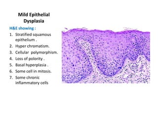
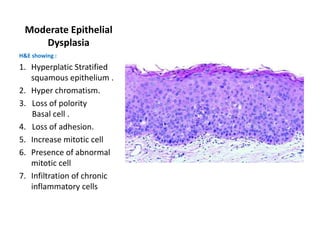
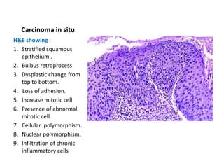
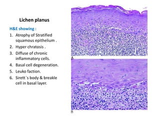
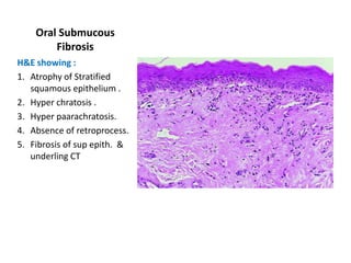

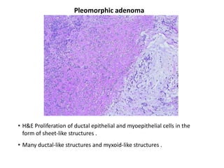
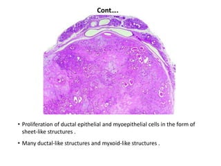
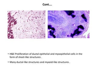
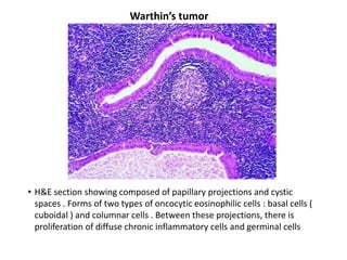
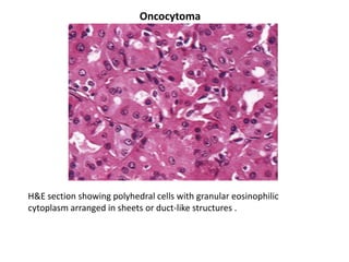
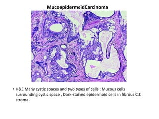
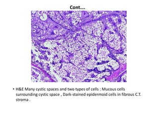
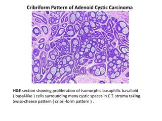
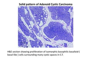
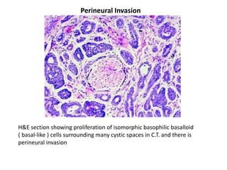
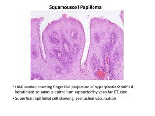


The document describes the histological features of various oral lesions seen on hematoxylin and eosin staining of biopsy specimens. These include premalignant lesions like mild and moderate epithelial dysplasia and carcinoma in situ. Other conditions described are lichen planus, oral submucous fibrosis, salivary gland diseases like necrotizing sialometaplasia and chronic sialadenitis, benign salivary gland tumors like pleomorphic adenoma, Warthin's tumor and oncocytoma. Malignant salivary gland tumors discussed include mucoepidermoid carcinoma and adenoid cystic carcinoma. Features of perineural invasion are also highlighted. Finally benign and malignant oral neoplasms like


![Neoplasia [part 1]](https://cdn.slidesharecdn.com/ss_thumbnails/neoplasiapart1-190918152450-thumbnail.jpg?width=640&height=640&fit=bounds)


