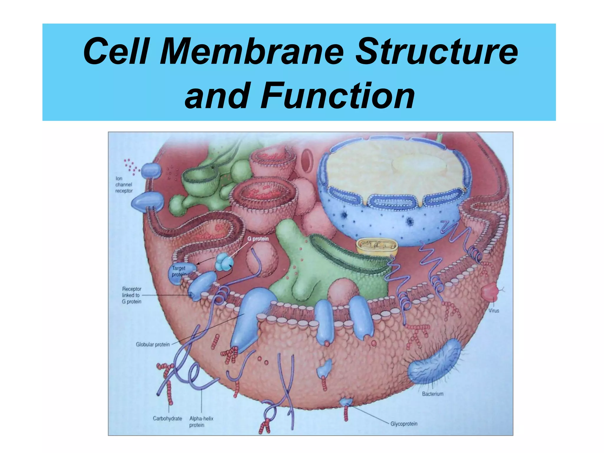
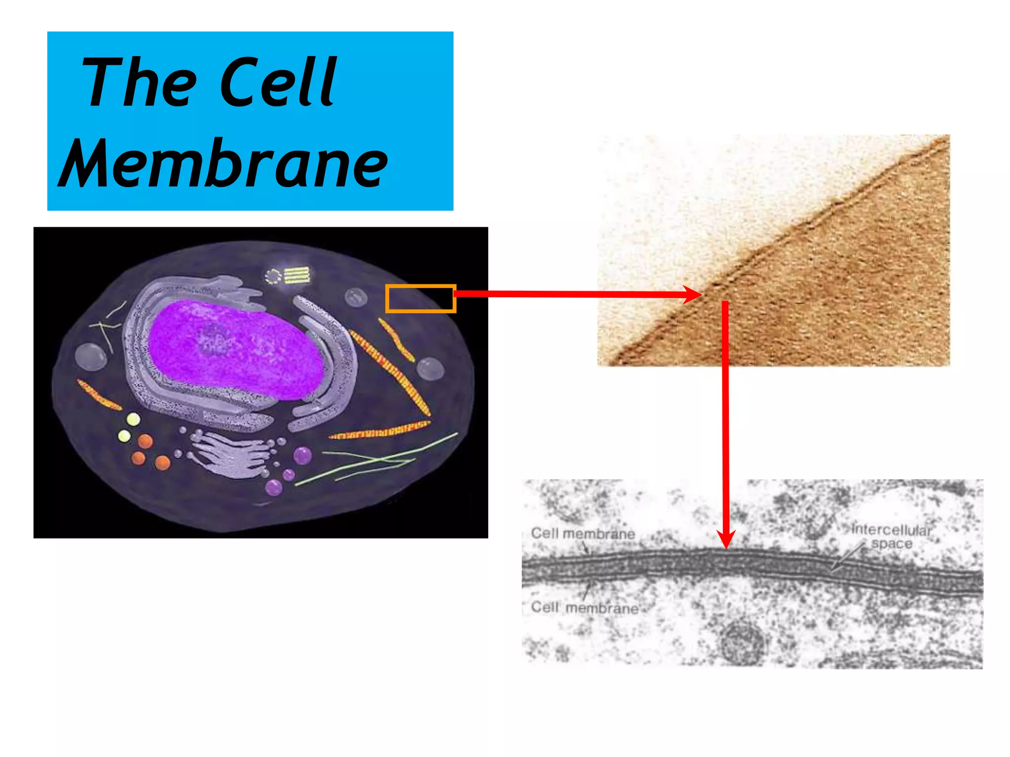
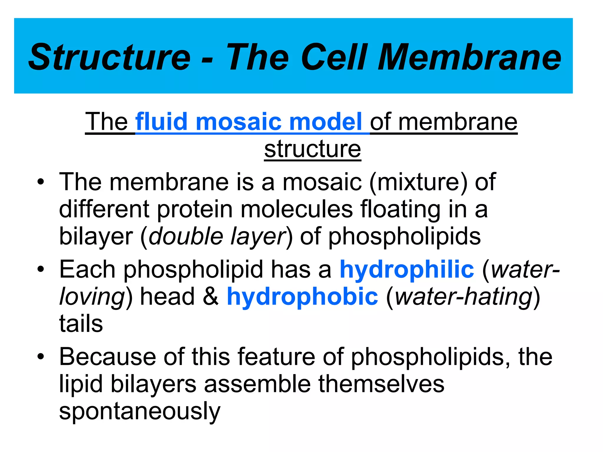
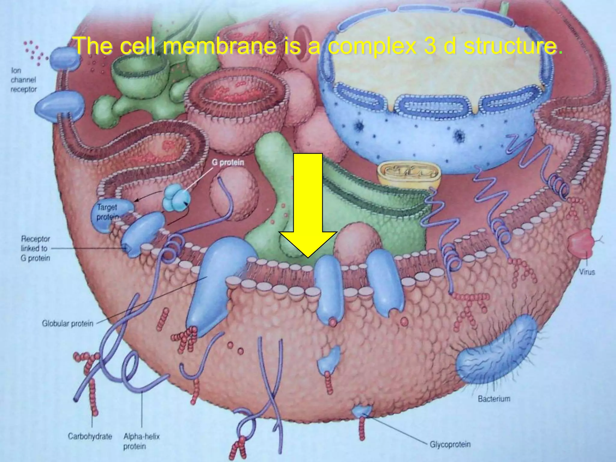
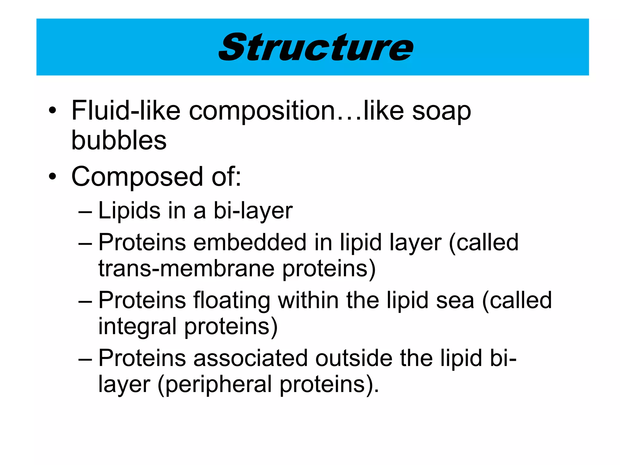
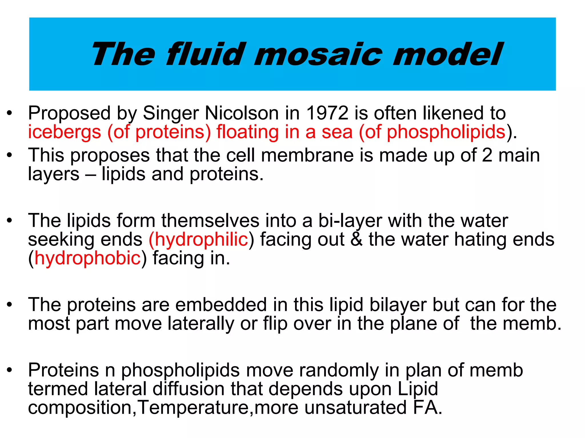
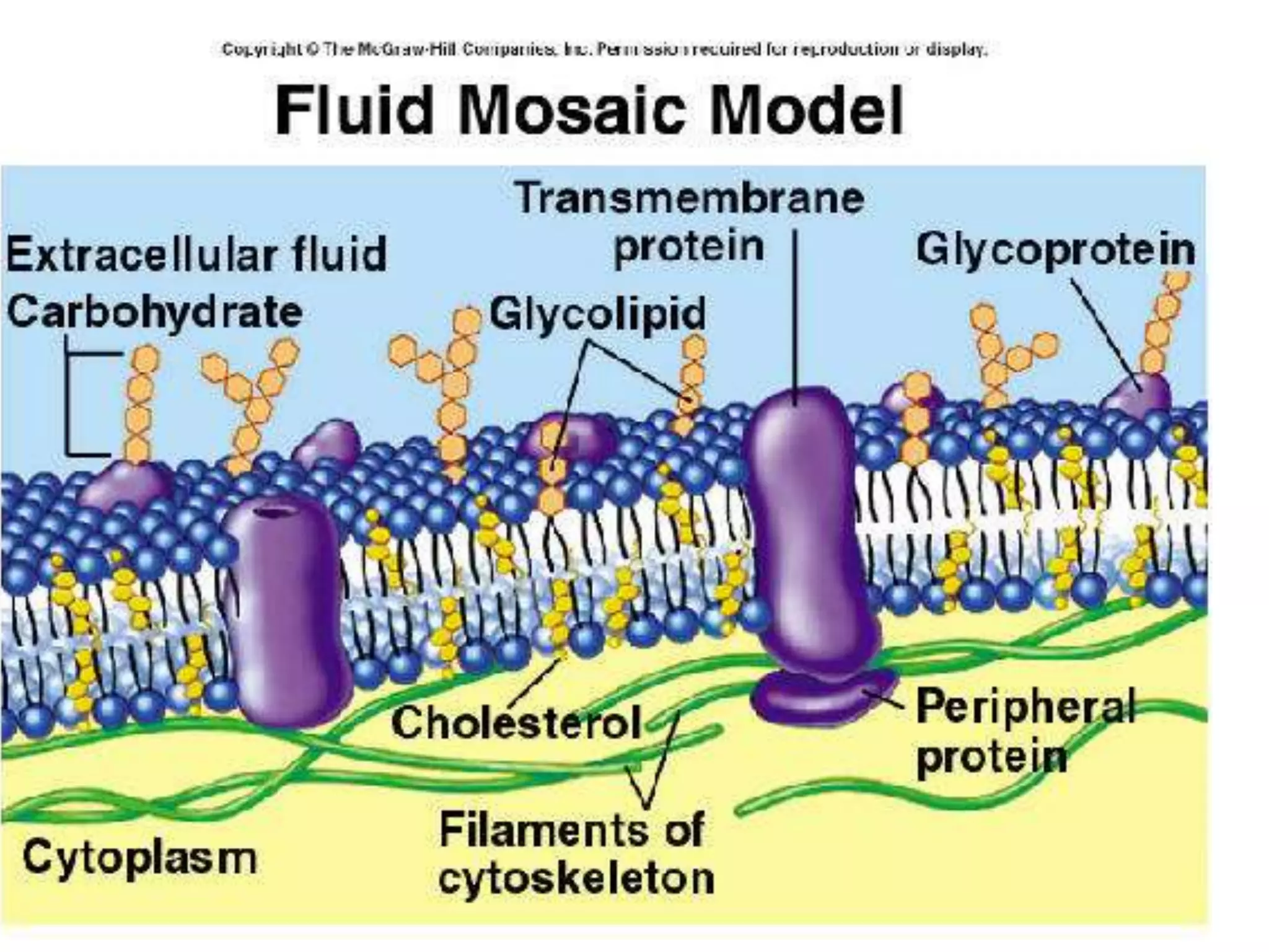
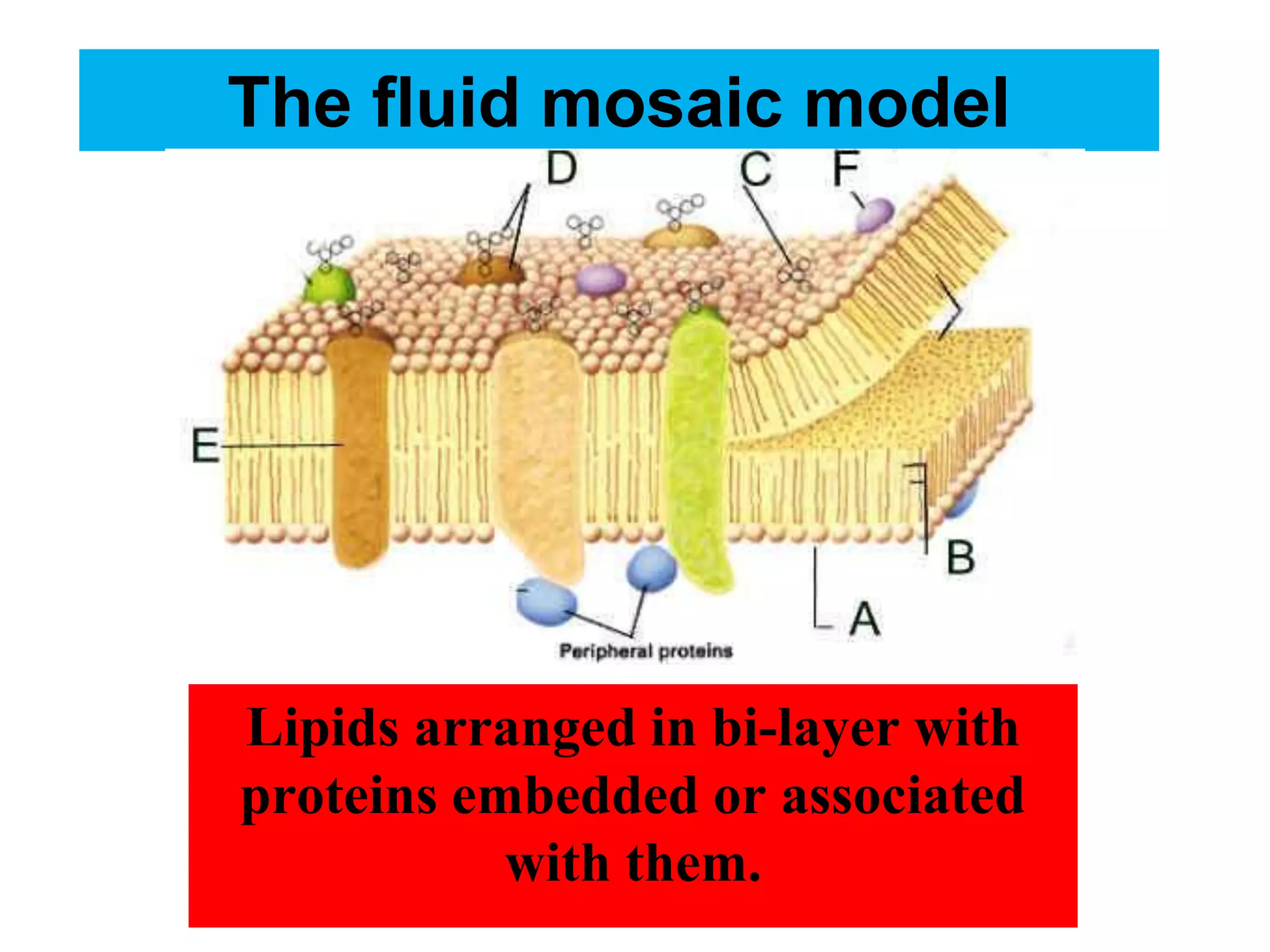
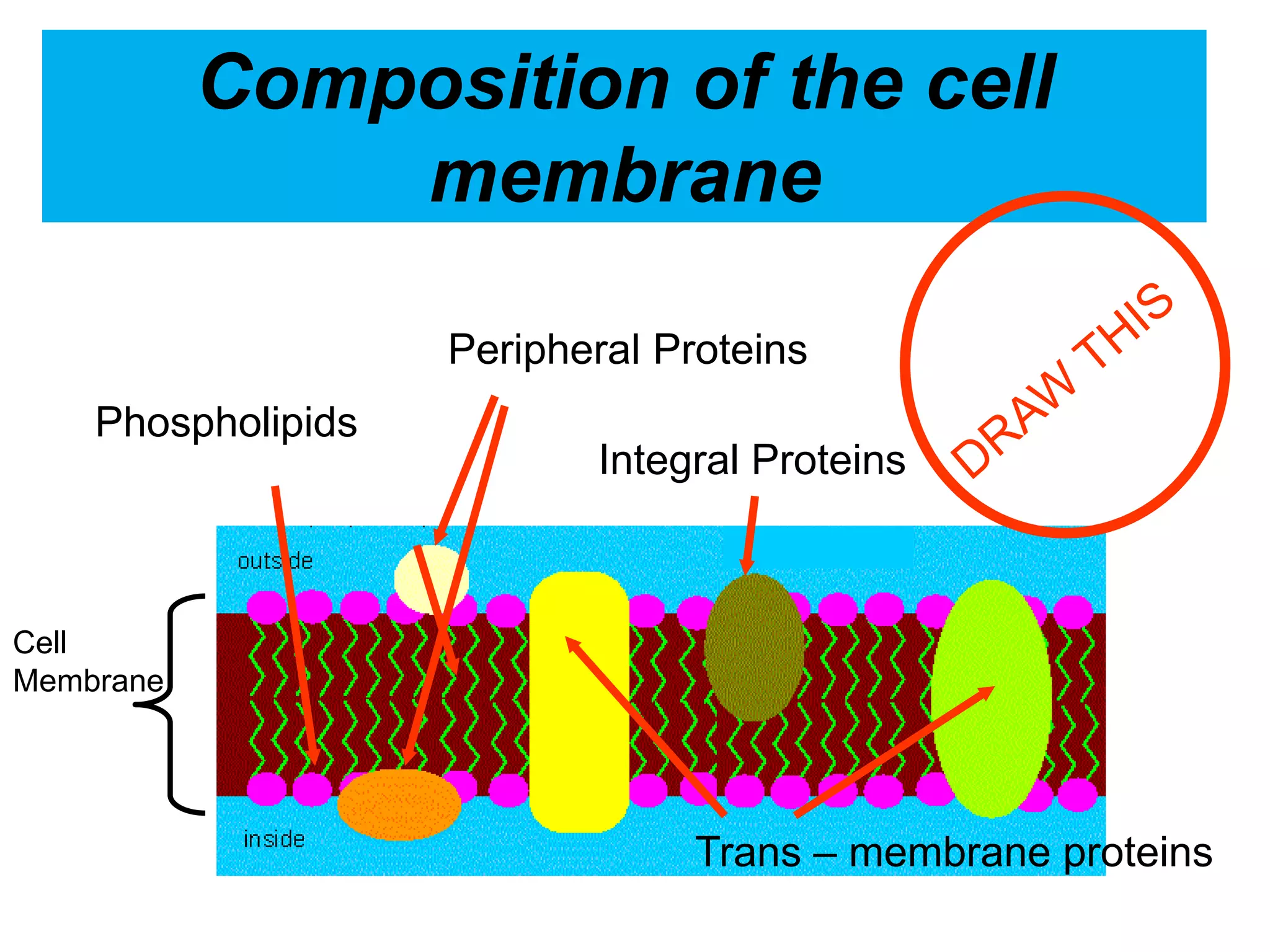
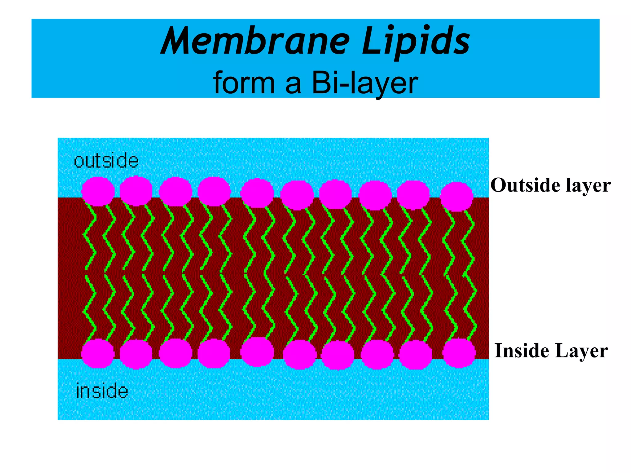
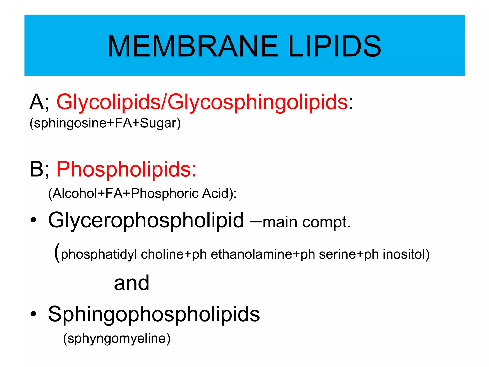
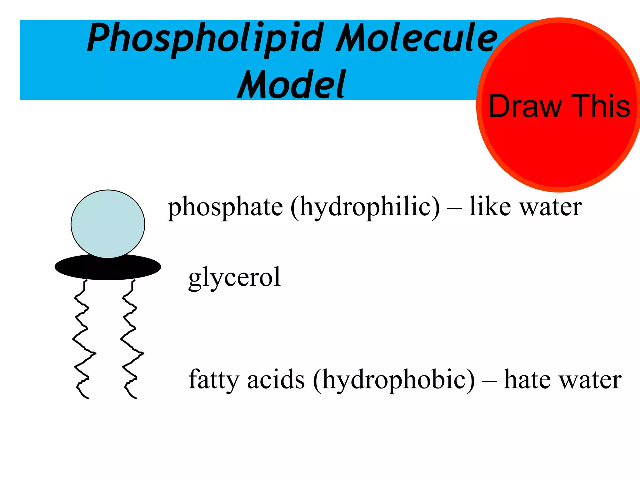
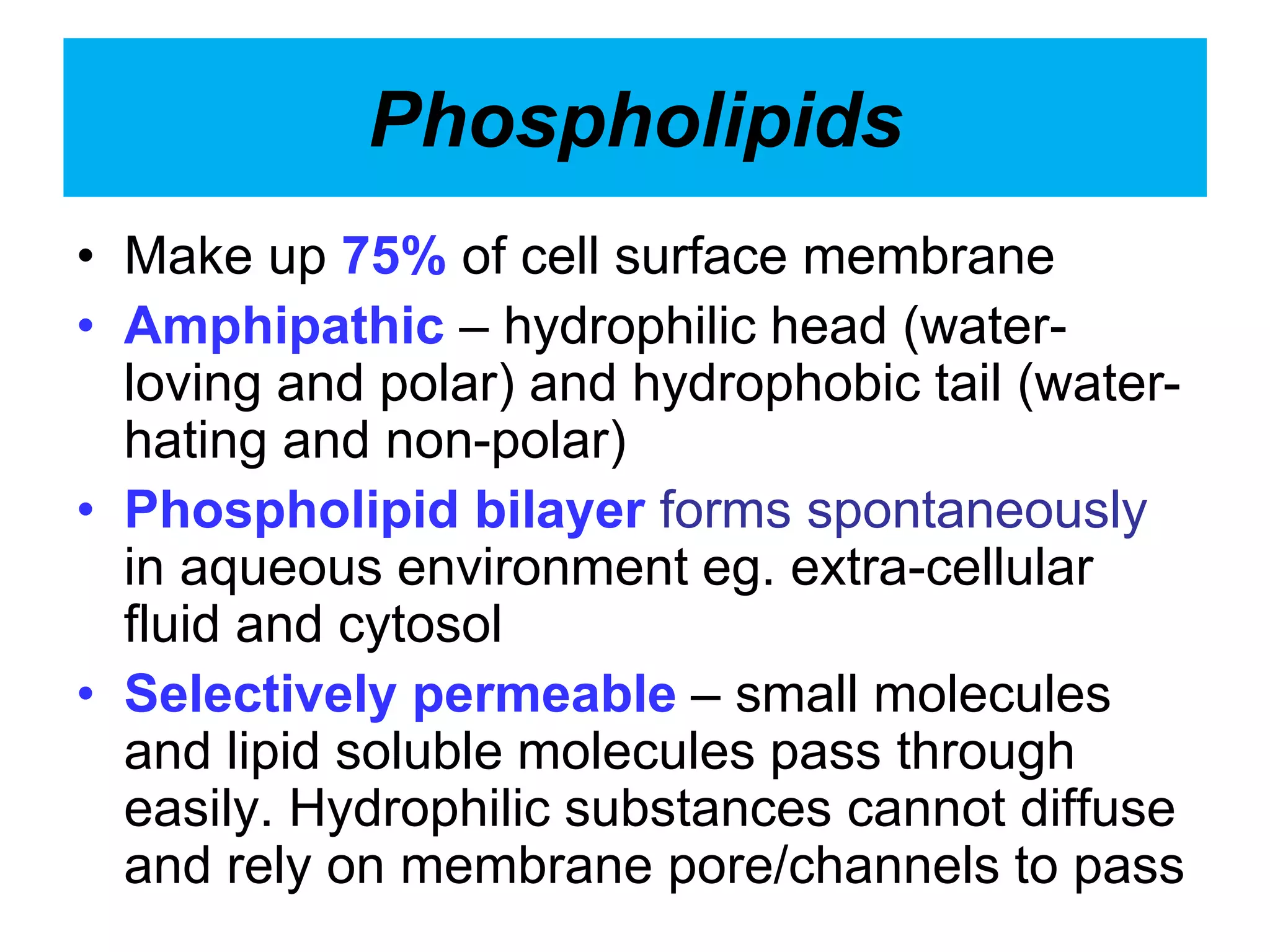
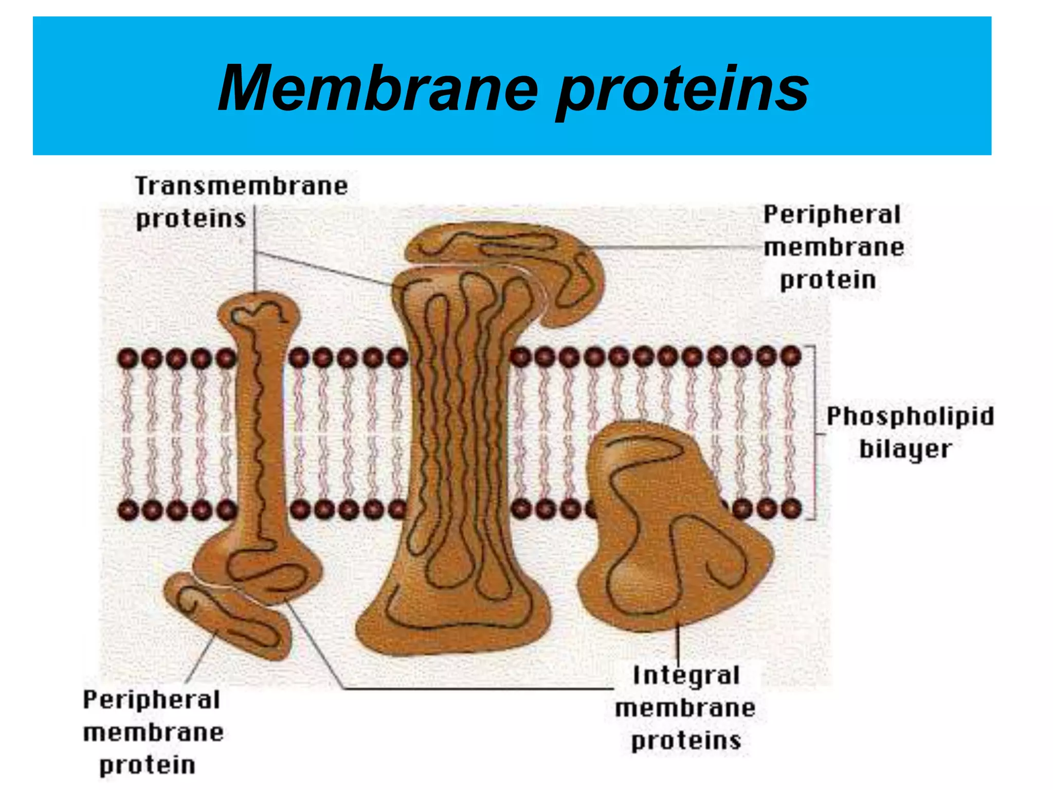
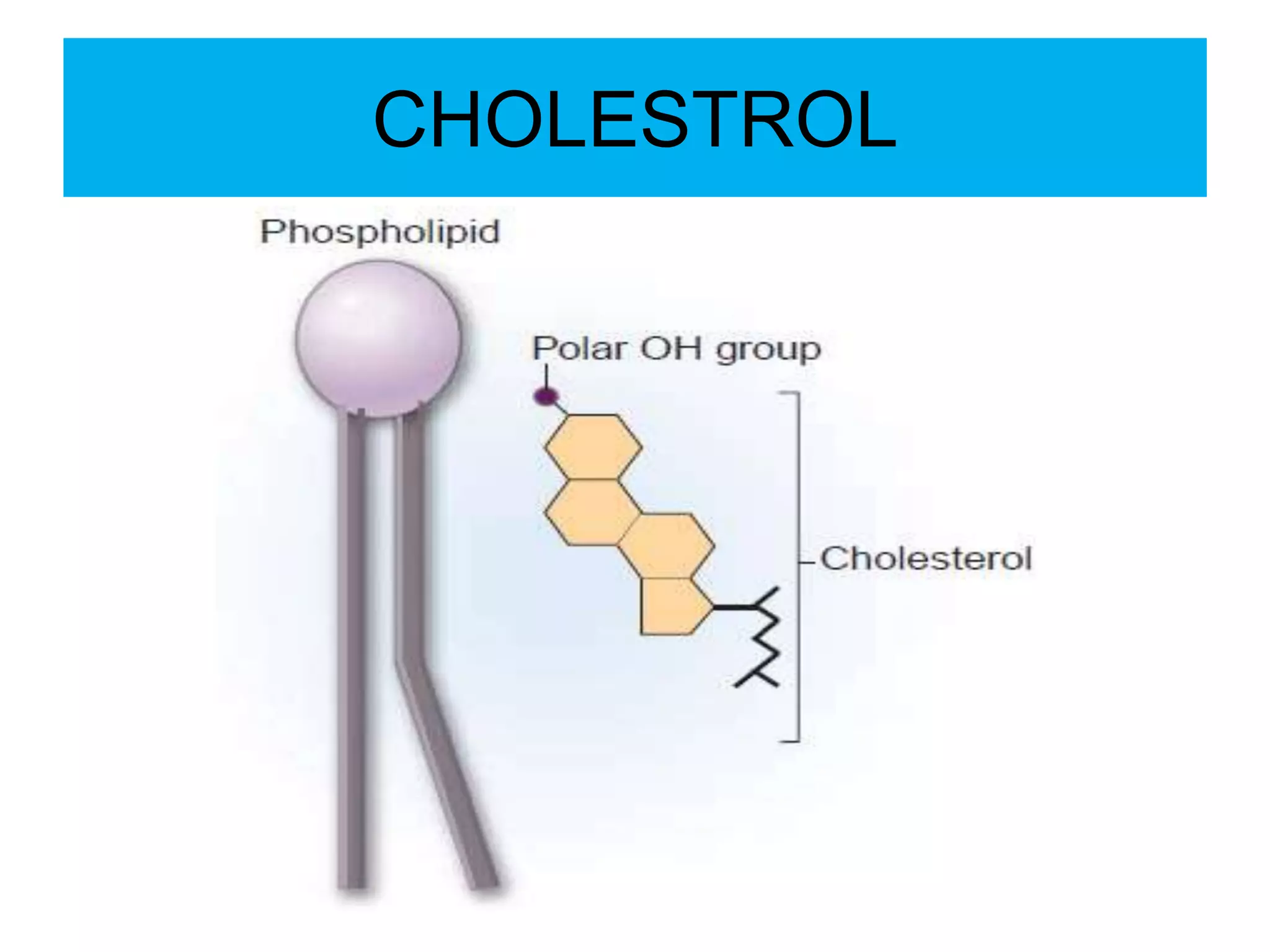
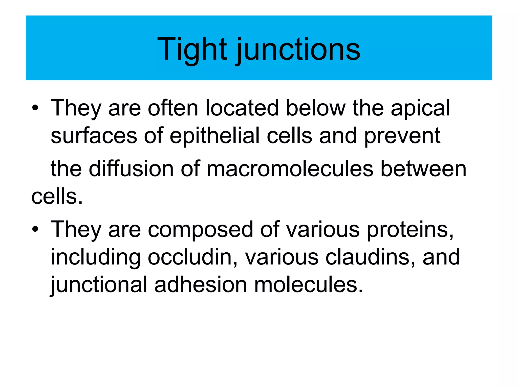
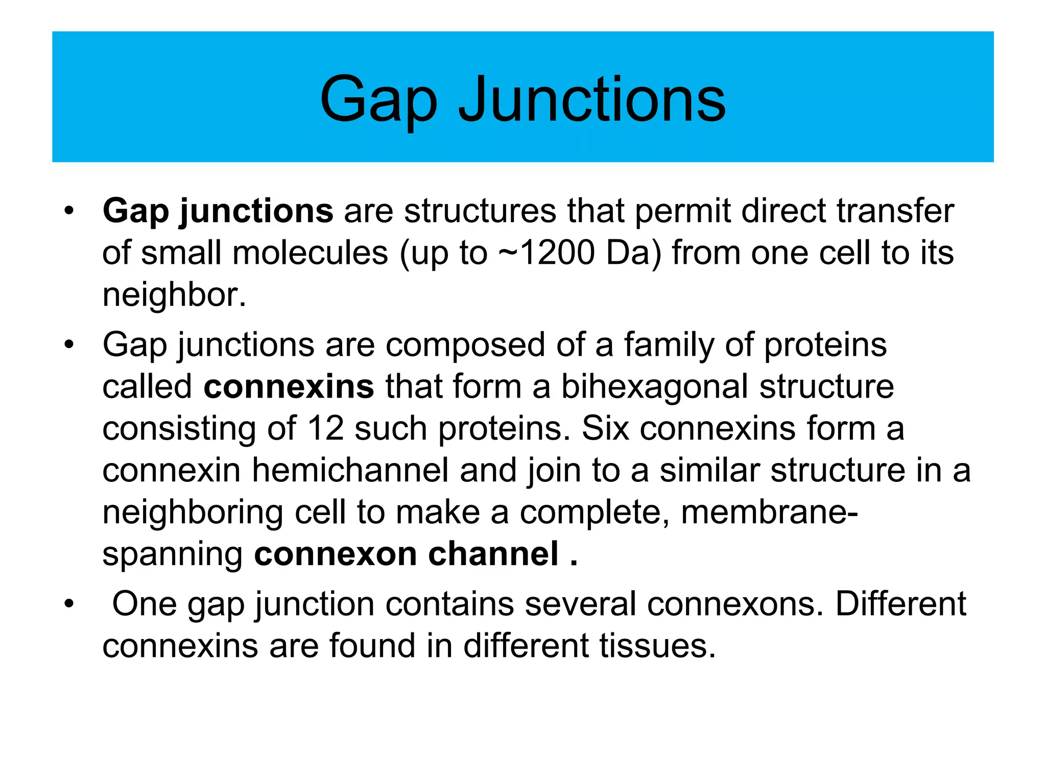
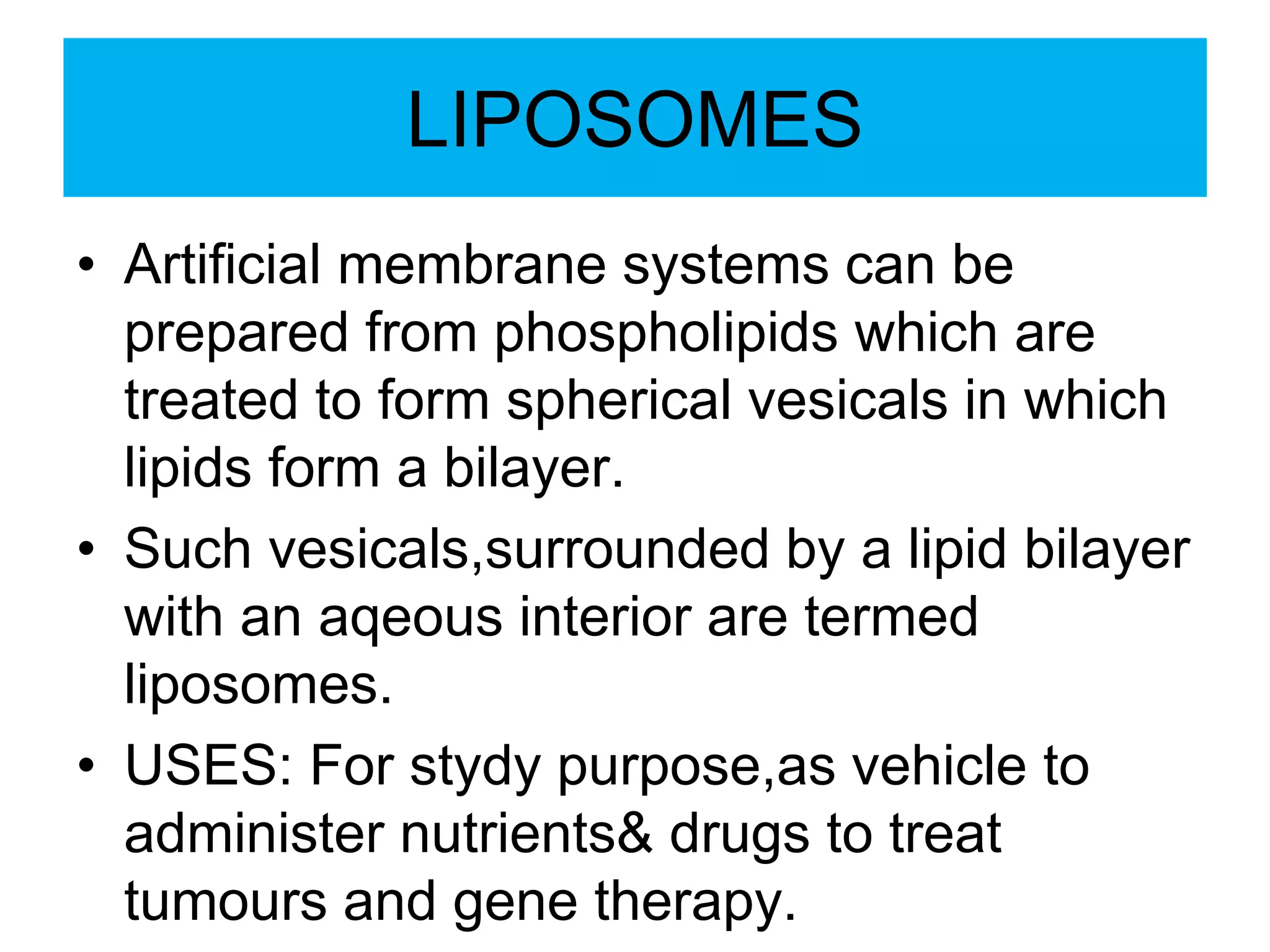
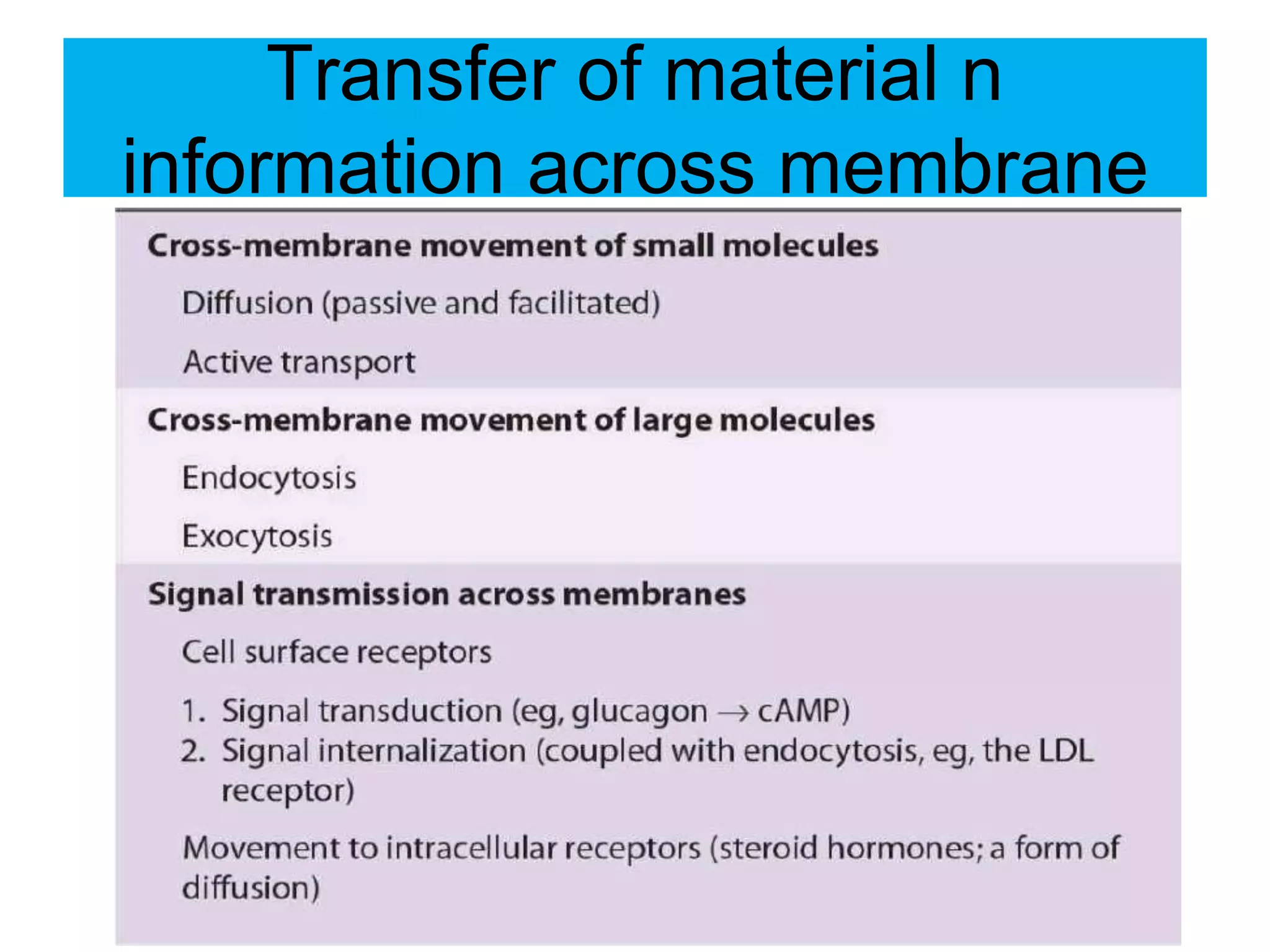
Cell Membrane Structure
and Function
Cell Membrane
• Every cell is encircled by a membrane and most cells
contain an extensive intracellular membrane system.
• Selective Barrier: Membranes fence off the cell's
interior from its surroundings (maintain extracellular
Na,Ca,Cl,HCO3& intracellular K.Mg,P,levels) .
• Transport: Membranes let in water, certain ions and
substrates and they excrete waste substances.
• Key role: in Cell-cell interaction and transmembrane
signalling
• Without a membrane: The cell contents would
diffuse into the surroundings, information containing
molecules would be lost and many metabolic
pathways would cease to work.
Membrane Function-transport
• Sometimes the elements bind to the
proteins, which flip over, thus transporting
the element into the cell.
• Some proteins form a ‘pore’ through which
the element can pass from the outside to
the inside of the cell membrane.
• The movement of the phospholipid and
protein components through the plasma
membrane permits the membrane to
change shape—this is known as fluidity.
• This flexibility is crucial to many different
types of cells.
Organelles Membrane
The cell is highly organized with many
functional units or organelles inside.
Each organelles is surrounded by a
membrane that contains specific proteins
and lipid components that enable it to
perform its unique roles.e.g.
Help shape organelles-
Site of energy transduction-mitochondria
Do excitation-response coupling-nerves
Localize enzymes-lysosomes
In essence membranes are essential for the
integrity and function of the cell.
The Cell
Membrane
Structure - The Cell Membrane
The fluid mosaic model of membrane
structure
• The membrane is a mosaic (mixture) of
different protein molecules floating in a
bilayer (double layer) of phospholipids
• Each phospholipid has a hydrophilic (water-
loving) head & hydrophobic (water-hating)
tails
• Because of this feature of phospholipids, the
lipid bilayers assemble themselves
spontaneously
The cell membrance is a complex
3d circular structure
The cell membrane is a complex 3 d structure.
Structure
• Fluid-like composition…like soap
bubbles
• Composed of:
– Lipids in a bi-layer
– Proteins embedded in lipid layer (called
trans-membrane proteins)
– Proteins floating within the lipid sea (called
integral proteins)
– Proteins associated outside the lipid bi-
layer (peripheral proteins).
The fluid mosaic model
• Proposed by Singer Nicolson in 1972 is often likened to
icebergs (of proteins) floating in a sea (of phospholipids).
• This proposes that the cell membrane is made up of 2 main
layers – lipids and proteins.
• The lipids form themselves into a bi-layer with the water
seeking ends (hydrophilic) facing out & the water hating ends
(hydrophobic) facing in.
• The proteins are embedded in this lipid bilayer but can for the
most part move laterally or flip over in the plane of the memb.
• Proteins n phospholipids move randomly in plan of memb
termed lateral diffusion that depends upon Lipid
composition,Temperature,more unsaturated FA.
The fluid mosaic model
Lipids arranged in bi-layer with
proteins embedded or associated
with them.
Composition of the cell
membrane
Phospholipids
Peripheral Proteins
Integral Proteins
Trans – membrane proteins
Cell
Membrane
Membrane Lipids
form a Bi-layer
Outside layer
Inside Layer
MEMBRANE LIPIDS
A; Glycolipids/Glycosphingolipids:
(sphingosine+FA+Sugar)
B; Phospholipids:
(Alcohol+FA+Phosphoric Acid):
• Glycerophospholipid –main compt.
(phosphatidyl choline+ph ethanolamine+ph serine+ph inositol)
and
• Sphingophospholipids
(sphyngomyeline)
Phospholipid Molecule
Model
glycerol
fatty acids (hydrophobic) – hate water
phosphate (hydrophilic) – like water
Draw This
• In outer memb mainly:
Phosphatidylcholin n Sphingomyeline
• In inner memb mainly:
Aminophospholipids
Phosphotidylinositol (transfer information)
Phospholipids
• Make up 75% of cell surface membrane
• Amphipathic – hydrophilic head (water-
loving and polar) and hydrophobic tail (water-
hating and non-polar)
• Phospholipid bilayer forms spontaneously
in aqueous environment eg. extra-cellular
fluid and cytosol
• Selectively permeable – small molecules
and lipid soluble molecules pass through
easily. Hydrophilic substances cannot diffuse
and rely on membrane pore/channels to pass
Membrane proteins
TRANSMEMBRANE PROTEIN
• Integral proteins/Transmembrane proteins
are most common,interact externally with
phospholipids n require detergents for their
separation.
These span the bilayer as a bundle of α-helical
once o or many times ,usually globular n
amphipathic with two hydrophylic ends protruding
on both surfaces of memb.and separated by an
intervening hydrophobic region that traverses
hydrophobic core of bilayer(GPCR-7pass R).
PERIPHERAL PROTEINS
• They do not interact directly with
hydrophobic cores of phospholipid bilayer
n don’t require detergent for their
release.They are bound to hydrophilic
region of specific integral proteins.They
can be released by salt soln. of high ion
strength(e.g.Ankyrin- RBCs).
Different Membranes Have Different
Protein Compositions
• Proteins are the major functional
molecules of membranes
• and consist of enzymes, pumps and
channels,
• structural components, antigens (eg, for
histocompatibility),
• and receptors for various molecules.
Proteins
• Variety of functions:
– Transport proteins for ions and polar
molecules
– Enzymes: eg ATPase in mitochondrial
membrane, chloroplast membrane, intestinal
wall cells (for hydrolysis of disaccharides)
Proteins - Glycoproteins
• transmembrane proteins span the entire
membrane & are usually glycoproteins:
• Four main functions
– Act as channels: Na+/K+ Pump to maintain ion
concentrations either side of the membrane
– Transporters: some proteins identify & attach to
specific substances eg. nutrients,
neurotransmitters
– Receptors: recognise & bind to target molecules
such as hormones
– Enzymes eg. ATPase
Glycoproteins
• Most proteins in plasma membrane have
short carbohydrate molecules attached
• H-bond with water to help stabilise
membrane structure
• May act as receptor for hormones,
neurotransmitters
• Antigens = glycoproteins that help cells
recognise each other. Each cell has it’s
own antigen
Glycolipids
• 5% of membrane lipids have short
carbohydrate chains attached
• Occur on external surface of membrane
• Maintain stability of cell membrane.
• Fascilitate cellular recognition for immune
response and for connection of cells to
form tissues.
Cholesterol
• In humans cholesterol (a steroid) is present in almost
same proportion as phospholipids.
• Also amphipathic – fit between phospholipids and
maintain membrane fluidity
• Important because it makes cell membrane more rigid
and prevents membrane being too fluid & breaking up
(37degrees = relatively high temp) = mechanical
stability
• Hydrophobic regions prevent ions & polar molecules
passing through – especially important in neurons
Cholestrol-fluidity of memb.
• Hydrophilic-OH part of cholesterol binds
with hydrophilic head of memb. lipids.
• Hydrophobic-HC chain of USFA-bent n
steroid nu bind with hydrophobic tails of
memb. lipids.
• It prevent hydrophobic chains from
packing too closely together as a result
protein molecules that are not bound can
rotate laterally in the plane of leaflet-so
maintaining fluidity of memb.
CHOLESTROL
GLYCOCALYX
• 5%of wt of cell memb. is carbohydrate part of
Glycoproteins and Glycolipids.It is well said that
all the cells are sugar coated.
• Their presence on outer surface of memb.(The
Glycocalyx) has been shown with use of plant
Lectins,proteins,that bind specific Glycosyl resid.
• Glycophorin is a major integral memb.
Glycoprotein of human erythrocytes.
Glycocalyx protect cell from digestion.
MEMBRANES ARE DYNAMIC
• Lipid and proteins in membrane undergo
turnover just as do in other compartments
of the cell.(in endocytosis)
• Lipids and certain Proteins show lateral
diffusion in the plane of their membranes,
slow Flip-Flop movement.
• Transverse movement of lipids across the
membrane is-extremely slow- flip-flop
movement.
Membranes Are Asymmetric
Structures (cho-lipids-
proteins,special structers)
(((• Outside surface: carbohydrats (glycolipids n
glycoproteins),specific proteins & phos,choline n
sphingomyeline
• Inside surface: specific proteins &
aminophospholipids, phos.inositol,
• Regional differences:
• Villous border of mucosal cell
• Lipid Rafts,Caveolae
• Gap junctions,Tight junctions n Synapsis
Lipid rafts
These are dynamic areas of the
exoplasmic leaflet of the lipid bilayer
enriched in
Lipids:cholesterol,sphingolipids,phospholipid
Proteins:Cytoplasmic proteins covalently
modified by saturated FA,e.g.Palmitoyl &
Myristoyl,others are Src kinases,G
proteins,GF Receptors,PKC.
they are involved in signal transduction and
possibly other processes.
Lipid rafts
Caveolae
• Caveolae may derive from lipid rafts. Many, if not all, contain the
• protein caveolin-1, which may be involved in their formation from
rafts.
• Caveolae are observable by electron microscopy as flask, or tube-
shaped indentations of the cell membrane into the cytosol .
• Proteins detected in caveolae include various components of the
signal transduction system (eg, the insulin receptor and some G
proteins;), the folate receptor, and endothelial nitric oxide synthase
(eNOS).
• Caveolae and lipid rafts are active areas of research
Caveolae
Caveolae
Tight junctions
• They are often located below the apical
surfaces of epithelial cells and prevent
the diffusion of macromolecules between
cells.
• They are composed of various proteins,
including occludin, various claudins, and
junctional adhesion molecules.
TIGHT JUNCTIONS
Desmosomes
• Desmosomes/macula adherens (binding
body)is a cell structure specialized for cell-
to-cell adhesion.It provide a connection
between intermediate filaments of cell
cytoskeleton of adjacent cells.
Desmosomes
Gap Junctions
• Gap junctions are structures that permit direct transfer
of small molecules (up to ~1200 Da) from one cell to its
neighbor.
• Gap junctions are composed of a family of proteins
called connexins that form a bihexagonal structure
consisting of 12 such proteins. Six connexins form a
connexin hemichannel and join to a similar structure in a
neighboring cell to make a complete, membrane-
spanning connexon channel .
• One gap junction contains several connexons. Different
connexins are found in different tissues.
CARDIOLIPIN
• Cardiolipin Is a Major Lipid of Mitochondrial
• Membranes
• Phosphatidic acid is a precursor of phosphatidylglycerol, which in
turn gives rise to cardiolipin
• This phospholipid is found only in mitochondrial membrane and is
essential for the mitochondrial function.
• Decreased cardiolipin levels or alterations in its structure or
metabolism cause mitochondrial dysfunction in aging and in
pathologic conditions including heart failure, hypothyroidism, and
Barth syndrome (cardioskeletal myopathy).
Cardiolipin and Syphlis
Cardiolipin is antigenic and is recognized
by antibodies raised against Treponema
pallidum in patients of syphilis causing
demage to all body cells.
LIPOSOMES
• Artificial membrane systems can be
prepared from phospholipids which are
treated to form spherical vesicals in which
lipids form a bilayer.
• Such vesicals,surrounded by a lipid bilayer
with an aqeous interior are termed
liposomes.
• USES: For stydy purpose,as vehicle to
administer nutrients& drugs to treat
tumours and gene therapy.
Transfer of material n
information across membrane

Cell membrane. structure AIMC

  • 1.
  • 2.
    Cell Membrane • Everycell is encircled by a membrane and most cells contain an extensive intracellular membrane system. • Selective Barrier: Membranes fence off the cell's interior from its surroundings (maintain extracellular Na,Ca,Cl,HCO3& intracellular K.Mg,P,levels) . • Transport: Membranes let in water, certain ions and substrates and they excrete waste substances. • Key role: in Cell-cell interaction and transmembrane signalling • Without a membrane: The cell contents would diffuse into the surroundings, information containing molecules would be lost and many metabolic pathways would cease to work.
  • 3.
    Membrane Function-transport • Sometimesthe elements bind to the proteins, which flip over, thus transporting the element into the cell. • Some proteins form a ‘pore’ through which the element can pass from the outside to the inside of the cell membrane. • The movement of the phospholipid and protein components through the plasma membrane permits the membrane to change shape—this is known as fluidity. • This flexibility is crucial to many different types of cells.
  • 4.
    Organelles Membrane The cellis highly organized with many functional units or organelles inside. Each organelles is surrounded by a membrane that contains specific proteins and lipid components that enable it to perform its unique roles.e.g. Help shape organelles- Site of energy transduction-mitochondria Do excitation-response coupling-nerves Localize enzymes-lysosomes In essence membranes are essential for the integrity and function of the cell.
  • 5.
  • 6.
    Structure - TheCell Membrane The fluid mosaic model of membrane structure • The membrane is a mosaic (mixture) of different protein molecules floating in a bilayer (double layer) of phospholipids • Each phospholipid has a hydrophilic (water- loving) head & hydrophobic (water-hating) tails • Because of this feature of phospholipids, the lipid bilayers assemble themselves spontaneously
  • 7.
    The cell membranceis a complex 3d circular structure The cell membrane is a complex 3 d structure.
  • 8.
    Structure • Fluid-like composition…likesoap bubbles • Composed of: – Lipids in a bi-layer – Proteins embedded in lipid layer (called trans-membrane proteins) – Proteins floating within the lipid sea (called integral proteins) – Proteins associated outside the lipid bi- layer (peripheral proteins).
  • 9.
    The fluid mosaicmodel • Proposed by Singer Nicolson in 1972 is often likened to icebergs (of proteins) floating in a sea (of phospholipids). • This proposes that the cell membrane is made up of 2 main layers – lipids and proteins. • The lipids form themselves into a bi-layer with the water seeking ends (hydrophilic) facing out & the water hating ends (hydrophobic) facing in. • The proteins are embedded in this lipid bilayer but can for the most part move laterally or flip over in the plane of the memb. • Proteins n phospholipids move randomly in plan of memb termed lateral diffusion that depends upon Lipid composition,Temperature,more unsaturated FA.
  • 10.
  • 12.
    The fluid mosaicmodel Lipids arranged in bi-layer with proteins embedded or associated with them.
  • 13.
    Composition of thecell membrane Phospholipids Peripheral Proteins Integral Proteins Trans – membrane proteins Cell Membrane
  • 14.
    Membrane Lipids form aBi-layer Outside layer Inside Layer
  • 15.
    MEMBRANE LIPIDS A; Glycolipids/Glycosphingolipids: (sphingosine+FA+Sugar) B;Phospholipids: (Alcohol+FA+Phosphoric Acid): • Glycerophospholipid –main compt. (phosphatidyl choline+ph ethanolamine+ph serine+ph inositol) and • Sphingophospholipids (sphyngomyeline)
  • 16.
    Phospholipid Molecule Model glycerol fatty acids(hydrophobic) – hate water phosphate (hydrophilic) – like water Draw This
  • 17.
    • In outermemb mainly: Phosphatidylcholin n Sphingomyeline • In inner memb mainly: Aminophospholipids Phosphotidylinositol (transfer information)
  • 18.
    Phospholipids • Make up75% of cell surface membrane • Amphipathic – hydrophilic head (water- loving and polar) and hydrophobic tail (water- hating and non-polar) • Phospholipid bilayer forms spontaneously in aqueous environment eg. extra-cellular fluid and cytosol • Selectively permeable – small molecules and lipid soluble molecules pass through easily. Hydrophilic substances cannot diffuse and rely on membrane pore/channels to pass
  • 19.
  • 20.
    TRANSMEMBRANE PROTEIN • Integralproteins/Transmembrane proteins are most common,interact externally with phospholipids n require detergents for their separation. These span the bilayer as a bundle of α-helical once o or many times ,usually globular n amphipathic with two hydrophylic ends protruding on both surfaces of memb.and separated by an intervening hydrophobic region that traverses hydrophobic core of bilayer(GPCR-7pass R).
  • 21.
    PERIPHERAL PROTEINS • Theydo not interact directly with hydrophobic cores of phospholipid bilayer n don’t require detergent for their release.They are bound to hydrophilic region of specific integral proteins.They can be released by salt soln. of high ion strength(e.g.Ankyrin- RBCs).
  • 22.
    Different Membranes HaveDifferent Protein Compositions • Proteins are the major functional molecules of membranes • and consist of enzymes, pumps and channels, • structural components, antigens (eg, for histocompatibility), • and receptors for various molecules.
  • 23.
    Proteins • Variety offunctions: – Transport proteins for ions and polar molecules – Enzymes: eg ATPase in mitochondrial membrane, chloroplast membrane, intestinal wall cells (for hydrolysis of disaccharides)
  • 24.
    Proteins - Glycoproteins •transmembrane proteins span the entire membrane & are usually glycoproteins: • Four main functions – Act as channels: Na+/K+ Pump to maintain ion concentrations either side of the membrane – Transporters: some proteins identify & attach to specific substances eg. nutrients, neurotransmitters – Receptors: recognise & bind to target molecules such as hormones – Enzymes eg. ATPase
  • 25.
    Glycoproteins • Most proteinsin plasma membrane have short carbohydrate molecules attached • H-bond with water to help stabilise membrane structure • May act as receptor for hormones, neurotransmitters • Antigens = glycoproteins that help cells recognise each other. Each cell has it’s own antigen
  • 26.
    Glycolipids • 5% ofmembrane lipids have short carbohydrate chains attached • Occur on external surface of membrane • Maintain stability of cell membrane. • Fascilitate cellular recognition for immune response and for connection of cells to form tissues.
  • 27.
    Cholesterol • In humanscholesterol (a steroid) is present in almost same proportion as phospholipids. • Also amphipathic – fit between phospholipids and maintain membrane fluidity • Important because it makes cell membrane more rigid and prevents membrane being too fluid & breaking up (37degrees = relatively high temp) = mechanical stability • Hydrophobic regions prevent ions & polar molecules passing through – especially important in neurons
  • 28.
    Cholestrol-fluidity of memb. •Hydrophilic-OH part of cholesterol binds with hydrophilic head of memb. lipids. • Hydrophobic-HC chain of USFA-bent n steroid nu bind with hydrophobic tails of memb. lipids. • It prevent hydrophobic chains from packing too closely together as a result protein molecules that are not bound can rotate laterally in the plane of leaflet-so maintaining fluidity of memb.
  • 29.
  • 30.
    GLYCOCALYX • 5%of wtof cell memb. is carbohydrate part of Glycoproteins and Glycolipids.It is well said that all the cells are sugar coated. • Their presence on outer surface of memb.(The Glycocalyx) has been shown with use of plant Lectins,proteins,that bind specific Glycosyl resid. • Glycophorin is a major integral memb. Glycoprotein of human erythrocytes. Glycocalyx protect cell from digestion.
  • 31.
    MEMBRANES ARE DYNAMIC •Lipid and proteins in membrane undergo turnover just as do in other compartments of the cell.(in endocytosis) • Lipids and certain Proteins show lateral diffusion in the plane of their membranes, slow Flip-Flop movement. • Transverse movement of lipids across the membrane is-extremely slow- flip-flop movement.
  • 32.
    Membranes Are Asymmetric Structures(cho-lipids- proteins,special structers) (((• Outside surface: carbohydrats (glycolipids n glycoproteins),specific proteins & phos,choline n sphingomyeline • Inside surface: specific proteins & aminophospholipids, phos.inositol, • Regional differences: • Villous border of mucosal cell • Lipid Rafts,Caveolae • Gap junctions,Tight junctions n Synapsis
  • 33.
    Lipid rafts These aredynamic areas of the exoplasmic leaflet of the lipid bilayer enriched in Lipids:cholesterol,sphingolipids,phospholipid Proteins:Cytoplasmic proteins covalently modified by saturated FA,e.g.Palmitoyl & Myristoyl,others are Src kinases,G proteins,GF Receptors,PKC. they are involved in signal transduction and possibly other processes.
  • 34.
  • 35.
    Caveolae • Caveolae mayderive from lipid rafts. Many, if not all, contain the • protein caveolin-1, which may be involved in their formation from rafts. • Caveolae are observable by electron microscopy as flask, or tube- shaped indentations of the cell membrane into the cytosol . • Proteins detected in caveolae include various components of the signal transduction system (eg, the insulin receptor and some G proteins;), the folate receptor, and endothelial nitric oxide synthase (eNOS). • Caveolae and lipid rafts are active areas of research
  • 36.
  • 37.
  • 38.
    Tight junctions • Theyare often located below the apical surfaces of epithelial cells and prevent the diffusion of macromolecules between cells. • They are composed of various proteins, including occludin, various claudins, and junctional adhesion molecules.
  • 39.
  • 40.
    Desmosomes • Desmosomes/macula adherens(binding body)is a cell structure specialized for cell- to-cell adhesion.It provide a connection between intermediate filaments of cell cytoskeleton of adjacent cells.
  • 41.
  • 42.
    Gap Junctions • Gapjunctions are structures that permit direct transfer of small molecules (up to ~1200 Da) from one cell to its neighbor. • Gap junctions are composed of a family of proteins called connexins that form a bihexagonal structure consisting of 12 such proteins. Six connexins form a connexin hemichannel and join to a similar structure in a neighboring cell to make a complete, membrane- spanning connexon channel . • One gap junction contains several connexons. Different connexins are found in different tissues.
  • 44.
    CARDIOLIPIN • Cardiolipin Isa Major Lipid of Mitochondrial • Membranes • Phosphatidic acid is a precursor of phosphatidylglycerol, which in turn gives rise to cardiolipin • This phospholipid is found only in mitochondrial membrane and is essential for the mitochondrial function. • Decreased cardiolipin levels or alterations in its structure or metabolism cause mitochondrial dysfunction in aging and in pathologic conditions including heart failure, hypothyroidism, and Barth syndrome (cardioskeletal myopathy).
  • 45.
    Cardiolipin and Syphlis Cardiolipinis antigenic and is recognized by antibodies raised against Treponema pallidum in patients of syphilis causing demage to all body cells.
  • 46.
    LIPOSOMES • Artificial membranesystems can be prepared from phospholipids which are treated to form spherical vesicals in which lipids form a bilayer. • Such vesicals,surrounded by a lipid bilayer with an aqeous interior are termed liposomes. • USES: For stydy purpose,as vehicle to administer nutrients& drugs to treat tumours and gene therapy.
  • 47.
    Transfer of materialn information across membrane