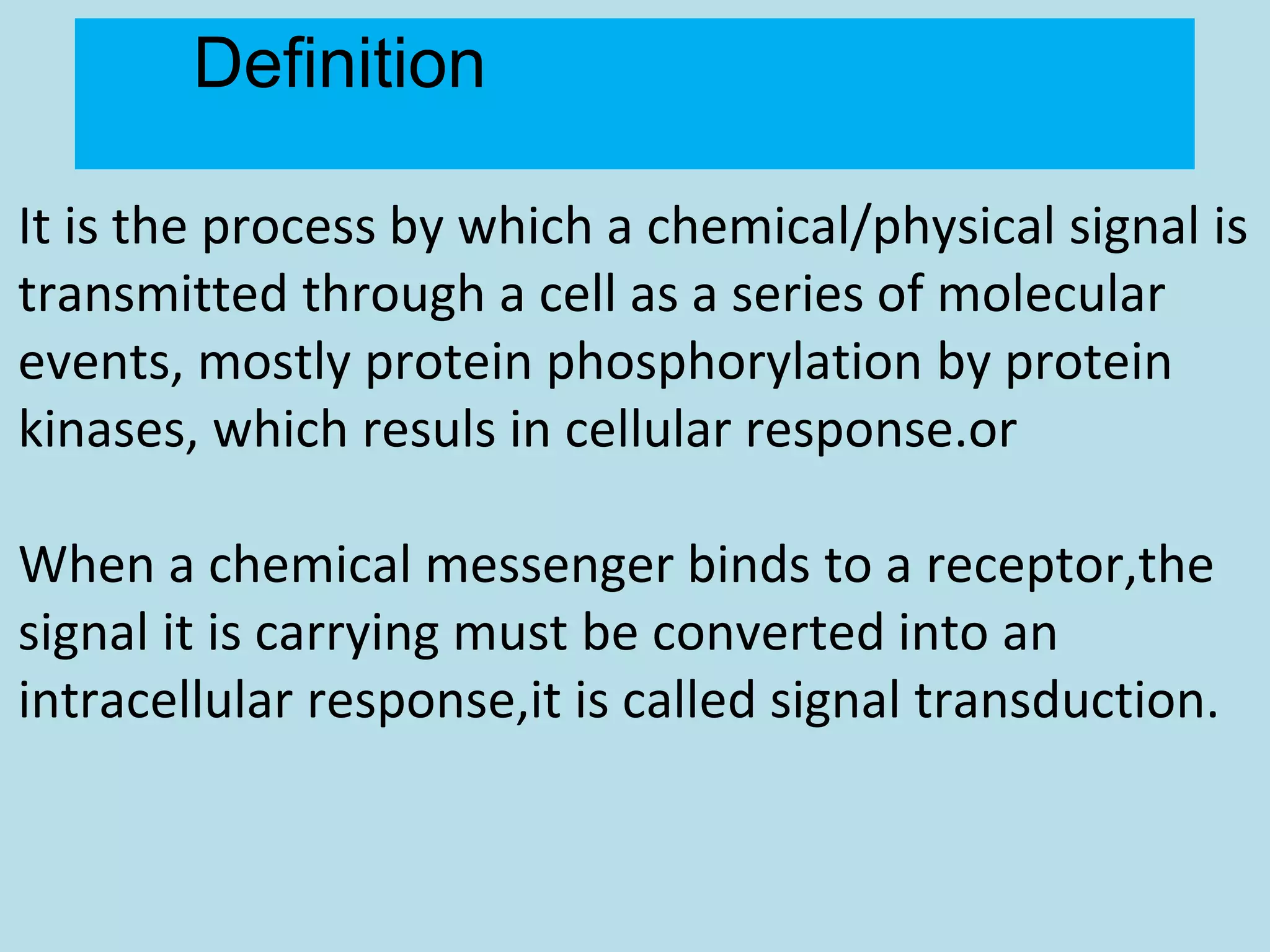
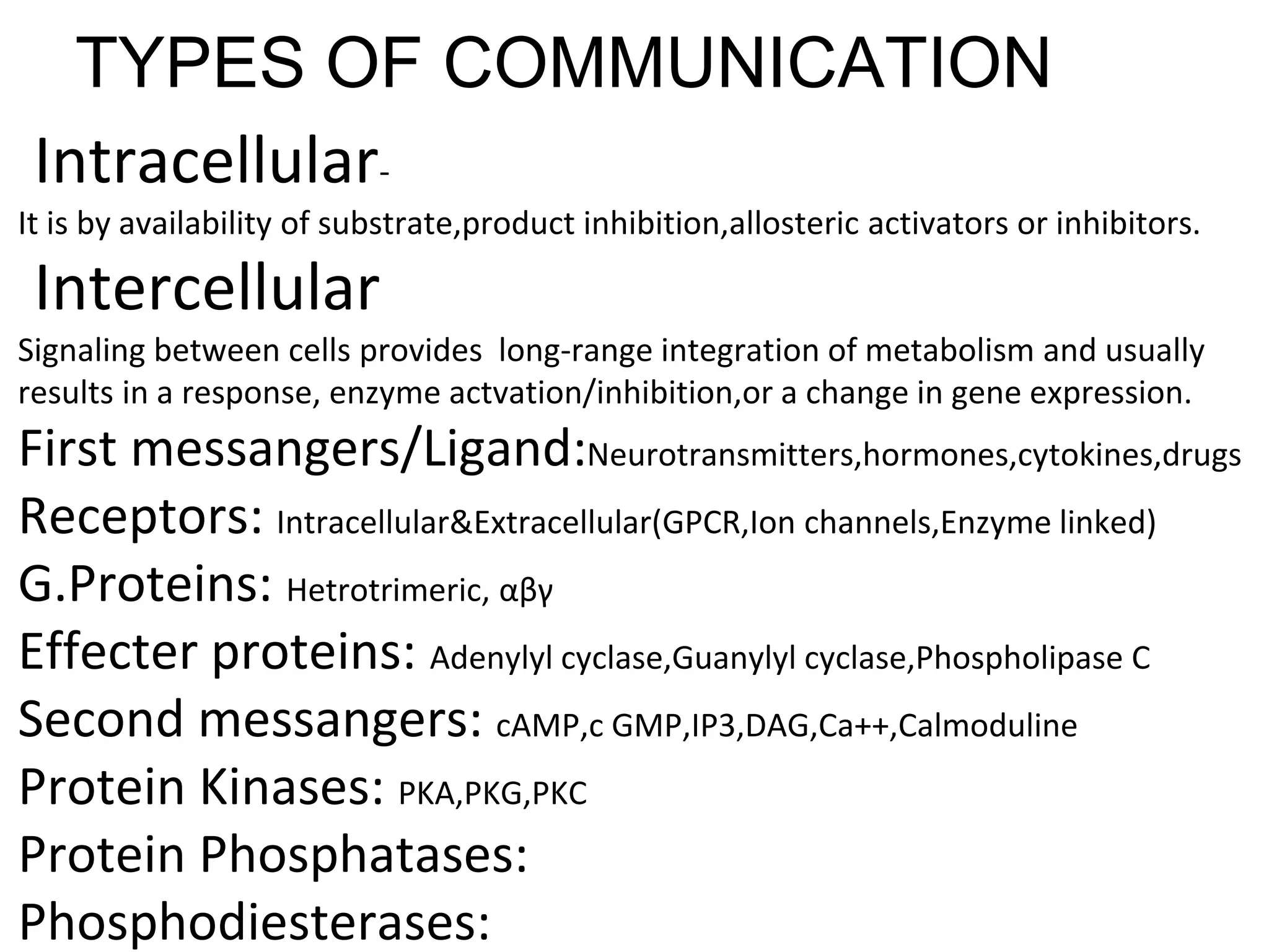
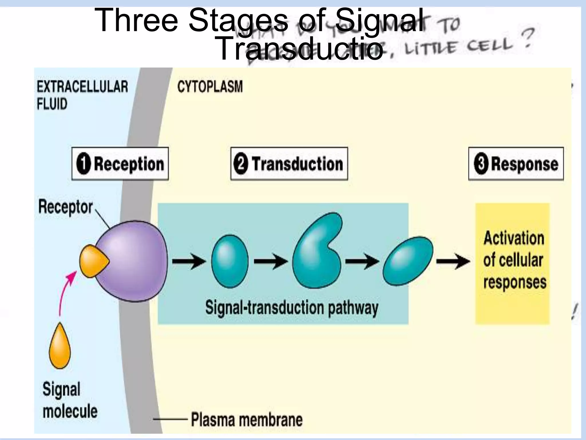
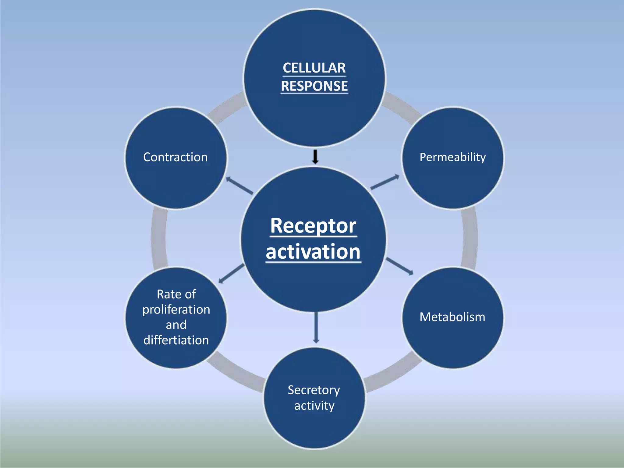
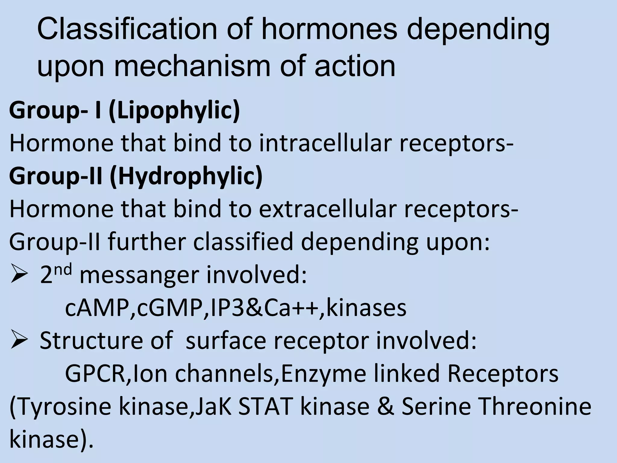
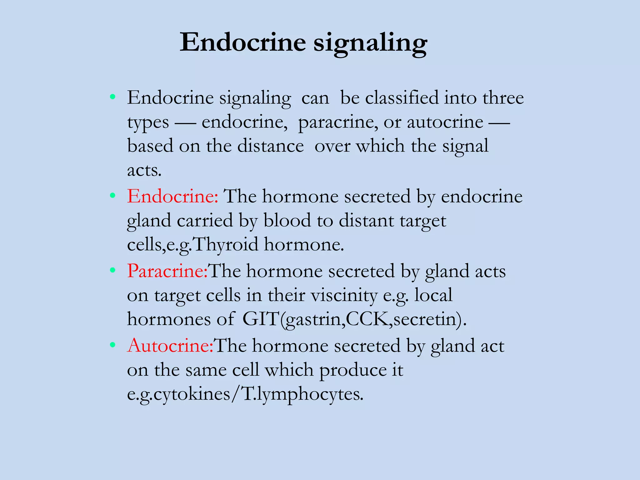
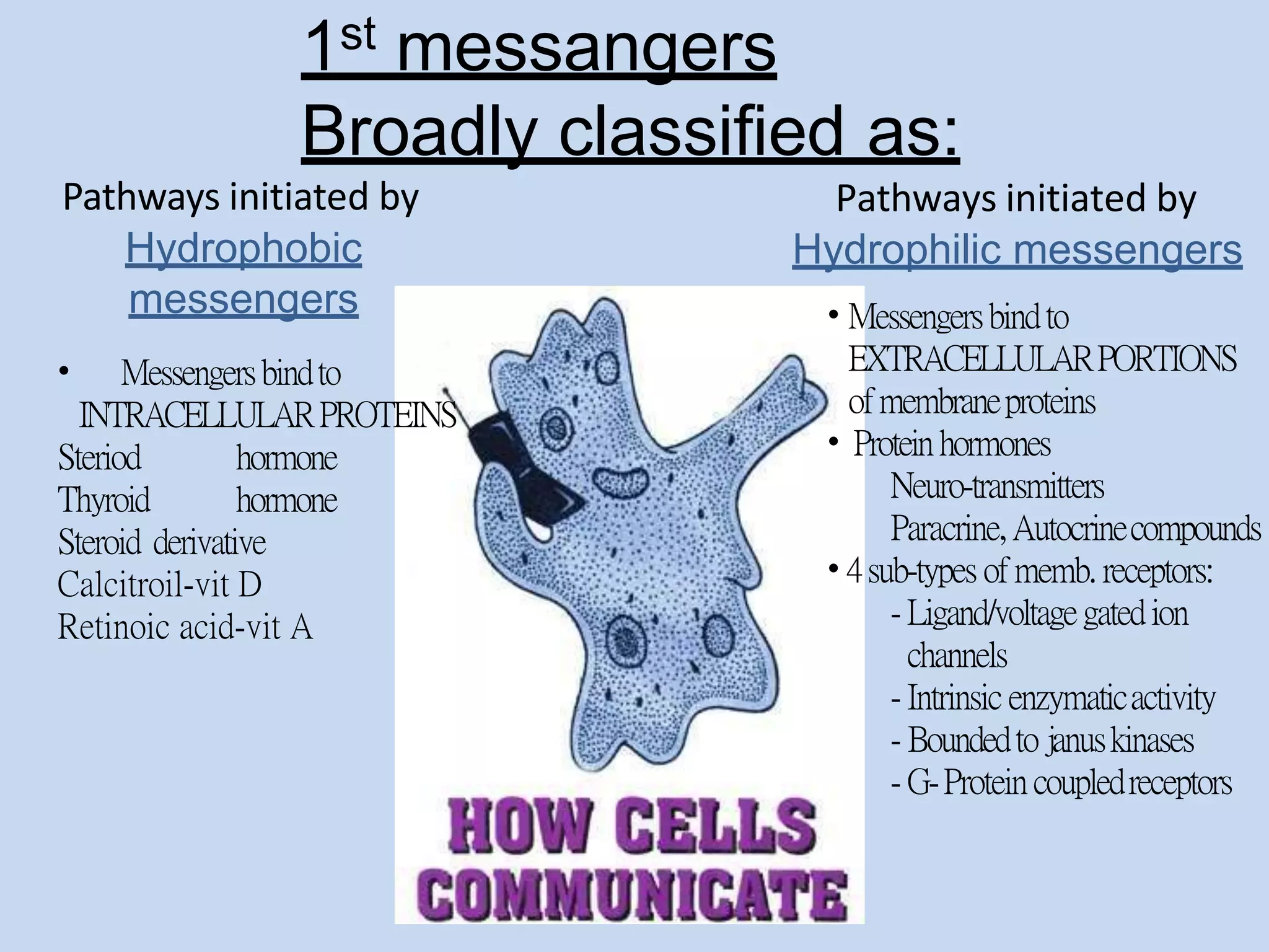
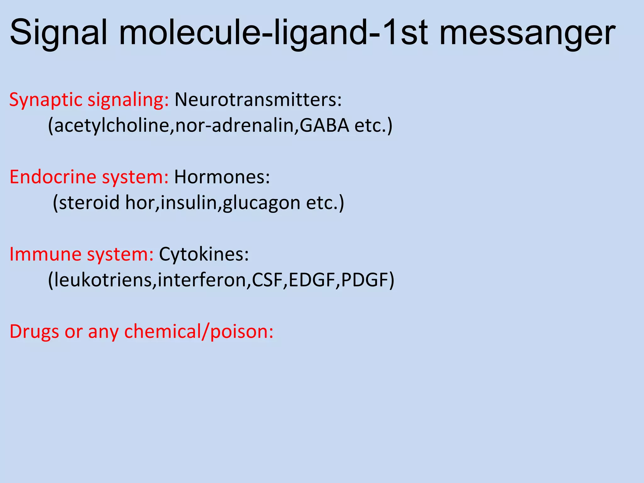
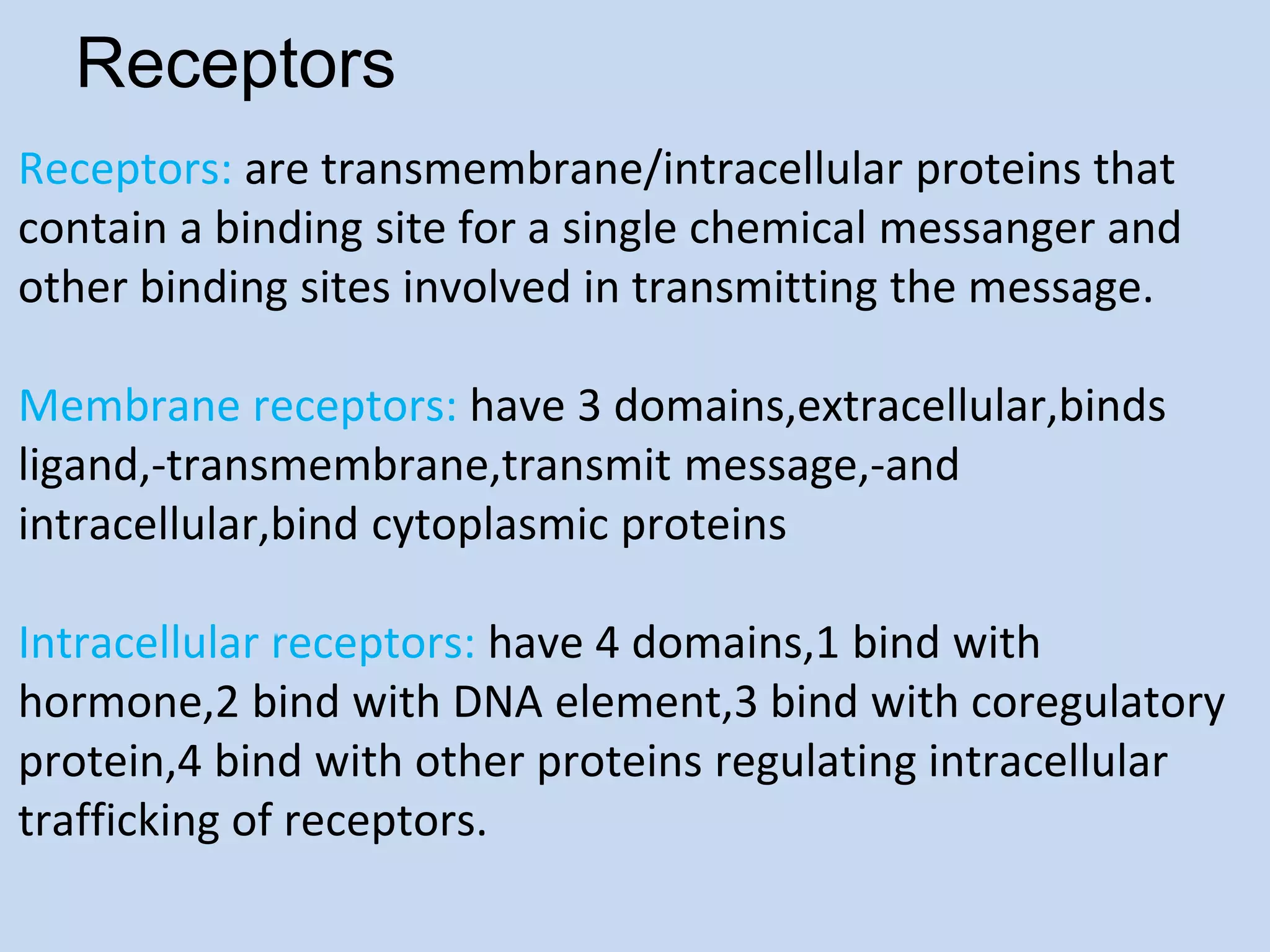
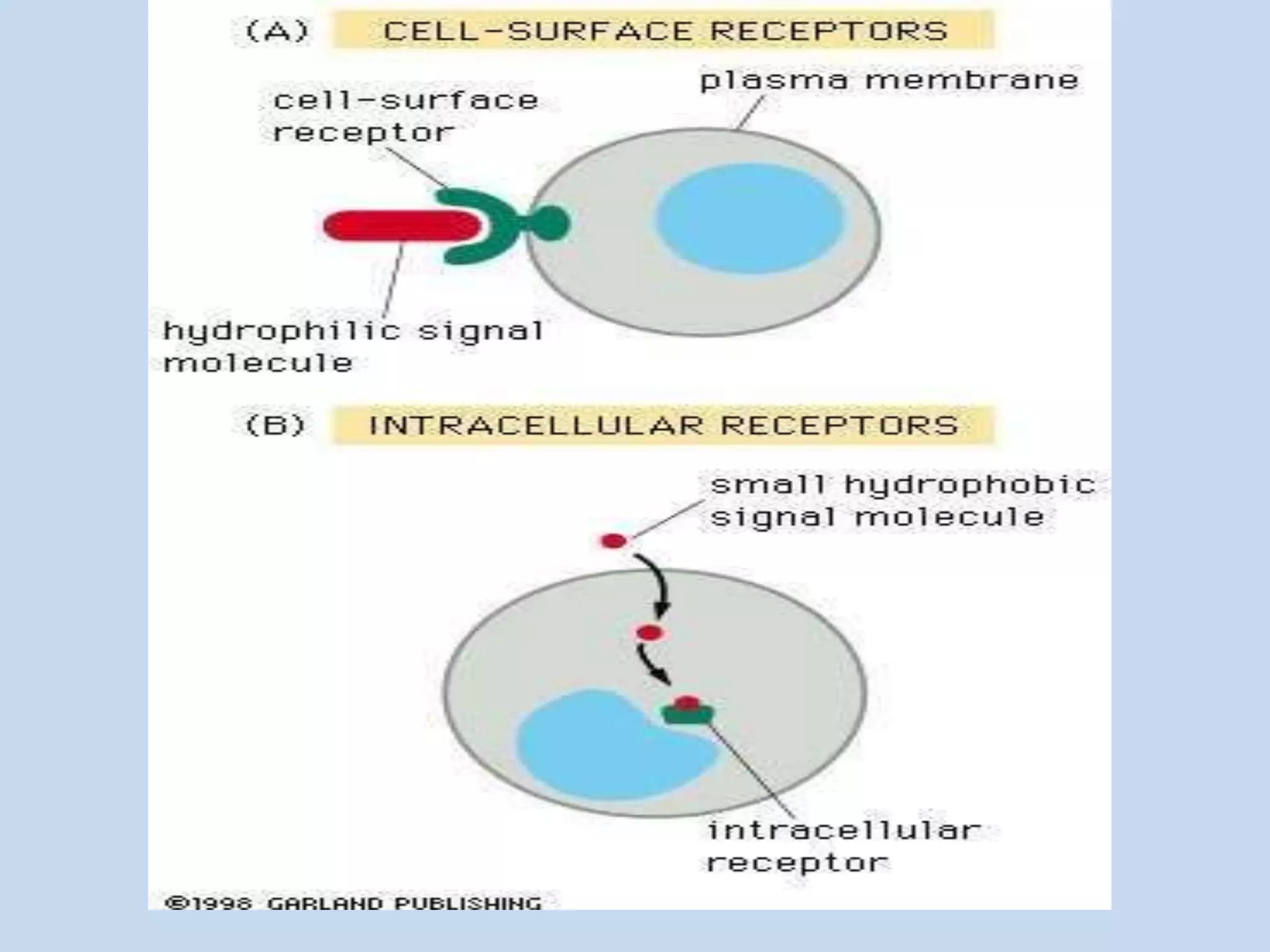
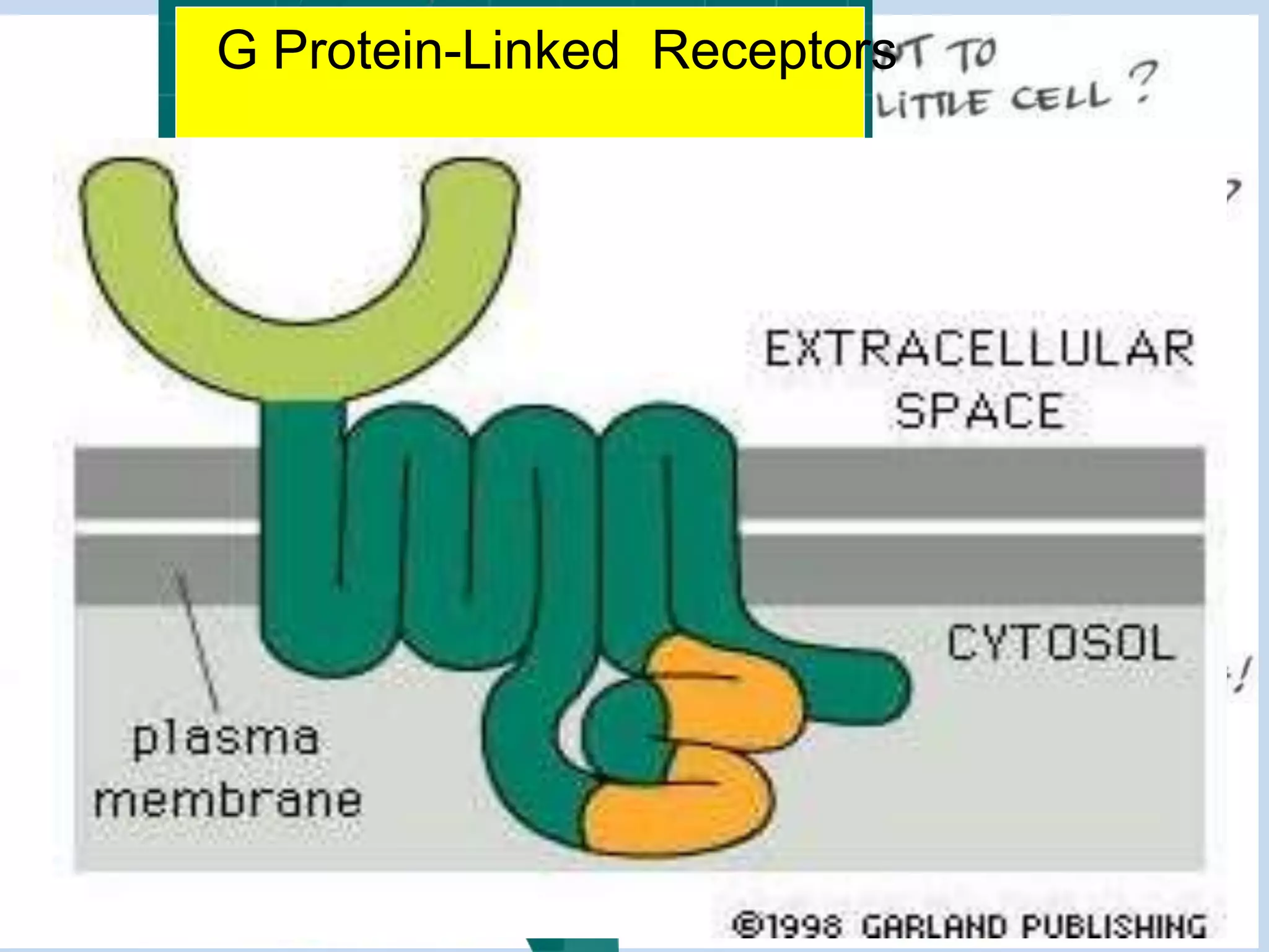
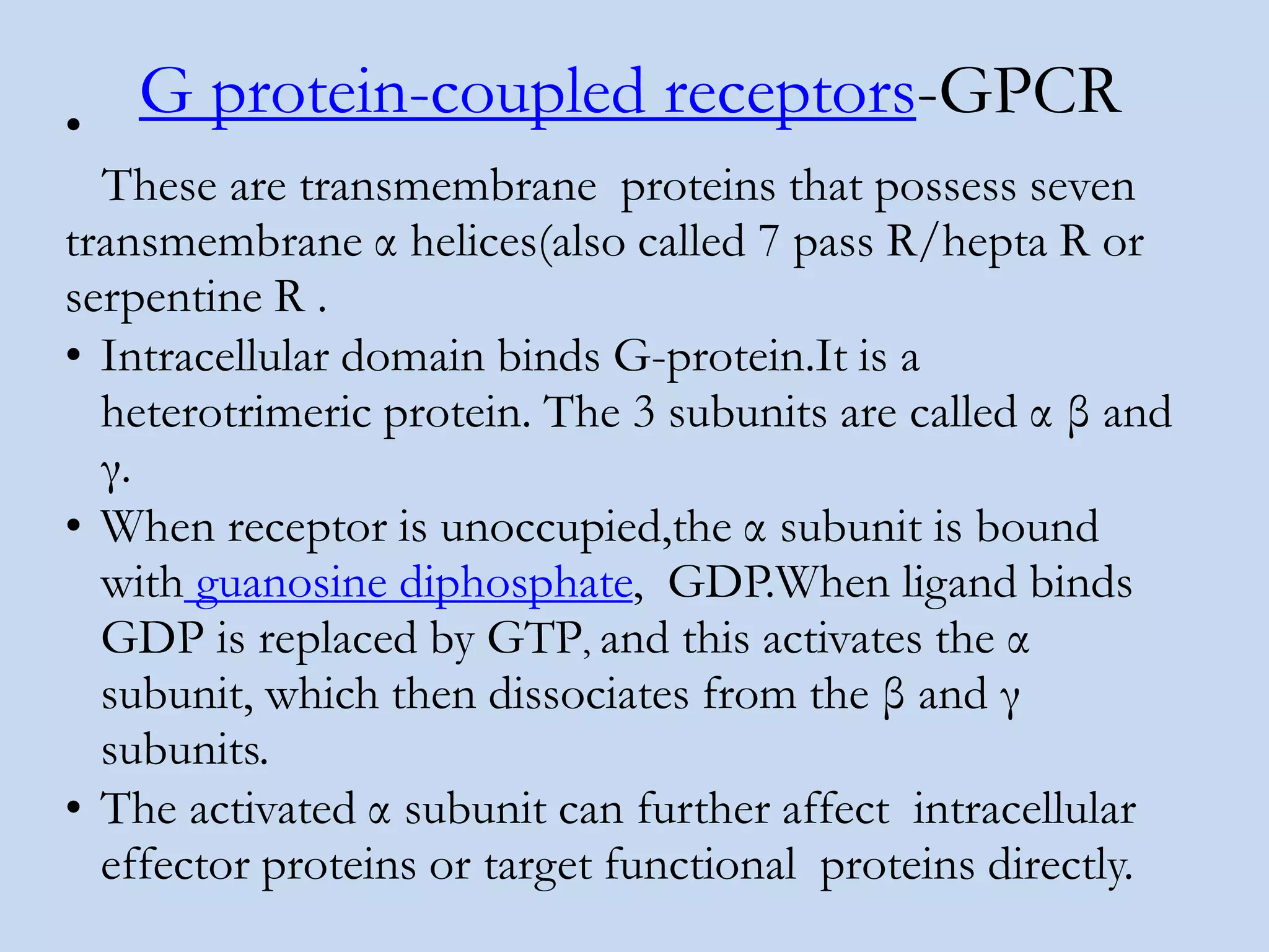
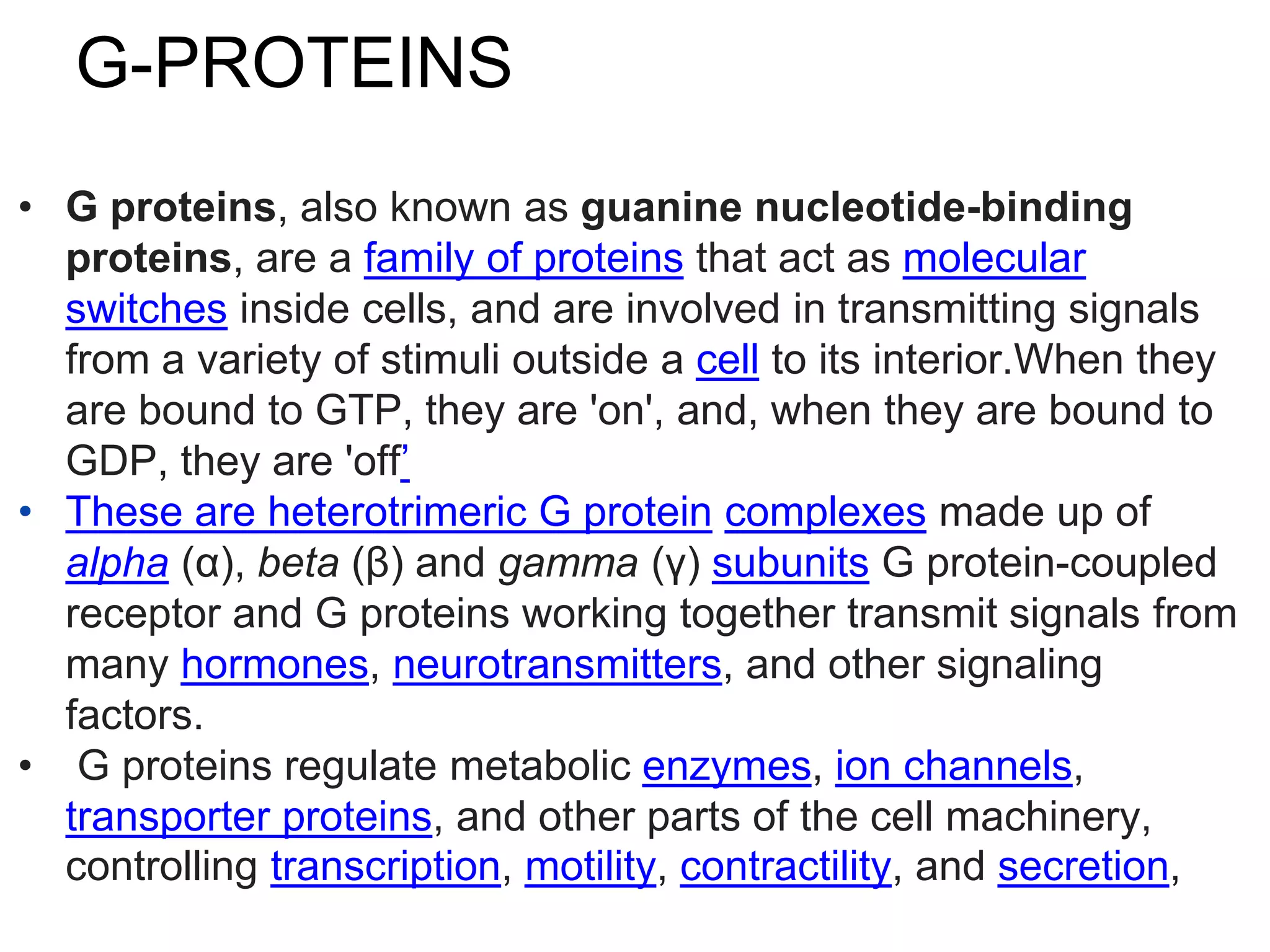
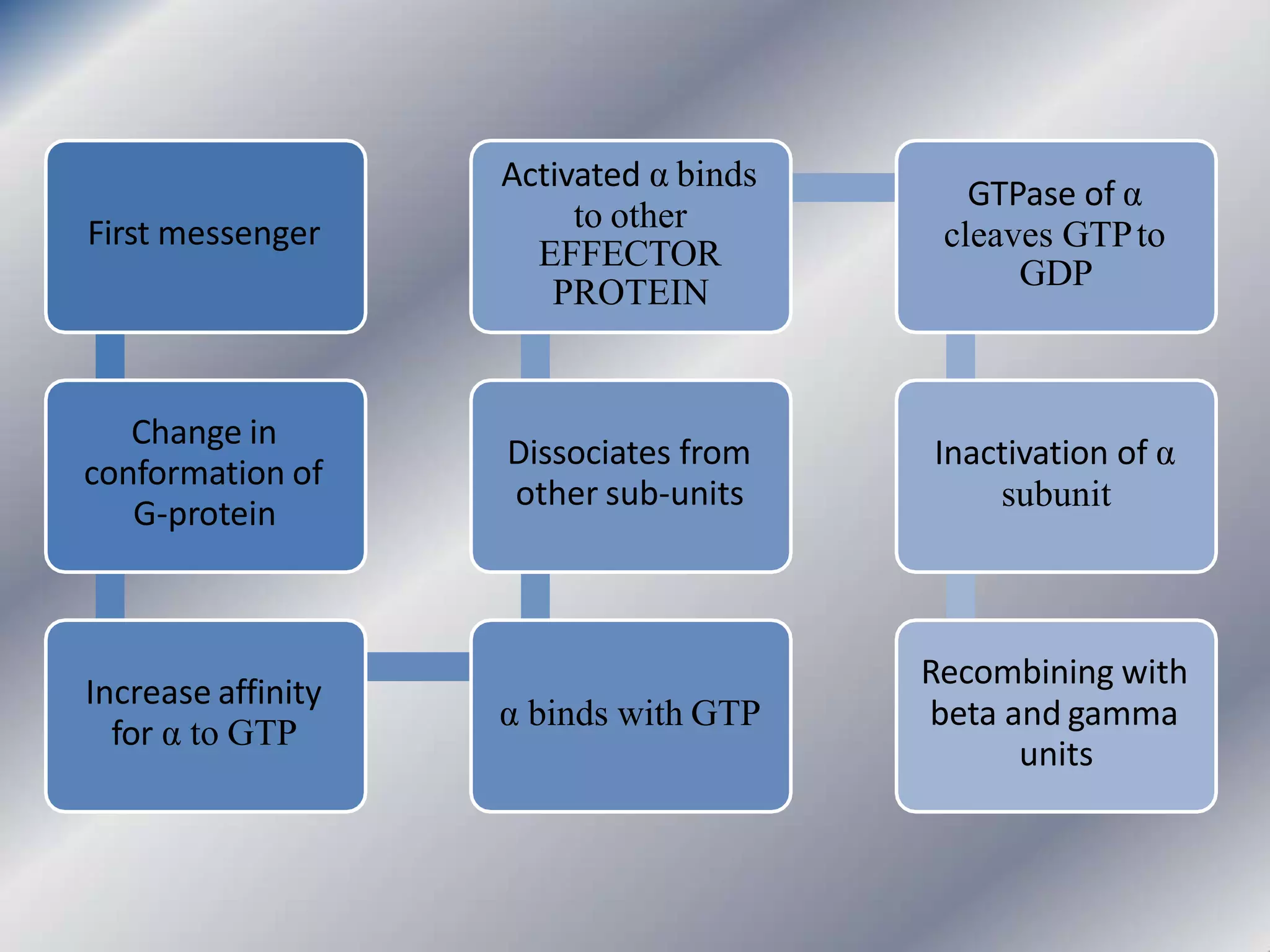
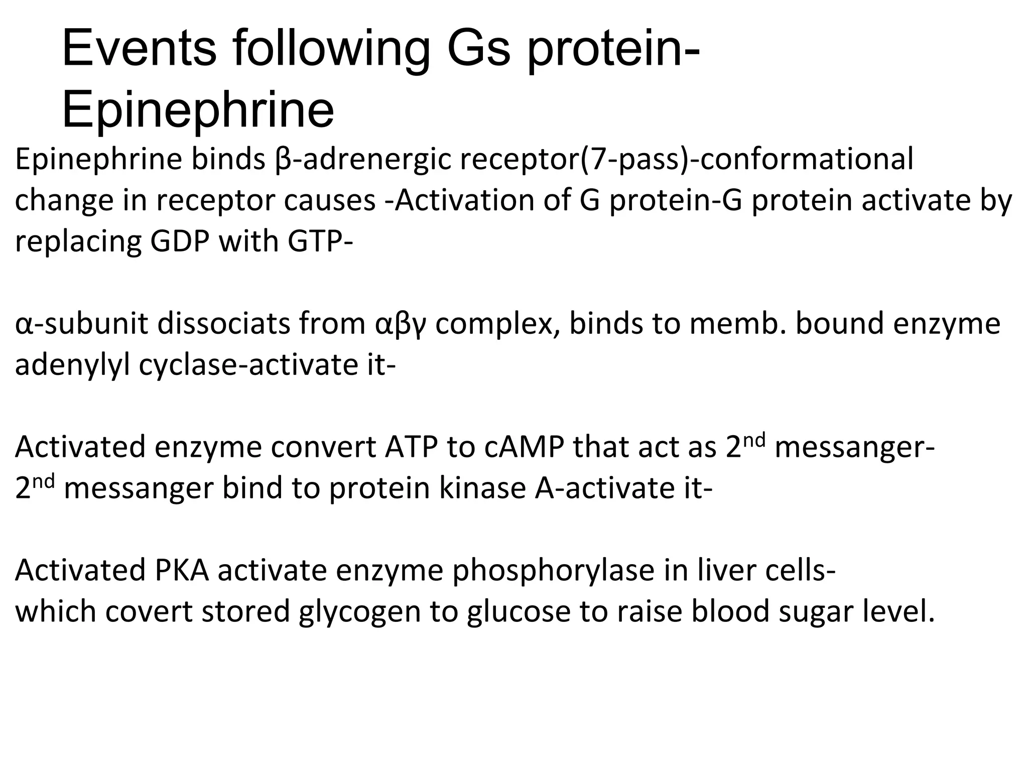
BY DR SHAMIM AKRAM AP BIOCHEM AIMC
BOOKS
Lippincott-p.94(GPCR),205(PLC),311(RTK)
HARPER-CH.41(Endo),42(signal T)
MARKS-CH.11(SECTION 2)(signal T)
Conten
t
• Definition
• Classification
• Pathways initiated by lipid soluble messengers
• Pathways initiated by water soluble messengers
• GPCR,G-PROTEINS,EFFECTOR
PROTEINS,P.KINASES
• 1ST MESSANGERS,2ND MESSANGERS
• TERMINATION OF SIGNALS
• Cellular Signaling
• Many living organisms contain billions of cells
that carry out diverse functions. In order for the
cells to cooperate, cells need to be able to
communicate with each other. Many of the
genes that cells are capable of synthesizing are
thought to be involved in cellular signaling.
Definition
It is the process by which a chemical/physical signal is
transmitted through a cell as a series of molecular
events, mostly protein phosphorylation by protein
kinases, which resuls in cellular response.or
When a chemical messenger binds to a receptor,the
signal it is carrying must be converted into an
intracellular response,it is called signal transduction.
TYPES OF COMMUNICATION
Intracellular-
It is by availability of substrate,product inhibition,allosteric activators or inhibitors.
Intercellular
Signaling between cells provides long-range integration of metabolism and usually
results in a response, enzyme actvation/inhibition,or a change in gene expression.
First messangers/Ligand:Neurotransmitters,hormones,cytokines,drugs
Receptors: Intracellular&Extracellular(GPCR,Ion channels,Enzyme linked)
G.Proteins: Hetrotrimeric, αβγ
Effecter proteins: Adenylyl cyclase,Guanylyl cyclase,Phospholipase C
Second messangers: cAMP,c GMP,IP3,DAG,Ca++,Calmoduline
Protein Kinases: PKA,PKG,PKC
Protein Phosphatases:
Phosphodiesterases:
Three Stages of Signal
Transductio
Receptor
activation
CELLULAR
RESPONSE
Permeability
Metabolism
Secretory
activity
Rate of
proliferation
and
differtiation
Contraction
Classification of hormones depending
upon mechanism of action
Group- I (Lipophylic)
Hormone that bind to intracellular receptors-
Group-II (Hydrophylic)
Hormone that bind to extracellular receptors-
Group-II further classified depending upon:
 2nd messanger involved:
cAMP,cGMP,IP3&Ca++,kinases
 Structure of surface receptor involved:
GPCR,Ion channels,Enzyme linked Receptors
(Tyrosine kinase,JaK STAT kinase & Serine Threonine
kinase).
Endocrine signaling
• Endocrine signaling can be classified into three
types — endocrine, paracrine, or autocrine —
based on the distance over which the signal
acts.
• Endocrine: The hormone secreted by endocrine
gland carried by blood to distant target
cells,e.g.Thyroid hormone.
• Paracrine:The hormone secreted by gland acts
on target cells in their viscinity e.g. local
hormones of GIT(gastrin,CCK,secretin).
• Autocrine:The hormone secreted by gland act
on the same cell which produce it
e.g.cytokines/T.lymphocytes.
1st messangers
Broadly classified as:
Pathways initiated by
Hydrophobic
messengers
• Messengersbindto
INTRACELLULARPROTEINS
Steriod hormone
Thyroid hormone
Steroid derivative
Calcitroil-vit D
Retinoic acid-vit A
Pathways initiated by
Hydrophilic messengers
• Messengersbindto
EXTRACELLULARPORTIONS
ofmembraneproteins
• Proteinhormones
Neuro-transmitters
Paracrine,Autocrinecompounds
• 4sub-types of memb. receptors:
- Ligand/voltagegatedion
channels
- Intrinsic enzymaticactivity
- Boundedto januskinases
- G-Proteincoupledreceptors
Signal molecule-ligand-1st messanger
Synaptic signaling: Neurotransmitters:
(acetylcholine,nor-adrenalin,GABA etc.)
Endocrine system: Hormones:
(steroid hor,insulin,glucagon etc.)
Immune system: Cytokines:
(leukotriens,interferon,CSF,EDGF,PDGF)
Drugs or any chemical/poison:
Receptors
Receptors: are transmembrane/intracellular proteins that
contain a binding site for a single chemical messanger and
other binding sites involved in transmitting the message.
Membrane receptors: have 3 domains,extracellular,binds
ligand,-transmembrane,transmit message,-and
intracellular,bind cytoplasmic proteins
Intracellular receptors: have 4 domains,1 bind with
hormone,2 bind with DNA element,3 bind with coregulatory
protein,4 bind with other proteins regulating intracellular
trafficking of receptors.
Types
• Receptors can be roughly divided into two major
classes: intracellular receptors and extracellular
receptors.
G Protein-Linked Receptors
PARTS OF EXTRACELLULAR
RECEPTOR-GPCR
Extracellular Domain
Transmembrane Domain
Intracellular Domain
The extracellular domain
• The extracellular domain is the part of the
receptor that sticks out of the membrane on
the outside of the cell or organelle.
• If the polypeptide chain of the receptor
crosses the bilayer several times, the external
domain can comprise several "loops" sticking
out of the membrane.
• The extracellular domain has binding site for
the ligand.
The transmembrane domains
• In the majority of receptors, transmembrane alpha helices
make up most of the transmembrane domain.It transmits
message carried by ligand to intra cellular domain.
In certain receptors, such as the nicotinic
acetylcholine receptor, the transmembrane domain
forms a protein-lined pore through the
membrane, or ion channel.
Upon activation of an extracellular domain by binding of
the appropriate ligand, the pore becomes accessible to ions,
which then pass through.
Intracellular (or cytoplasmic) domain
• The intracellular (or cytoplasmic) domain of the
receptor interacts with the interior of the cell or
organelle, relaying the signal. There are two
fundamentally different ways for this interaction:
• The intracellular domain communicates via specific protein-
G-protein that interact with effector proteins, which in turn
send the signal along a signal chain to its destination.
• With enzyme-linked receptors, the intracellular domain has
enzymatic activity. Often, this is a tyrosine kinase activity. The
enzymatic activity can also be located on an enzyme
associated with the intracellular domain.
TYPES OF MEMBRANE
RECEPTORS
ION CHANNELLS -Liganed/voltage gated-
Neurotransmitters(epinephrine,Acetylcholin),Cation/Anion(Na,K,Ca,Cl)
ENZYME LINKED RECEPTORS: RTK,JAK-STAT,Serine-threonine.
(insulin,GH,cytokines)
GPCR-G-protein coupled receptors:
Hormones (Epinephrine,glucagon)
1. Ligand gated ion channels
• Receptor proteinacts as anionchannel
• Seen in;
- Nerve-nerve junctions
- Neuro-muscularjunctions
Ion channel linked receptors
• Ion channel linked receptors are ion-channels
(including cation-channels and anion-channels)
themselves and constitute a large family of
multipass transmembrane proteins.
• They are involved in rapid signaling events
most generally found in electrically excitable
cells such as neurons and are also called
ligand-gated ion channels. Opening and closing
of Ion channels are controlled by
neurotransmitters.
Ligand binds
to ion
channel
Opening of
ion channels
Increase
diffusion
across
membrane
Change in
electrical
charge
Cell response
Contraction/
conduction
Enzyme-linked receptors
• Enzyme-linked receptors are either
enzymes themselves, or are directly
associated with the enzymes that they
activate. These are usually single-pass
transmembrane receptors, with the
enzymatic portion of the receptor being
intracellular. The majority of enzyme-
lined receptors are protein kinases, or
associate with protein kinases.
• Receptors with kinase activity has
amino acid residues tyrosine,serine or
threonine with OH gp.which provide
site for binding PO4 gp.for activation
of other proteins.
G protein-coupled receptors-GPCR•
These are transmembrane proteins that possess seven
transmembrane α helices(also called 7 pass R/hepta R or
serpentine R .
• Intracellular domain binds G-protein.It is a
heterotrimeric protein. The 3 subunits are called α β and
γ.
• When receptor is unoccupied,the α subunit is bound
with guanosine diphosphate, GDP.When ligand binds
GDP is replaced by GTP, and this activates the α
subunit, which then dissociates from the β and γ
subunits.
• The activated α subunit can further affect intracellular
effector proteins or target functional proteins directly.
G-PROTEINS
• G proteins, also known as guanine nucleotide-binding
proteins, are a family of proteins that act as molecular
switches inside cells, and are involved in transmitting signals
from a variety of stimuli outside a cell to its interior.When they
are bound to GTP, they are 'on', and, when they are bound to
GDP, they are 'off’
• These are heterotrimeric G protein complexes made up of
alpha (α), beta (β) and gamma (γ) subunits G protein-coupled
receptor and G proteins working together transmit signals from
many hormones, neurotransmitters, and other signaling
factors.
• G proteins regulate metabolic enzymes, ion channels,
transporter proteins, and other parts of the cell machinery,
controlling transcription, motility, contractility, and secretion,
First messenger
Change in
conformation of
G-protein
Increase affinity
for α to GTP
α binds with GTP
Dissociates from
other sub-units
Activated α binds
to other
EFFECTOR
PROTEIN
GTPase of α
cleaves GTPto
GDP
Inactivation of α
subunit
Recombining with
beta and gamma
units
Events following Gs protein-
Epinephrine
Epinephrine binds β-adrenergic receptor(7-pass)-conformational
change in receptor causes -Activation of G protein-G protein activate by
replacing GDP with GTP-
α-subunit dissociats from αβγ complex, binds to memb. bound enzyme
adenylyl cyclase-activate it-
Activated enzyme convert ATP to cAMP that act as 2nd messanger-
2nd messanger bind to protein kinase A-activate it-
Activated PKA activate enzyme phosphorylase in liver cells-
which covert stored glycogen to glucose to raise blood sugar level.
Induction of enzyme synthesis
Glucagon increases transcription of thegene for PEPCK and G6P for
gluconeogenesis.
It is via the transcription factor cAMP response element–binding (CREB)
protein, thereby increasing the availability of this enzyme as levels of
its substrate rise during fasting.
[Note: Cortisol (a glucocorticoid) also increases expression of the gene,
whereas insulin decreases expression.]
G-Stimulatory & G-Inhibitory
Different peptide hormones can either stimulate (s) or inhibit (i)
the production of cAMP from adenylyl cyclase through the action
of the G proteins.
Two parallel systems, a stimulatory (s) one and an inhibitory (i)
one,converge upon a catalytic molecule (C). Each consists of a
receptor, Rs or Ri, and a regulatory G-protein complex termed Gs
and Gi.
Both Gs and Gi are heterotrimeric G-proteins composed of α,
β, and γ subunits.
Since the α subunit in Gs differs from that in Gi, the proteins, which
are distinct gene products, are designated αs and αi.
• Effecter proteins could be
- adenylyl cyclase
- Guanylyl cyclase
- Phospholipase C
- Ion channels
• 2nd messangers are
- cAMP, cGMP,
- IP3,DAG
- Ca++,Calmodulin
Events following activation of Gi
protein by Acetylecholine
Acetylecholine binds Ri receptor-
Conformational change in recepto activate associated Gi proteins-
Gi protein inhibit Adenylyl Cyclase-no formation of c AMP-no muscle
contraction.
Events following activation of Gq
and phospholipase C by oxytocin
Oxytocin binds GPCR-receptor on cell membrane-Conformational change
in GPCR activate G proteins,when GDP is replaced by GTP-
Activated G protein α-subunit binds memb bound enzyme phospholipase
C to activate it-activated phospholipase C cleaves PIP2 to DAG & IP3-
IP3 binds Ca receptor on ER,which open with release of ca in
myoepithelial cells of breast with ejection of milk.
Ca & DAG activate PKC,which actvate other kinases to get other cellular
responses.
TERMINATION OF SIGNALS
No chemical or stimulus-no signal-ligand catabolized
Receptor desensitized to messanger by
phosphorylation,internalization or degraded
G protein have GTPase activity, automatically
hydrolyze GTP to GDP
2nd messanger degraded(e.g.c AMP by
phosphodiesterase).
Protein Kinases deactivated by phosphatases.
Celltocellcommunicationis acomplexmechanismthat hasbeen
classifiedtostudyandtounderstand. Acellularresponse is generally
notbecauseofasinglemediatorormessengerbutvarious
biochemical processesareinvolvedintransductionpathways.
Theseprocesses arethepart of thecomplexnetworkofcommunication
that governbasiccellularactivities andcoordinatetheactionofcells
Thank
you…

signal transduction AIMC

  • 1.
    BY DR SHAMIMAKRAM AP BIOCHEM AIMC
  • 2.
  • 3.
    Conten t • Definition • Classification •Pathways initiated by lipid soluble messengers • Pathways initiated by water soluble messengers • GPCR,G-PROTEINS,EFFECTOR PROTEINS,P.KINASES • 1ST MESSANGERS,2ND MESSANGERS • TERMINATION OF SIGNALS
  • 4.
    • Cellular Signaling •Many living organisms contain billions of cells that carry out diverse functions. In order for the cells to cooperate, cells need to be able to communicate with each other. Many of the genes that cells are capable of synthesizing are thought to be involved in cellular signaling.
  • 5.
    Definition It is theprocess by which a chemical/physical signal is transmitted through a cell as a series of molecular events, mostly protein phosphorylation by protein kinases, which resuls in cellular response.or When a chemical messenger binds to a receptor,the signal it is carrying must be converted into an intracellular response,it is called signal transduction.
  • 6.
    TYPES OF COMMUNICATION Intracellular- Itis by availability of substrate,product inhibition,allosteric activators or inhibitors. Intercellular Signaling between cells provides long-range integration of metabolism and usually results in a response, enzyme actvation/inhibition,or a change in gene expression. First messangers/Ligand:Neurotransmitters,hormones,cytokines,drugs Receptors: Intracellular&Extracellular(GPCR,Ion channels,Enzyme linked) G.Proteins: Hetrotrimeric, αβγ Effecter proteins: Adenylyl cyclase,Guanylyl cyclase,Phospholipase C Second messangers: cAMP,c GMP,IP3,DAG,Ca++,Calmoduline Protein Kinases: PKA,PKG,PKC Protein Phosphatases: Phosphodiesterases:
  • 8.
    Three Stages ofSignal Transductio
  • 10.
  • 11.
    Classification of hormonesdepending upon mechanism of action Group- I (Lipophylic) Hormone that bind to intracellular receptors- Group-II (Hydrophylic) Hormone that bind to extracellular receptors- Group-II further classified depending upon:  2nd messanger involved: cAMP,cGMP,IP3&Ca++,kinases  Structure of surface receptor involved: GPCR,Ion channels,Enzyme linked Receptors (Tyrosine kinase,JaK STAT kinase & Serine Threonine kinase).
  • 14.
    Endocrine signaling • Endocrinesignaling can be classified into three types — endocrine, paracrine, or autocrine — based on the distance over which the signal acts. • Endocrine: The hormone secreted by endocrine gland carried by blood to distant target cells,e.g.Thyroid hormone. • Paracrine:The hormone secreted by gland acts on target cells in their viscinity e.g. local hormones of GIT(gastrin,CCK,secretin). • Autocrine:The hormone secreted by gland act on the same cell which produce it e.g.cytokines/T.lymphocytes.
  • 17.
    1st messangers Broadly classifiedas: Pathways initiated by Hydrophobic messengers • Messengersbindto INTRACELLULARPROTEINS Steriod hormone Thyroid hormone Steroid derivative Calcitroil-vit D Retinoic acid-vit A Pathways initiated by Hydrophilic messengers • Messengersbindto EXTRACELLULARPORTIONS ofmembraneproteins • Proteinhormones Neuro-transmitters Paracrine,Autocrinecompounds • 4sub-types of memb. receptors: - Ligand/voltagegatedion channels - Intrinsic enzymaticactivity - Boundedto januskinases - G-Proteincoupledreceptors
  • 18.
    Signal molecule-ligand-1st messanger Synapticsignaling: Neurotransmitters: (acetylcholine,nor-adrenalin,GABA etc.) Endocrine system: Hormones: (steroid hor,insulin,glucagon etc.) Immune system: Cytokines: (leukotriens,interferon,CSF,EDGF,PDGF) Drugs or any chemical/poison:
  • 19.
    Receptors Receptors: are transmembrane/intracellularproteins that contain a binding site for a single chemical messanger and other binding sites involved in transmitting the message. Membrane receptors: have 3 domains,extracellular,binds ligand,-transmembrane,transmit message,-and intracellular,bind cytoplasmic proteins Intracellular receptors: have 4 domains,1 bind with hormone,2 bind with DNA element,3 bind with coregulatory protein,4 bind with other proteins regulating intracellular trafficking of receptors.
  • 20.
    Types • Receptors canbe roughly divided into two major classes: intracellular receptors and extracellular receptors.
  • 21.
  • 22.
    PARTS OF EXTRACELLULAR RECEPTOR-GPCR ExtracellularDomain Transmembrane Domain Intracellular Domain
  • 23.
    The extracellular domain •The extracellular domain is the part of the receptor that sticks out of the membrane on the outside of the cell or organelle. • If the polypeptide chain of the receptor crosses the bilayer several times, the external domain can comprise several "loops" sticking out of the membrane. • The extracellular domain has binding site for the ligand.
  • 25.
    The transmembrane domains •In the majority of receptors, transmembrane alpha helices make up most of the transmembrane domain.It transmits message carried by ligand to intra cellular domain. In certain receptors, such as the nicotinic acetylcholine receptor, the transmembrane domain forms a protein-lined pore through the membrane, or ion channel. Upon activation of an extracellular domain by binding of the appropriate ligand, the pore becomes accessible to ions, which then pass through.
  • 26.
    Intracellular (or cytoplasmic)domain • The intracellular (or cytoplasmic) domain of the receptor interacts with the interior of the cell or organelle, relaying the signal. There are two fundamentally different ways for this interaction: • The intracellular domain communicates via specific protein- G-protein that interact with effector proteins, which in turn send the signal along a signal chain to its destination. • With enzyme-linked receptors, the intracellular domain has enzymatic activity. Often, this is a tyrosine kinase activity. The enzymatic activity can also be located on an enzyme associated with the intracellular domain.
  • 27.
    TYPES OF MEMBRANE RECEPTORS IONCHANNELLS -Liganed/voltage gated- Neurotransmitters(epinephrine,Acetylcholin),Cation/Anion(Na,K,Ca,Cl) ENZYME LINKED RECEPTORS: RTK,JAK-STAT,Serine-threonine. (insulin,GH,cytokines) GPCR-G-protein coupled receptors: Hormones (Epinephrine,glucagon)
  • 28.
    1. Ligand gatedion channels • Receptor proteinacts as anionchannel • Seen in; - Nerve-nerve junctions - Neuro-muscularjunctions
  • 29.
    Ion channel linkedreceptors • Ion channel linked receptors are ion-channels (including cation-channels and anion-channels) themselves and constitute a large family of multipass transmembrane proteins. • They are involved in rapid signaling events most generally found in electrically excitable cells such as neurons and are also called ligand-gated ion channels. Opening and closing of Ion channels are controlled by neurotransmitters.
  • 33.
    Ligand binds to ion channel Openingof ion channels Increase diffusion across membrane Change in electrical charge Cell response Contraction/ conduction
  • 34.
    Enzyme-linked receptors • Enzyme-linkedreceptors are either enzymes themselves, or are directly associated with the enzymes that they activate. These are usually single-pass transmembrane receptors, with the enzymatic portion of the receptor being intracellular. The majority of enzyme- lined receptors are protein kinases, or associate with protein kinases. • Receptors with kinase activity has amino acid residues tyrosine,serine or threonine with OH gp.which provide site for binding PO4 gp.for activation of other proteins.
  • 37.
    G protein-coupled receptors-GPCR• Theseare transmembrane proteins that possess seven transmembrane α helices(also called 7 pass R/hepta R or serpentine R . • Intracellular domain binds G-protein.It is a heterotrimeric protein. The 3 subunits are called α β and γ. • When receptor is unoccupied,the α subunit is bound with guanosine diphosphate, GDP.When ligand binds GDP is replaced by GTP, and this activates the α subunit, which then dissociates from the β and γ subunits. • The activated α subunit can further affect intracellular effector proteins or target functional proteins directly.
  • 38.
    G-PROTEINS • G proteins,also known as guanine nucleotide-binding proteins, are a family of proteins that act as molecular switches inside cells, and are involved in transmitting signals from a variety of stimuli outside a cell to its interior.When they are bound to GTP, they are 'on', and, when they are bound to GDP, they are 'off’ • These are heterotrimeric G protein complexes made up of alpha (α), beta (β) and gamma (γ) subunits G protein-coupled receptor and G proteins working together transmit signals from many hormones, neurotransmitters, and other signaling factors. • G proteins regulate metabolic enzymes, ion channels, transporter proteins, and other parts of the cell machinery, controlling transcription, motility, contractility, and secretion,
  • 42.
    First messenger Change in conformationof G-protein Increase affinity for α to GTP α binds with GTP Dissociates from other sub-units Activated α binds to other EFFECTOR PROTEIN GTPase of α cleaves GTPto GDP Inactivation of α subunit Recombining with beta and gamma units
  • 43.
    Events following Gsprotein- Epinephrine Epinephrine binds β-adrenergic receptor(7-pass)-conformational change in receptor causes -Activation of G protein-G protein activate by replacing GDP with GTP- α-subunit dissociats from αβγ complex, binds to memb. bound enzyme adenylyl cyclase-activate it- Activated enzyme convert ATP to cAMP that act as 2nd messanger- 2nd messanger bind to protein kinase A-activate it- Activated PKA activate enzyme phosphorylase in liver cells- which covert stored glycogen to glucose to raise blood sugar level.
  • 44.
    Induction of enzymesynthesis Glucagon increases transcription of thegene for PEPCK and G6P for gluconeogenesis. It is via the transcription factor cAMP response element–binding (CREB) protein, thereby increasing the availability of this enzyme as levels of its substrate rise during fasting. [Note: Cortisol (a glucocorticoid) also increases expression of the gene, whereas insulin decreases expression.]
  • 46.
    G-Stimulatory & G-Inhibitory Differentpeptide hormones can either stimulate (s) or inhibit (i) the production of cAMP from adenylyl cyclase through the action of the G proteins. Two parallel systems, a stimulatory (s) one and an inhibitory (i) one,converge upon a catalytic molecule (C). Each consists of a receptor, Rs or Ri, and a regulatory G-protein complex termed Gs and Gi. Both Gs and Gi are heterotrimeric G-proteins composed of α, β, and γ subunits. Since the α subunit in Gs differs from that in Gi, the proteins, which are distinct gene products, are designated αs and αi.
  • 48.
    • Effecter proteinscould be - adenylyl cyclase - Guanylyl cyclase - Phospholipase C - Ion channels • 2nd messangers are - cAMP, cGMP, - IP3,DAG - Ca++,Calmodulin
  • 51.
    Events following activationof Gi protein by Acetylecholine Acetylecholine binds Ri receptor- Conformational change in recepto activate associated Gi proteins- Gi protein inhibit Adenylyl Cyclase-no formation of c AMP-no muscle contraction.
  • 54.
    Events following activationof Gq and phospholipase C by oxytocin Oxytocin binds GPCR-receptor on cell membrane-Conformational change in GPCR activate G proteins,when GDP is replaced by GTP- Activated G protein α-subunit binds memb bound enzyme phospholipase C to activate it-activated phospholipase C cleaves PIP2 to DAG & IP3- IP3 binds Ca receptor on ER,which open with release of ca in myoepithelial cells of breast with ejection of milk. Ca & DAG activate PKC,which actvate other kinases to get other cellular responses.
  • 57.
    TERMINATION OF SIGNALS Nochemical or stimulus-no signal-ligand catabolized Receptor desensitized to messanger by phosphorylation,internalization or degraded G protein have GTPase activity, automatically hydrolyze GTP to GDP 2nd messanger degraded(e.g.c AMP by phosphodiesterase). Protein Kinases deactivated by phosphatases.
  • 58.
    Celltocellcommunicationis acomplexmechanismthat hasbeen classifiedtostudyandtounderstand.Acellularresponse is generally notbecauseofasinglemediatorormessengerbutvarious biochemical processesareinvolvedintransductionpathways. Theseprocesses arethepart of thecomplexnetworkofcommunication that governbasiccellularactivities andcoordinatetheactionofcells
  • 59.