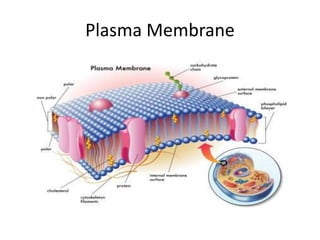
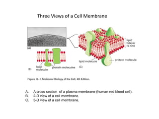
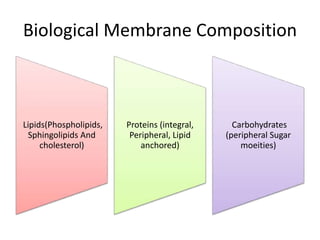
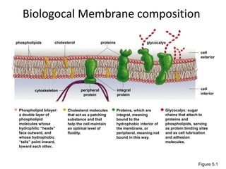
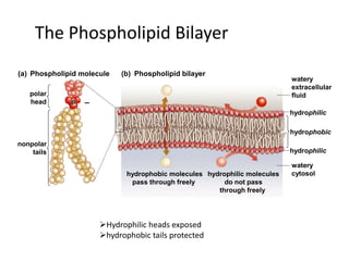
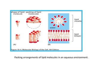
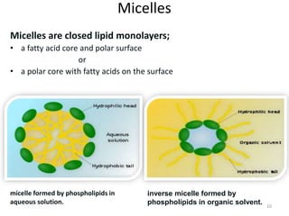
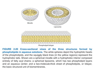
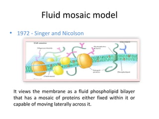
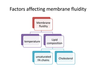
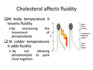
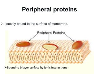
Plasma Membrane
Three Views of a Cell Membrane
A. A cross section of a plasma membrane (human red blood cell).
B. 2-D view of a cell membrane.
C. 3-D view of a cell membrane.
Membrane Functions
3. transport
4. Signal detection
5. Cell-cell
communication
2. Localize
specific
functions
1. boundaries
6. Cell-cell
adhesion
Biological Membrane Composition
Lipids(Phospholipids,
Sphingolipids And
cholesterol)
Proteins (integral,
Peripheral, Lipid
anchored)
Carbohydrates
(peripheral Sugar
moeities)
Biologocal Membrane composition
Figure 5.1
glycocalyx
proteins
integral
protein
peripheral
protein
cytoskeleton
cholesterol
Phospholipid bilayer:
a double layer of
phospholipid
molecules whose
hydrophilic “heads”
face outward, and
whose hydrophobic
“tails” point inward,
toward each other.
Glycocalyx: sugar
chains that attach to
proteins and
phospholipids, serving
as protein binding sites
and as cell lubrication
and adhesion
molecules.
Cholesterol molecules
that act as a patching
substance and that
help the cell maintain
an optimal level of
fluidity.
Proteins, which are
integral, meaning
bound to the
hydrophobic interior of
the membrane, or
peripheral, meaning not
bound in this way.
phospholipids
cell
interior
cell
exterior
The Phospholipid Bilayer
• The fatty acid chains are hydrophobic,
meaning they avoid water, while the
phosphate group is hydrophilic, meaning it
readily bonds with water.
The Phospholipid Bilayer
polar
head
nonpolar
tails
watery
extracellular
fluid
watery
cytosol
hydrophilic
hydrophobic
hydrophobic molecules
pass through freely
hydrophilic molecules
do not pass
through freely
hydrophilic
(a) (b)
Phospholipid molecule Phospholipid bilayer
–
Hydrophilic heads exposed
hydrophobic tails protected
Packing arrangements of lipid molecules in an aqueous environment.
Micelles
Micelles are closed lipid monolayers;
• a fatty acid core and polar surface
or
• a polar core with fatty acids on the surface
10
micelle formed by phospholipids in
aqueous solution.
inverse micelle formed by
phospholipids in organic solvent.
Liposomes
11
' LIPO ' meaning fat
' Soma ' meaning body
concentric bilayered vesicles in which an
aqueous volume is entirely enclosed by a
membraneous lipid bilayer.
FIGURE 2-20 Cross-sectional views of the three structures formed by
phospholipids in aqueous solutions. The white spheres depict the hydrophilic heads
of the phospholipids, and the squiggly black lines (in the yellow regions) represent the
hydrophobic tails. Shown are a spherical micelle with a hydrophobic interior composed
entirely of fatty acyl chains; a spherical liposome, which has two phospholipid layers
and an aqueous center; and a two-molecule-thick sheet of phospholipids, or bilayer,
the basic structural unit of biomembranes.
Membrane Structure
• Not all membranes are identical
– Membranes with different functions differ in their
chemical composition and structure
• Fluid Mosaic Model best describes our current
understanding of membrane structure
Fluid mosaic model
• 1972 - Singer and Nicolson
It views the membrane as a fluid phospholipid bilayer
that has a mosaic of proteins either fixed within it or
capable of moving laterally across it.
Mosaic quality of the membrane
• Membranes have their own unique collection of proteins
• Proteins form different patterns
• Combination of proteins makes membrane unique
• So, the membrane is said to be mosaic
Fluidity
• Fluidity is defined as “easy of flow”.
• Membranes are in motion with fast drifting
lipids and slower drifting proteins
Factors affecting membrane fluidity
Membrane
fluidity
temperature
Lipid
composition
unsaturated
FA chains
Cholesterol
Presence of unsaturated FA chains
Lipids bilayer can be converted
to rigid crystalline (or gel) state
at characteristic freezing point.
Phase transition can be
avoided
 if the hydrocarbon chains
are short/ have double bonds.
Lower
fluidity
Higher
fluidity
Presence of unsaturated FA chains
Reduces the tendency of the hydrocarbon
tails to interact with one another.
Cis-double bonds produce kinks in the chains
that make them more difficult to pack together.
 so that the membrane remains fluid at lower
temperatures.
Cholesterol affects fluidity
At body temperature it
lessens fluidity
By restraining the
movement of
phospholipids
 At colder temperatures
it adds fluidity
 By not allowing
phospholipids to pack
close together.
Lipid Mobility
 Lipids are free to move
laterally in a lipid bilayer
Flip-flop movement is
very energetically
unfavorable
 catalyzed by flippases.
Membrane Proteins
Integral proteins
Peripheral proteins
Lipid anchored proteins
Integral proteins
The hydrophobic region
23
Outside of cell
Hydrophilic region
Peripheral proteins
 loosely bound to the surface of membrane.
24
Bound to bilayer surface by ionic interactions
Lipid Anchored Proteins
Proteins of the Plasma Membrane Provide 6
Membrane Functions:
1) Transport Proteins
2) Receptor Proteins
3) Enzymatic Proteins
4) Cell Recognition Proteins
5) Attachment Proteins
6) Intercellular Junction
Proteins
1) Transport Proteins
Carrier Proteins – bind to a
substance and carry it
across membrane,
change shape in process
Channel Proteins – channel
for lipid insoluble
molecules and ions to
pass freely through
2) Receptor Proteins
– Bind to chemical
messengers (Ex.
hormones) which sends
a message into the cell
causing cellular reaction.
3) Enzymatic Proteins
– Carry out enzymatic
reactions right at the
membrane when a
substrate binds to the
active site.
4) Cell Recognition Proteins
– Glycoproteins (and
glycolipids) on
extracellular surface
serve as ID tags
(which species, type
of cell, individual).
5) Attachment Proteins
- Attach to cytoskeleton
(to maintain cell shape
and stabilize proteins)
and/or the extracellular
matrix (integrins connect
to both).
6) Intercellular Junction Proteins
– Bind cells together
Carbohydrates diversify membranes
• Membrane carbohydrates
are only found on the
outside (external) face of
membranes
• Attach to lipids or protein
(glycolipid/ glycoprotein)
• Enable cells to
distinguish/ recognize
one another
Membrane trafficking
• Materials must move in
and out of the cell
through the plasma
membrane.
• Some materials move
between the
phospholipids.
• Some materials move
through the proteins.
Membrane trafficking
simple
diffusion
facilitated
diffusion
active
transport
ATP

plasma membrane.pptx

  • 1.
  • 2.
    Three Views ofa Cell Membrane A. A cross section of a plasma membrane (human red blood cell). B. 2-D view of a cell membrane. C. 3-D view of a cell membrane.
  • 3.
    Membrane Functions 3. transport 4.Signal detection 5. Cell-cell communication 2. Localize specific functions 1. boundaries 6. Cell-cell adhesion
  • 4.
    Biological Membrane Composition Lipids(Phospholipids, SphingolipidsAnd cholesterol) Proteins (integral, Peripheral, Lipid anchored) Carbohydrates (peripheral Sugar moeities)
  • 5.
    Biologocal Membrane composition Figure5.1 glycocalyx proteins integral protein peripheral protein cytoskeleton cholesterol Phospholipid bilayer: a double layer of phospholipid molecules whose hydrophilic “heads” face outward, and whose hydrophobic “tails” point inward, toward each other. Glycocalyx: sugar chains that attach to proteins and phospholipids, serving as protein binding sites and as cell lubrication and adhesion molecules. Cholesterol molecules that act as a patching substance and that help the cell maintain an optimal level of fluidity. Proteins, which are integral, meaning bound to the hydrophobic interior of the membrane, or peripheral, meaning not bound in this way. phospholipids cell interior cell exterior
  • 6.
    The Phospholipid Bilayer •The fatty acid chains are hydrophobic, meaning they avoid water, while the phosphate group is hydrophilic, meaning it readily bonds with water.
  • 7.
    The Phospholipid Bilayer polar head nonpolar tails watery extracellular fluid watery cytosol hydrophilic hydrophobic hydrophobicmolecules pass through freely hydrophilic molecules do not pass through freely hydrophilic (a) (b) Phospholipid molecule Phospholipid bilayer – Hydrophilic heads exposed hydrophobic tails protected
  • 8.
    Packing arrangements oflipid molecules in an aqueous environment.
  • 9.
    Micelles Micelles are closedlipid monolayers; • a fatty acid core and polar surface or • a polar core with fatty acids on the surface 10 micelle formed by phospholipids in aqueous solution. inverse micelle formed by phospholipids in organic solvent.
  • 10.
    Liposomes 11 ' LIPO 'meaning fat ' Soma ' meaning body concentric bilayered vesicles in which an aqueous volume is entirely enclosed by a membraneous lipid bilayer.
  • 11.
    FIGURE 2-20 Cross-sectionalviews of the three structures formed by phospholipids in aqueous solutions. The white spheres depict the hydrophilic heads of the phospholipids, and the squiggly black lines (in the yellow regions) represent the hydrophobic tails. Shown are a spherical micelle with a hydrophobic interior composed entirely of fatty acyl chains; a spherical liposome, which has two phospholipid layers and an aqueous center; and a two-molecule-thick sheet of phospholipids, or bilayer, the basic structural unit of biomembranes.
  • 12.
    Membrane Structure • Notall membranes are identical – Membranes with different functions differ in their chemical composition and structure • Fluid Mosaic Model best describes our current understanding of membrane structure
  • 13.
    Fluid mosaic model •1972 - Singer and Nicolson It views the membrane as a fluid phospholipid bilayer that has a mosaic of proteins either fixed within it or capable of moving laterally across it.
  • 14.
    Mosaic quality ofthe membrane • Membranes have their own unique collection of proteins • Proteins form different patterns • Combination of proteins makes membrane unique • So, the membrane is said to be mosaic
  • 15.
    Fluidity • Fluidity isdefined as “easy of flow”. • Membranes are in motion with fast drifting lipids and slower drifting proteins
  • 16.
    Factors affecting membranefluidity Membrane fluidity temperature Lipid composition unsaturated FA chains Cholesterol
  • 17.
    Presence of unsaturatedFA chains Lipids bilayer can be converted to rigid crystalline (or gel) state at characteristic freezing point. Phase transition can be avoided  if the hydrocarbon chains are short/ have double bonds. Lower fluidity Higher fluidity
  • 18.
    Presence of unsaturatedFA chains Reduces the tendency of the hydrocarbon tails to interact with one another. Cis-double bonds produce kinks in the chains that make them more difficult to pack together.  so that the membrane remains fluid at lower temperatures.
  • 19.
    Cholesterol affects fluidity Atbody temperature it lessens fluidity By restraining the movement of phospholipids  At colder temperatures it adds fluidity  By not allowing phospholipids to pack close together.
  • 20.
    Lipid Mobility  Lipidsare free to move laterally in a lipid bilayer Flip-flop movement is very energetically unfavorable  catalyzed by flippases.
  • 21.
    Membrane Proteins Integral proteins Peripheralproteins Lipid anchored proteins
  • 22.
    Integral proteins The hydrophobicregion 23 Outside of cell Hydrophilic region
  • 23.
    Peripheral proteins  looselybound to the surface of membrane. 24 Bound to bilayer surface by ionic interactions
  • 24.
  • 25.
    Proteins of thePlasma Membrane Provide 6 Membrane Functions: 1) Transport Proteins 2) Receptor Proteins 3) Enzymatic Proteins 4) Cell Recognition Proteins 5) Attachment Proteins 6) Intercellular Junction Proteins
  • 26.
    1) Transport Proteins CarrierProteins – bind to a substance and carry it across membrane, change shape in process Channel Proteins – channel for lipid insoluble molecules and ions to pass freely through
  • 27.
    2) Receptor Proteins –Bind to chemical messengers (Ex. hormones) which sends a message into the cell causing cellular reaction.
  • 28.
    3) Enzymatic Proteins –Carry out enzymatic reactions right at the membrane when a substrate binds to the active site.
  • 29.
    4) Cell RecognitionProteins – Glycoproteins (and glycolipids) on extracellular surface serve as ID tags (which species, type of cell, individual).
  • 30.
    5) Attachment Proteins -Attach to cytoskeleton (to maintain cell shape and stabilize proteins) and/or the extracellular matrix (integrins connect to both).
  • 31.
    6) Intercellular JunctionProteins – Bind cells together
  • 32.
    Carbohydrates diversify membranes •Membrane carbohydrates are only found on the outside (external) face of membranes • Attach to lipids or protein (glycolipid/ glycoprotein) • Enable cells to distinguish/ recognize one another
  • 33.
    Membrane trafficking • Materialsmust move in and out of the cell through the plasma membrane. • Some materials move between the phospholipids. • Some materials move through the proteins.
  • 34.