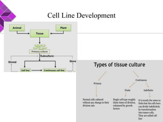
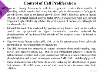
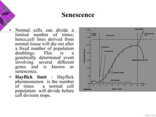
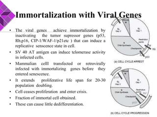
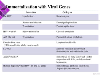
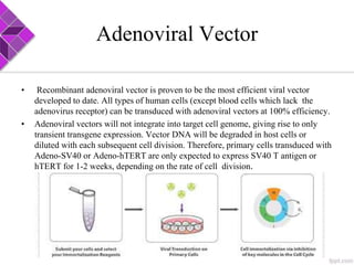
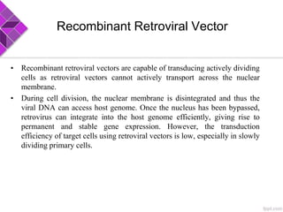
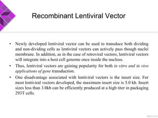
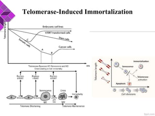
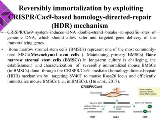
This document discusses various methods for selecting and developing cell lines for research. It covers primary culture isolation, subculture propagation to establish a cell line, control of cell proliferation through growth factors and intracellular regulators, senescence limits on cell divisions, differentiation inhibiting proliferation, and the need for continuous cell lines. Methods to immortalize cell lines include viral genes like SV40 LT and HPV E6/E7 to inactivate tumor suppressors, adenoviral and retroviral vectors, telomerase induction with hTERT, use of oncogenes, and cell hybridization. The goal is to generate stable, consistent cell lines that proliferate indefinitely while maintaining similar phenotype to the original tissue.
This document discusses cell line development and characterization. It describes how primary cell cultures can be taken directly from tissue but have a limited lifespan, while cell lines can proliferate indefinitely through random mutations or genetic modifications. The document outlines techniques for isolating and culturing cells, establishing cell lines, identifying cell lines, and subculturing cells. Immortal cell lines divide rapidly and do not require attachment for growth, making them useful models for studying biology and testing compounds.
In vitro toxicity testing methods can more cost-effectively identify potentially hazardous chemicals compared to animal testing. Cell culture models are commonly used to screen for general toxicity through estimation of cell viability, morphology, and growth. Both qualitative and quantitative cytotoxicity assays are employed, such as membrane integrity assays measuring LDH leakage and viability assays measuring mitochondrial or lysosomal activity via dyes like MTT or neutral red. While 2D cell cultures have limitations, 3D tissue models better maintain native cell morphology and functionality for improved prediction of drug toxicity and efficacy compared to animal models.
Biology and characterization of the cell cultureKAUSHAL SAHU
This document provides an overview of cell culture techniques. It discusses the history of cell culture beginning in the late 1800s. Key terms like cell line, normal cell line, and continuous cell line are defined. The document outlines the biology of cultured cells including adhesion, proliferation, differentiation, and energy metabolism. It also discusses the origin of cultured cells and characterization techniques for authenticating cell lines such as morphology, chromosome content, and DNA analysis. The applications of animal cell culture and conclusions are presented.
This document provides information about cell lines. It defines different types of cell lines including primary cell cultures, continuous cell lines, and transient cell lines. It discusses the HeLa cell line which was the first human cancer cell line. It outlines the process for generating stable cell lines including transfection, selection, cloning, and screening for stability. Steps for freezing and storing cell lines are described. Considerations for choosing an appropriate cell line for experiments are presented. Details are given about specific commonly used cell lines like 3T3, HEK293, PC12, CHO, and BC1. References for further information on cell line generation and characterization are provided.
Cell lines need to be routinely maintained and stored long-term to preserve their valuable characteristics. Routine maintenance involves periodic medium changes and subculturing depending on the growth rate of the specific cell line. Long-term storage is achieved through cryopreservation, where cells are frozen at temperatures below -100°C. This process aims to minimize ice crystal formation and associated cell damage through slow freezing in the presence of cryoprotectants like DMSO or glycerol. Proper freezing and storage methods help preserve cell lines for future use and distribution.
Organ culture involves maintaining small fragments of whole organs or tissues in culture media while retaining their three-dimensional structure and spatial distribution of cells. There are several methods of organ culture including culturing on plasma clots, agar, liquid media, or raft methods. Organ culture has various applications and allows studying cell interactions in a way that mimics the in vivo organ. It is currently being used to develop replacement organs and tissues for applications such as growing bladders, lungs, and heart patches. While progress is being made, developing fully functional human organs remains a challenge.
Introduction
Terminologies
Types of tissue culture
Applications
Culturing
Sub-culturing
Cryopreservation
Detection of contaminants
In vitro transformation of cells
Cell viability
Rules for working in the Lab
Advantages
Limitations
This document discusses different types of mammalian cell culture. It describes primary cell culture, which uses cells directly from tissue that can undergo a limited number of divisions before senescing. Finite and continuous cell lines can proliferate for extended periods through transformation or immortalization. Common cell lines include HeLa cells and other tumor-derived lines. The document also covers techniques for attachment and suspension cell culture, and factors that influence cell growth in vitro.
Transfection involves introducing foreign DNA into host cells to produce a new phenotype. There are two main methods of transfection - vector-mediated and non-vector mediated. Vector-mediated transfection uses bacteriophage, retroviral, cosmid, baculovirus, and plasmid vectors to introduce DNA. Non-vector mediated methods include direct techniques like microinjection, electroporation, and particle bombardment, and indirect techniques like calcium phosphate precipitation and DEAE-dextran. Retroviral vectors are modified retroviruses that can introduce foreign DNA into host chromosomal DNA. Microinjection involves injecting DNA directly into cells using a micropipette under a microscope. Electroporation uses electric pulses to create temporary
RETROVIRUS MEDIATED GENE TRANSFER AND EXPRESSION CLONINGSrishtiRoy10
- The retroviral virion is a spherical particle 80-100 nm in diameter composed of a lipid bilayer envelope containing glycoproteins and a capsid containing two copies of the viral RNA genome and enzymes.
- Retroviruses replicate by reverse transcribing their RNA genome into DNA which is then integrated into the host cell genome by an integrase enzyme to become a provirus, allowing transcription of viral genes.
- Retrovirus mediated gene transfer involves the virus producing a DNA copy of its genome using reverse transcriptase, with the DNA then integrating randomly into the host cell genome, allowing investigation of gene function.
Introduction
History
Scale up in suspension:Stirred culture,Continuous flow culture,Air- lift culture,Nasa bioreactor
Scale up in monolayer culture: Roller bottle culture , multisurface culture,fixed -bed culture
Other type of culture for scaling up: HARV Vessels,STLV vessels
Monitoring of scale up
Conclusion
References
The document discusses various characteristics of cells in culture. It describes how primary cultures are established directly from animal tissue, while cell lines come from established cultures. Primary cultures have cells taken directly from tissue and placed in growth medium. The document also discusses how to isolate a single cell type, factors that allow differentiation, and how normal cells differ from transformed cells that can grow indefinitely.
The document discusses methods for genetically engineering animal cells, including transferring genes into cells using viral or non-viral methods, and selecting and amplifying transfected cells. Common methods to introduce genes include calcium phosphate transfection, lipofection, electroporation, and viral transduction. Genetic markers like DHFR or antibiotic resistance genes allow selection of cells containing the introduced gene.
Cell lines are permanently established cell cultures that can proliferate indefinitely. They are derived from primary cell cultures which have a finite lifespan. Continuous cell lines have undergone transformation to proliferate indefinitely, unlike finite primary cultures. Common types of cell lines include normal and transformed cell lines of epithelial, fibroblast, or lymphoblast morphology derived from human or animal tissues. Primary cultures are used to establish cell lines which are then used for applications like drug screening, toxicity testing, and cancer research.
The document summarizes key aspects of culturing cells outside of their native biological environment. Cultured cells experience changes to their microenvironment, cell-cell interactions, and exposure to stimuli. Their growth is influenced by factors like the substrate, medium composition, temperature, and gas phase. Most cells require attachment to a substrate to proliferate. Adhesion molecules like integrins and cadherins mediate attachment and formation of intercellular junctions. The extracellular matrix and cytoskeleton also influence cell behavior. Control of the cell cycle, proliferation, differentiation, motility, and response to the culture environment are described at a high level. Challenges like dedifferentiation and evolution of cell lines over multiple passages are also covered.
Introduction
Primary Culture
Steps In Primary Culture
Isolation Of Tissue
Dissection And/Or Disaggregation
Types Of Primary Culture
Primary Explant Culture
Enzymatic Disaggregation
Mechanical Disaggregation
Cell Line( Finite & Continuous)
Naming A Cell Line
Choosing A Cell Line
Maintenance Of Cell Line
Conclusion
reference
Primary and established cell line cultureKAUSHAL SAHU
Introduction
Primary Culture
Steps of Primary Culture
Isolation Of Tissue
Dissection And Disaggregation
Types Of Primary Culture
Primary Explants Culture
Enzymatic Disaggregation
Mechanical Disaggregation
Cell Line( Finite & Continuous)
Naming A Cell Line
Choosing A Cell Line
Maintenance Of Cell Line
Conclusion
Reference
Types of animal cell culture, characterization and preservationSantosh Kumar Sahoo
Animal cell culture involves growing cells outside their natural environment under controlled conditions. There are two main types of cell culture: primary cell culture which uses cells directly from an animal, and secondary cell culture which uses cell lines that can be propagated repeatedly. Cells may be adherent, attaching to culture surfaces, or in suspension. Characterization of cell lines assesses identity, purity and suitability for use. Cryopreservation allows long-term storage of cells by freezing them at very low temperatures.
Cell culture involves growing cells from tissue or organ samples in artificial environments outside of the original organism. There are several stages of cell culture, beginning with isolating tissues through enzymatic or mechanical means. Primary cell cultures have a limited lifespan, while continuous cell lines can proliferate indefinitely. Proper culture conditions require appropriate media, substrates, gases, and temperature/humidity control. Cells may be grown as adherent monolayers or in suspension. Cell culture has many applications including drug development, cancer research, and production of therapeutic products.
Cell viability and proliferation assays measure aspects of cellular health and function, such as membrane integrity, metabolic activity, and DNA synthesis. Common assays include MTT, which measures mitochondrial activity; ATP assays, which measure ATP concentration as a marker of viability; Sulforhodamine B, which binds cellular proteins to measure biomass; and propidium iodide staining, which detects compromised membranes. These assays are useful for screening drug toxicity and effects on cell growth.
Organ culture technique in synthetic media- animal tissue culture neeru02
Organ culture is a development from tissue culture that allows for the culture of pieces of organs on artificial media to accurately model organ functions in various states. Special culture methods are required as organs require high oxygen levels. Organ pieces can be cultured on plasma clots, agar, raft methods using lens paper or rayon, grid methods, or in liquid media using supports like gauze or rafts. Organ culture faces limitations as results may not match whole animal studies due to lack of in vivo drug metabolism.
This document discusses basic concepts of animal cell culture. It begins with an introduction and brief history of cell culture. Some key advancements include the use of antibiotics to reduce contamination, trypsin to subculture adherent cells, and chemically defined culture media. Current applications of cell culture include cancer research, genetic engineering, and gene therapy. The document then covers various cell culture types (primary vs cell line), requirements, techniques, and factors affecting growth. It distinguishes between adherent and suspension cultures as well as finite and continuous cell lines.
Introduction
History
Cell culture techniques
Species cloned
Approaches of cell cloning
Monolayer culture- Dilution cloning
Microtitration plate
Suspension culture- Cloning in agar
Cloning in methocel
Isolation of clone
By clonal rings
By suspension clone
Application of cell cloning
Conclusion
Reference
Cell synchronization helps in obtaining distinct sub population of cells representing different stages of cell cycle.It helps in collecting population wide data of cells progressing through various stages of cell cycle. Immortalization, refers to cells having capability of undergoing cell division infinitely. Immortal cells are particularly preferred in cell culture to enable long time storage and use. This presentation teaches about cell synchronization, methods of cell synchronization, cellular transformation, immortalization and mechanism of immortalization.
The document discusses cell lines, including their origins from primary cell cultures. A cell line is permanently established and will proliferate indefinitely, unlike a cell strain which has finite divisions. Cell lines can be finite or continuous. Continuous cell lines are immortalized through spontaneous or induced genetic changes. Common sources of cell lines include ATCC, ECACC, and NCCS. Selection of cell lines considers species, growth characteristics, stability, and availability. Cell lines must be maintained under controlled conditions like temperature and pH. Applications include drug screening and cancer research. Commonly used cell lines include HEK293 derived from embryonic kidney cells.
The document discusses the eight hallmarks of cancer identified by Hanahan and Weinberg: 1) sustaining proliferative signaling, 2) evading growth suppressors, 3) resisting cell death, 4) enabling replicative immortality, 5) inducing angiogenesis, 6) activating invasion and metastasis, 7) evading immune destruction, and 8) deregulating cellular metabolism. It provides details on the molecular mechanisms cancer cells use to acquire these hallmark capabilities, such as generating their own growth signals, inactivating tumor suppressors, increasing anti-apoptotic factors, maintaining telomeres, secreting angiogenic factors, enhancing proteases, and adapting metabolism.
This document discusses in vitro transformation, which is the alteration of cells in culture that results in a continuous cell line. In vitro transformation can occur spontaneously or be induced by viruses, transfection, carcinogens, or radiation. Transformed cells exhibit immortalization, aberrant growth control, and malignancy. Immortalization involves infinite lifespan and loss of contact inhibition and density-dependent growth control. Aberrant growth control includes loss of serum dependence and anchorage independence. Malignancy is characterized by tumorigenicity, invasiveness, angiogenesis, and metastasis.
The document outlines the eight hallmarks of cancer that enable tumor growth and metastatic spread. These hallmarks include sustaining proliferative signaling, evading growth suppressors, resisting cell death, enabling replicative immortality, inducing angiogenesis, activating invasion and metastasis, reprogramming energy metabolism, and evading immune destruction. The hallmarks represent biological capabilities that are acquired during the development and progression of cancer and provide an organizing framework for understanding the complexities of the disease.
The document discusses the hallmarks of cancer, which are the essential biological capabilities that enable tumor growth and metastatic spread. Originally, there were six hallmarks proposed, including self-sufficiency in growth signals, evading growth suppressors, resisting cell death, unlimited replication, sustained angiogenesis, and tissue invasion/metastasis. Later, two more hallmarks were added: evading immune destruction and deregulating cellular metabolism. The document provides details on how cancer cells acquire each of these hallmark capabilities through genetic and epigenetic changes.
The document discusses the hallmarks of cancer, which are the essential biological capabilities that enable tumor growth and metastatic spread. Originally, there were six hallmarks proposed, including self-sufficiency in growth signals, evading growth suppressors, resisting cell death, unlimited replication, sustained angiogenesis, and tissue invasion/metastasis. Later, two more were added: evading immune destruction and deregulating cellular metabolism. The hallmarks describe common functional properties of cancer cells and provide a conceptual framework for understanding the biological basis of cancer.
There are several types of immortalized cell lines that are important research tools. Some cell lines were derived from cancers and underwent mutations that allowed unlimited proliferation, similar to cancerous cells. Other normal cell lines can be immortalized through intentional induction of mutations. The best known immortalized cell line is HeLa, which was derived from a cervical cancer in 1951 and was the first human cells successfully cloned. Vero cells come from monkey kidney cells and are widely used as hosts for growing viruses and parasites. Immortalized cell lines have undergone genetic changes that allow unlimited division, unlike primary cells which eventually senesce and stop dividing.
This presentation will help to understand the basics of mammalian cell culture. I have also covered the difference between adherent and suspension cell lines. I have also included the advantages and disadvantages of the cell line.
This document discusses induced pluripotent stem cells (iPSCs). iPSCs are adult cells that have been genetically reprogrammed to an embryonic stem cell-like state. This allows them to differentiate into various cell types. The document outlines the properties and characterization of iPSCs, the genes involved in reprogramming, potential applications for modeling diseases and regenerative medicine, and advantages and limitations of iPSCs compared to embryonic stem cells.
Malignant tumors are cancerous and can invade nearby tissues, spread to other parts of the body through the bloodstream, and form new tumors (metastasis). Benign tumors are not cancerous, do not invade tissues or spread, and can be surgically removed without threat to life. Cancer cells have characteristics like sustained growth signaling, evading growth suppression, resisting cell death, increased replication ability, inducing angiogenesis, and spreading to other areas (metastasis). These characteristics arise through genetic mutations that alter the functions of oncogenes and tumor suppressor genes.
The document summarizes the hallmarks of cancer, which are the key capabilities that enable tumor growth and metastasis. The hallmarks include sustaining proliferative signaling, evading growth suppressors, resisting cell death, enabling replicative immortality, inducing angiogenesis, activating invasion and metastasis, deregulating cellular energetics, and evading immune destruction. Genomic instability and mutation, and tumor-promoting inflammation also enable cancer's capabilities by altering DNA and creating an inflammatory microenvironment. Targeting these hallmark capabilities is important for cancer therapeutics.
Cell culture involves growing cells under controlled conditions outside of their natural environment. A cell line is a permanently established cell culture that will proliferate indefinitely. Primary cell cultures have a finite lifespan before senescence, while continuous cell lines are immortalized, aneuploid, and tumorigenic. When selecting a cell line, factors like species, growth characteristics, stability, and phenotypic expression should be considered depending on the experimental purpose. Common cell lines are derived from tissues like liver, kidney, lung, and ovary and are used for applications such as drug screening, bioassays, and production of vaccines and therapeutic proteins.
This document discusses the requirements and procedures for virus isolation through cell culture. The four main requirements are a cell culture facility, sterile technique, quality reagents, and knowledge of cell culture techniques. Primary and continuous cell lines differ in their lifespan and ability to produce growth factors. Cell cultures provide a convenient way to isolate viruses compared to eggs or animals. Successful virus isolation relies on appropriate sample collection, processing, and detection of cytopathic effects in cell monolayers through microscopic examination. Quality control measures like reagent screening and aseptic technique help prevent microbial contamination of cell cultures.
Basis of viral oncogenesis and the most common viruses causing cancer and their mechanism of causing cancer. Helpful for undergraduate and postgraduate teaching.
Infectious bursal disease (IBD) is a highly contagious viral disease affecting young chickens. It is caused by infectious bursal disease virus (IBDV), which destroys lymphocytes in the bursa of Fabricius, impairing the immune system. Clinical signs include diarrhea, lethargy, and immunosuppression. At necropsy, the bursa appears swollen and hemorrhagic. Diagnosis relies on detecting viral antigens or nucleic acids in the bursa. Vaccination is the main control method, with live attenuated and in ovo vaccines available.
Coccidiosis is caused by parasitic protozoa of the genus Eimeria that infect the intestinal tract of poultry. There are seven species that commonly infect chickens. The parasite undergoes asexual reproduction within intestinal cells causing damage before being shed in feces. Clinical signs include diarrhea, poor growth, and decreased egg production. Post-mortem examination reveals damage to the intestinal lining. Diagnosis involves finding oocysts in feces. Control is through vaccination, anticoccidial drugs, and biosecurity measures to prevent transmission between flocks.
Chicken Infectious Anaemia, also known as Chicken anaemia virus syndrome, is caused by the Chicken anaemia virus (CAV). It affects young chickens less than 3 weeks old. CAV is transmitted both vertically from hen to egg and horizontally between chickens. Affected chickens appear depressed, pale and have reduced weight gain. Post mortem findings include reduced thymus and bone marrow sizes, fatty liver and haemorrhages. Diagnosis is through ELISA, PCR and virus isolation from tissues. Treatment focuses on supportive care and secondary infections. Control relies on vaccination of breeders and monitoring flocks for antibodies.
This document discusses aflatoxicosis in poultry. It begins by introducing aflatoxicosis and its etiology, caused by fungi such as Aspergillus flavus that produce aflatoxins. These mycotoxins are heat stable and immunosuppressive, affecting young birds more than adults. Clinical signs include reduced growth and increased susceptibility to other diseases. Post mortem findings can include liver damage and hemorrhaging. Diagnosis involves identifying aflatoxins in feed. Prevention focuses on proper storage and removal of contaminated feed, while treatment requires replacing toxic feed and supplementing vitamins and minerals.
Exosomes - Diagnostics and TherapeuticsSumedhaBobade
This document discusses exosomes, which are extracellular vesicles that originate inside cells and are released outside. It provides background on the discovery of exosomes and their structure. Exosomes are 40-150nm in size and have a phospholipid bilayer. They are secreted by various immune cells, epithelial cells, and are present in many body fluids. The document outlines their biogenesis pathway and composition. It discusses the many potential roles and applications of exosomes in diagnostics and therapeutics for diseases like cancer, neurological disorders, and infectious diseases. Exosomes show promise as novel biomarkers for diagnosis and as vehicles for drug delivery.
Magnetoferritin is a novel magnetic protein nanocarrier synthesized using the H-chain ferritin protein as a template. Ferritin self-assembles into a spherical cage-like structure with an inner diameter of 8 nm that can encapsulate magnetic iron oxide nanoparticles. Magnetoferritin combines the biocompatibility and programmability of ferritin with magnetic properties and has applications as a contrast agent, drug delivery vehicle, and nanomaterial template. The document discusses the structure and properties of ferritin, methods for synthesizing magnetoferritin, and its potential applications in areas such as MRI contrasting, drug delivery, quantum dots, catalysis, and biosensing.
The document discusses purification of recombinant proteins using affinity tags. It describes immobilized metal affinity chromatography (IMAC) as a widely used method to purify recombinant proteins fused to tags like histidine, GST or MBP. The document outlines the steps involved, including gene amplification, cloning, expression in bacteria or yeast, and purification. It focuses on using histidine tags and nickel-chelate affinity chromatography, noting the advantages of tags for simplifying purification and detection of recombinant proteins.
Genomic imprinting refers to genes whose expression depends on whether they are inherited maternally or paternally. Imprinted genes are regulated by epigenetic mechanisms like DNA methylation and histone modifications. Disruption of imprinting can cause diseases in both humans and animals. In humans, imprinting disorders include Prader-Willi and Angelman syndromes, which result from deletions on chromosome 15 and can be paternal or maternal in origin. In animals, disruption of imprinting can cause conditions like large offspring syndrome.
Current Ms word generated power point presentation covers major details about the micronuclei test. It's significance and assays to conduct it. It is used to detect the micronuclei formation inside the cells of nearly every multicellular organism. It's formation takes place during chromosomal sepration at metaphase.
Travis Hills of MN is Making Clean Water Accessible to All Through High Flux ...Travis Hills MN
By harnessing the power of High Flux Vacuum Membrane Distillation, Travis Hills from MN envisions a future where clean and safe drinking water is accessible to all, regardless of geographical location or economic status.
ESA/ACT Science Coffee: Diego Blas - Gravitational wave detection with orbita...Advanced-Concepts-Team
Presentation in the Science Coffee of the Advanced Concepts Team of the European Space Agency on the 07.06.2024.
Speaker: Diego Blas (IFAE/ICREA)
Title: Gravitational wave detection with orbital motion of Moon and artificial
Abstract:
In this talk I will describe some recent ideas to find gravitational waves from supermassive black holes or of primordial origin by studying their secular effect on the orbital motion of the Moon or satellites that are laser ranged.
The binding of cosmological structures by massless topological defectsSérgio Sacani
Assuming spherical symmetry and weak field, it is shown that if one solves the Poisson equation or the Einstein field
equations sourced by a topological defect, i.e. a singularity of a very specific form, the result is a localized gravitational
field capable of driving flat rotation (i.e. Keplerian circular orbits at a constant speed for all radii) of test masses on a thin
spherical shell without any underlying mass. Moreover, a large-scale structure which exploits this solution by assembling
concentrically a number of such topological defects can establish a flat stellar or galactic rotation curve, and can also deflect
light in the same manner as an equipotential (isothermal) sphere. Thus, the need for dark matter or modified gravity theory is
mitigated, at least in part.
Mending Clothing to Support Sustainable Fashion_CIMaR 2024.pdfSelcen Ozturkcan
Ozturkcan, S., Berndt, A., & Angelakis, A. (2024). Mending clothing to support sustainable fashion. Presented at the 31st Annual Conference by the Consortium for International Marketing Research (CIMaR), 10-13 Jun 2024, University of Gävle, Sweden.
The cost of acquiring information by natural selectionCarl Bergstrom
This is a short talk that I gave at the Banff International Research Station workshop on Modeling and Theory in Population Biology. The idea is to try to understand how the burden of natural selection relates to the amount of information that selection puts into the genome.
It's based on the first part of this research paper:
The cost of information acquisition by natural selection
Ryan Seamus McGee, Olivia Kosterlitz, Artem Kaznatcheev, Benjamin Kerr, Carl T. Bergstrom
bioRxiv 2022.07.02.498577; doi: https://doi.org/10.1101/2022.07.02.498577
Immersive Learning That Works: Research Grounding and Paths ForwardLeonel Morgado
We will metaverse into the essence of immersive learning, into its three dimensions and conceptual models. This approach encompasses elements from teaching methodologies to social involvement, through organizational concerns and technologies. Challenging the perception of learning as knowledge transfer, we introduce a 'Uses, Practices & Strategies' model operationalized by the 'Immersive Learning Brain' and ‘Immersion Cube’ frameworks. This approach offers a comprehensive guide through the intricacies of immersive educational experiences and spotlighting research frontiers, along the immersion dimensions of system, narrative, and agency. Our discourse extends to stakeholders beyond the academic sphere, addressing the interests of technologists, instructional designers, and policymakers. We span various contexts, from formal education to organizational transformation to the new horizon of an AI-pervasive society. This keynote aims to unite the iLRN community in a collaborative journey towards a future where immersive learning research and practice coalesce, paving the way for innovative educational research and practice landscapes.
The technology uses reclaimed CO₂ as the dyeing medium in a closed loop process. When pressurized, CO₂ becomes supercritical (SC-CO₂). In this state CO₂ has a very high solvent power, allowing the dye to dissolve easily.
3. Points to be considered during the selection of
cell line:
• Primary culture is the first procedure employed for selection of cell line
for development.
• After the first subculture, or passage , the primary culture becomes known
as a cell line and may be propagated and subcultured several times.
• Isolation : Mechanical damage Enzymatic damage
• Primary culture: Adhesion of explant; outgrowth (migration), cell
• Proliferation Cell adhesion and spreading, cell proliferation
• First subculture: Trypsinization , nutrient, hormone, and substrate
limitations; proliferative ability
• Propagation as a cell line : Relative growth rates of different cells;
selective overgrowth of one lineage
• Senescence; transformation: Normal cells die out; transformed cells
overgrow
4. Control of Cell Proliferation
• Low cell density leaves cells with free edges and renders them capable of
spreading, which permits their entry into the cycle in the presence of mitogenic
growth factors, such as epidermal growth factor (EGF), fibroblast growth factors
(FGFs), or platelet-derived growth factor (PDGF) interacting with cell surface
receptors. High cell density inhibits the proliferation of normal cells (though not
transformed cells).
• Intracellular control is mediated by positive-acting factors, such as the cyclins
,which are upregulated by signal transduction cascades activated by
phosphorylation of the intracellular domain of the receptor when it is bound to
growth factor.
• Negative-acting factors such as p53, p16 , or the Rb gene product block cell cycle
progression at restriction points or checkpoints .
• The link between the extracellular control elements (both positiveacting, e.g.,
PDGF, and negative-acting, e.g., TGF-β) and intracellular effectors is made by
cell membrane receptors and signal transduction pathways, often involving protein
phosphorylation and second messengers such as cAMP, Ca2+, and diacylglycerol
• These studieshave had other benefits as well, including the identification of genes
that enhance cell proliferation, some of which can be used to immortalize finite
cell lines
5. Senescence
• Normal cells can divide a
limited number of times;
hence,cell lines derived from
normal tissue will die out after
a fixed number of population
doublings. This is a
genetically determined event
involving several different
genes and is known as
senescence.
• Hayflick limit : Hayflick
phemnomenon is the number
of times a normal cell
population will devide before
cell division stops.
6. Differentiation
• Differentiation is the process leading to the
expression of phenotypic properties
characteristic of the functionally mature cell in
vivo.
• As differentiation progresses, cell division is
reduced and eventually ceases.
• In most cell systems, cell proliferation is
incompatible with the expression of
differentiated properties.
7. Need of establishment of Continuous cell line
• As primary cells reach senescence after a limited number of population
doublings, researchers frequently need to re-establish fresh cultures from
explanted tissue a tedious process which can also add significant variation
from one preparation to another. In order to have consistent material
throughout a research project, researchers need primary cells with an
extended replicative capacity, or immortalized cells. The ideal
immortalized cells are cells that are not only capable of extended
proliferation, but also possess similar or identical genotype and phenotype
to their parental tissue.
8. The Development of Continuous
Cell Lines/Transformed Cell Line
• When the finite cell line undergoes transformation and ability
to divide indifinately ,it becomes a continious cell line.
• The alteration in a culture that gives rise to a continuous cell
line is commonly called in vitro transformation and may occur
spontaneously or be chemically or virally induced
• The word transformation is used rather loosely and can mean
different things to different people. .
• Immortalization means the acquisition of an infinite life span
and transformation implies an additional alteration in growth
characteristics (anchorage independence, loss of contact
inhibition and density limitation of growth) that will often, but
not necessarily, correlate with tumorigenicity.
9. Transformation ,Transfection and imortalization
• Transformation: implies a change in phenotype
that is dependent on the uptake of new genetic
material.
• achievable artificially in mammalian cells it is
called transfection or DNA transfer in this case to
distinguish it from transformation.
Transformation (Three major phenotypic change)
• immortalization, the acquisition of an infinite life span,
• Aberrant growth control, the loss of contact inhibition of cell
motility, density limitation of cell proliferation, and anchorage
dependence,
• Malignancy, as evidenced by the growth of invasive tumors in
vivo.
10. Biological method
• The finite life span of cells in culture is regulated by a
group of 10 or more dominantly acting senescence
genes, the products of which negatively regulate cell
cycle progression immortalization is a multistep
process involving the inactivation of a number of cell
cycle regulatory genes, such as Rb and p53. The
SV40 LT gene is often used to induce
immortalization.
11. Immortalization with Viral Genes
• The viral genes achieve immortalization by
inactivating the tumor supressor genes (p53,
Rb,p16, CIP-1/WAF-1/p21etc ) that can induce a
replicative senscence state in cell.
• SV 40 AT antigen can induce telomerase activity
in infected cells.
• Mammalian celll transfected or retrovirally
infected with immortalizing genes before they
entered senescence.
• It extends proliferative life span for 20-30
population doubling.
• Cell ceases proliferation and enter crisis.
• Fraction of immortal cell obtained.
• These can cause little dedifferentiation.
12. Immortalization with Viral Genes
Insertion Cell type
SV 40LT Lipofection Keratinocytes
Adenovirus infection Easophgeal epithelium
Transfection Prostate epithelium
HPV 16 e6/e7 Retroviral transfer Cervical epithelium
Ad5 E1a htrt Transfection Pigmented retinal epithelium
Epstein–Barr virus
(EBV; usually the whole virus is used)
Lymphoblastoid cells
SV40LT adherent cells such as fibroblast
skeratinocytes and endothelial cells.
Adenovirus E1A immortalize rat baby kidney cell and in
conjuction with E1b ,rat differentiated
hepatocute.
Human Papilomavirus (HPV) E6 and E7 genes immoratalized epithelial ,endothelial
,hepatocytes,melanocytes.
13. Adenoviral Vector
• Recombinant adenoviral vector is proven to be the most efficient viral vector
developed to date. All types of human cells (except blood cells which lack the
adenovirus receptor) can be transduced with adenoviral vectors at 100% efficiency.
• Adenoviral vectors will not integrate into target cell genome, giving rise to only
transient transgene expression. Vector DNA will be degraded in host cells or
diluted with each subsequent cell division. Therefore, primary cells transduced with
Adeno-SV40 or Adeno-hTERT are only expected to express SV40 T antigen or
hTERT for 1-2 weeks, depending on the rate of cell division.
14. Recombinant Retroviral Vector
• Recombinant retroviral vectors are capable of transducing actively dividing
cells as retroviral vectors cannot actively transport across the nuclear
membrane.
• During cell division, the nuclear membrane is disintegrated and thus the
viral DNA can access host genome. Once the nucleus has been bypassed,
retrovirus can integrate into the host genome efficiently, giving rise to
permanent and stable gene expression. However, the transduction
efficiency of target cells using retroviral vectors is low, especially in slowly
dividing primary cells.
15. Recombinant Lentiviral Vector
• Newly developed lentiviral vector can be used to transduce both dividing
and non-dividing cells as lentiviral vectors can actively pass though nuclei
membrane. In addition, as in the case of retroviral vectors, lentiviral vectors
will integrate into a host cell genome once inside the nucleus.
• Thus, lentiviral vectors are gaining popularity for both in vitro and in vivo
applications of gene transduction.
• One disadvantage associated with lentiviral vectors is the insert size. For
most lentiviral vectors developed, the maximum insert size is 5.0 kb. Insert
sizes less than 3.0kb can be efficiently produced at a high titer in packaging
293T cells.
16. Telomerase-Induced
Immortalization
• Telomeres play an essential role in chromosome stability and
determining cellular life span.
• Telomerase or terminal transferase is composed of two main
subunits,
• RNA component (hTR) and a protein catalytic subunit (hTERT).
• The primary cause of senescence appears to be telomeric shortening.
• Transfecting cells with the telomerase gene htrt extends the life span
of the cell line and a proportion of these cells become immortal but
not malignantly transformed..
• Keratinocytes ,myocytes are the lineages immortalized with this
technique.
• hTERT for mesenchymal stem cells and as number of other cells.
• Endothelial cells have also been immortalized by irradiation
18. Oncogenes
• Autonomous growth control is also achieved in transformed cells by
oncogenes, expressed as modified receptors, such as the erb-B2 oncogene
product, and the modified G protein, such as mutant ras, or by the
overexpression of genes regulating stages in signal transduction (e.g., src
kinase) or transcriptional control (e.g., myc, fos, and jun) .
• In many cases, the gene product is permanently active and is unable to be
regulated.
• Oncogenes such as myc,ras,and p53 has been used to establish several cell
line
19. Hybrid Cell Line
• The somatic fusion between finite and immortal cell lines
have shown tht ususally te immortalized phenotype is
recessive and that the senscence genes are dominant although
there are exception not limited to hybridomas.
• Hybridoma
• Hybridomas are the result of fusion of neoplastic B cell with
splenocytes from an immunized animal creating an immortal
hybrid cell line that produces monoclonal
antibodies.Auxotropic strains are used for selection on HAT
medium.
20. Reversibly immortalization by exploiting
CRISPR/Cas9-based homology-directed-repair
(HDR) mechanism
• CRISPR/Cas9 system induces DNA double-strand breaks at specific sites of
genomic DNA, which should allow safer and targeted gene delivery of the
immortalizing genes.
• Bone marrow stromal stem cells (BMSCs) represent one of the most commonly-
used MSCs(Mesenchymal stem cells ). Maintaining primary BMSCs( Bone
marrow stromal stem cells (BMSCs) in long-term culture is challeging, the
establishment and characterization of reversibly immortalized mouse BMSCs
(imBMSCs) done through the CRISPR/Cas9- mediated homology-directed-repair
(HDR) mechanism by targeting SV40T to mouse Rosa26 locus and efficiently
immortalize mouse BMSCs (i.e., imBMSCs). (Hu et al., 2017).
21. References
• Hu X. et al., 2017 CRISPR/Cas9-mediated reversibly immortalized mouse
bone marrow stromal stem cells (BMSCs) retain multipotent features of
mesenchymal stem cells (MSCs) .
• Oncotarget, 2017, Vol. 8, (No. 67), pp: 111847-111865
• Patrick Salmon José Oberholzer Teresa Occhiodoro Philippe Morel Jinning
Lou Didier Trono Reversible Immortalization of Human Primary Cells by
Lentivector-Mediated Transfer of Specific Genes. Molecular Therapy
Volu. 2, Issue 4, p404–414, October 2000.
• R.I.Freshney Culture of Animal cell –A manual of basis technique and
Specialized application 6th Ed. ,Wiley –Blackwell.