
Download as PDF, PPTX


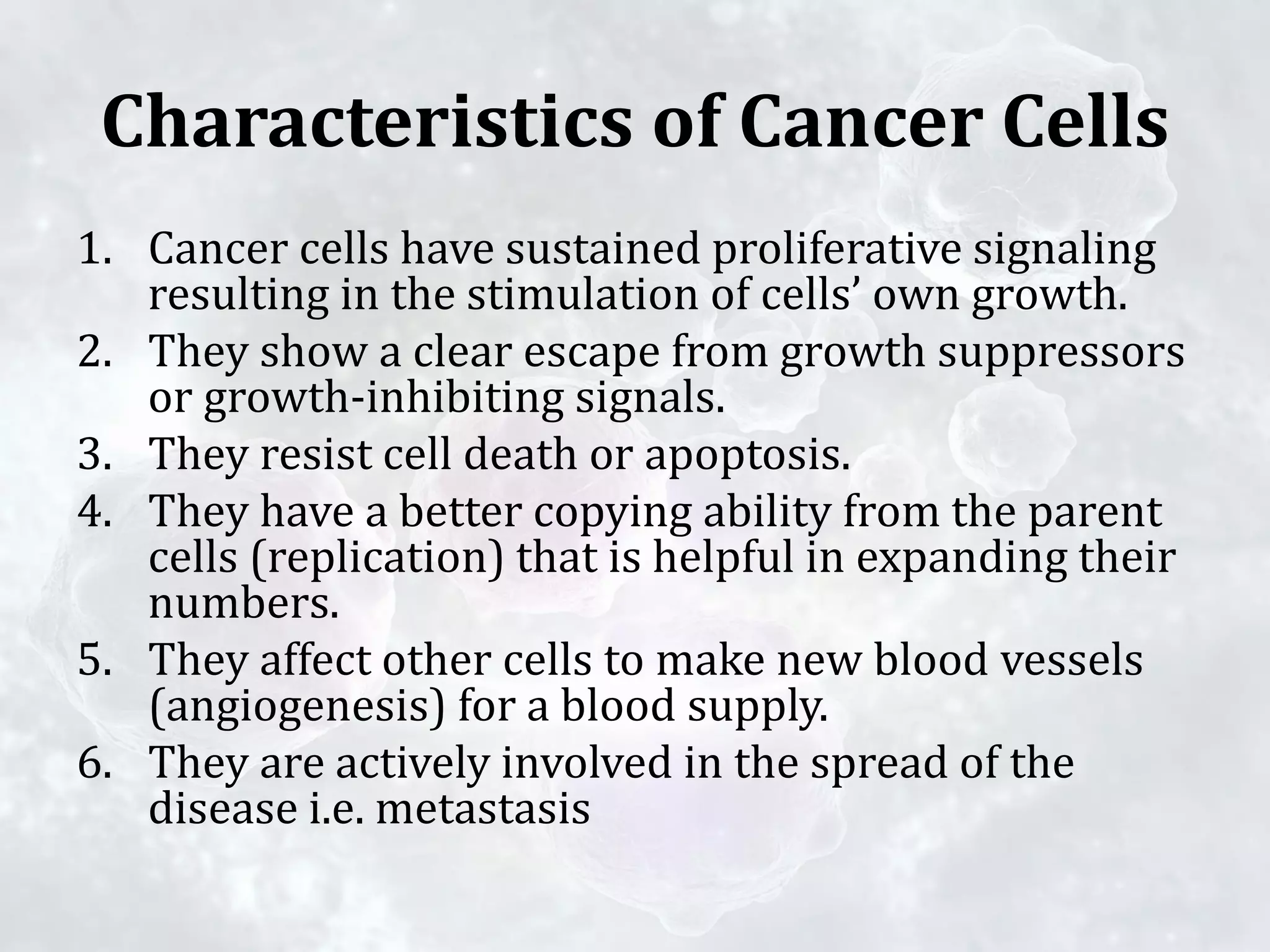


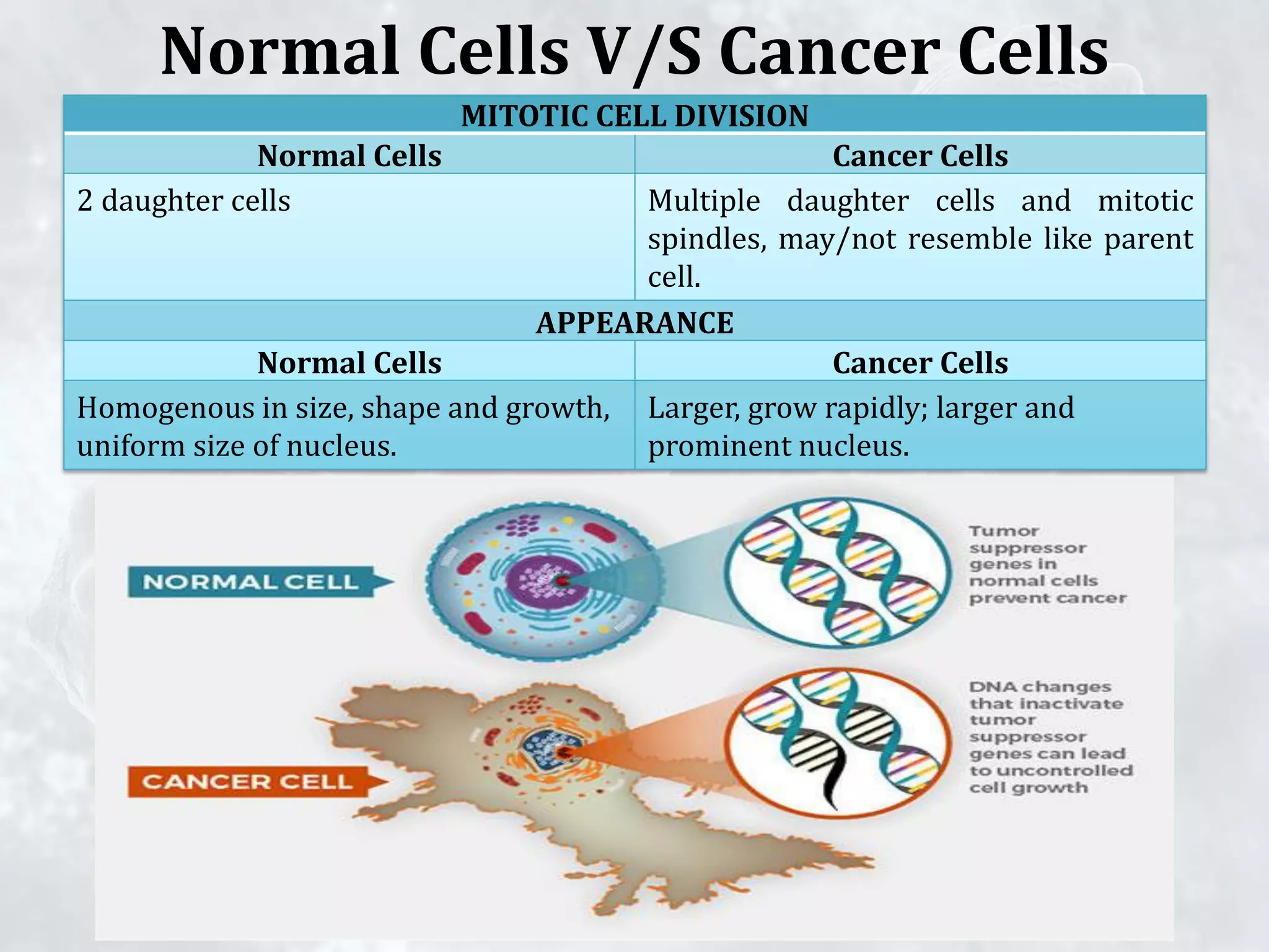


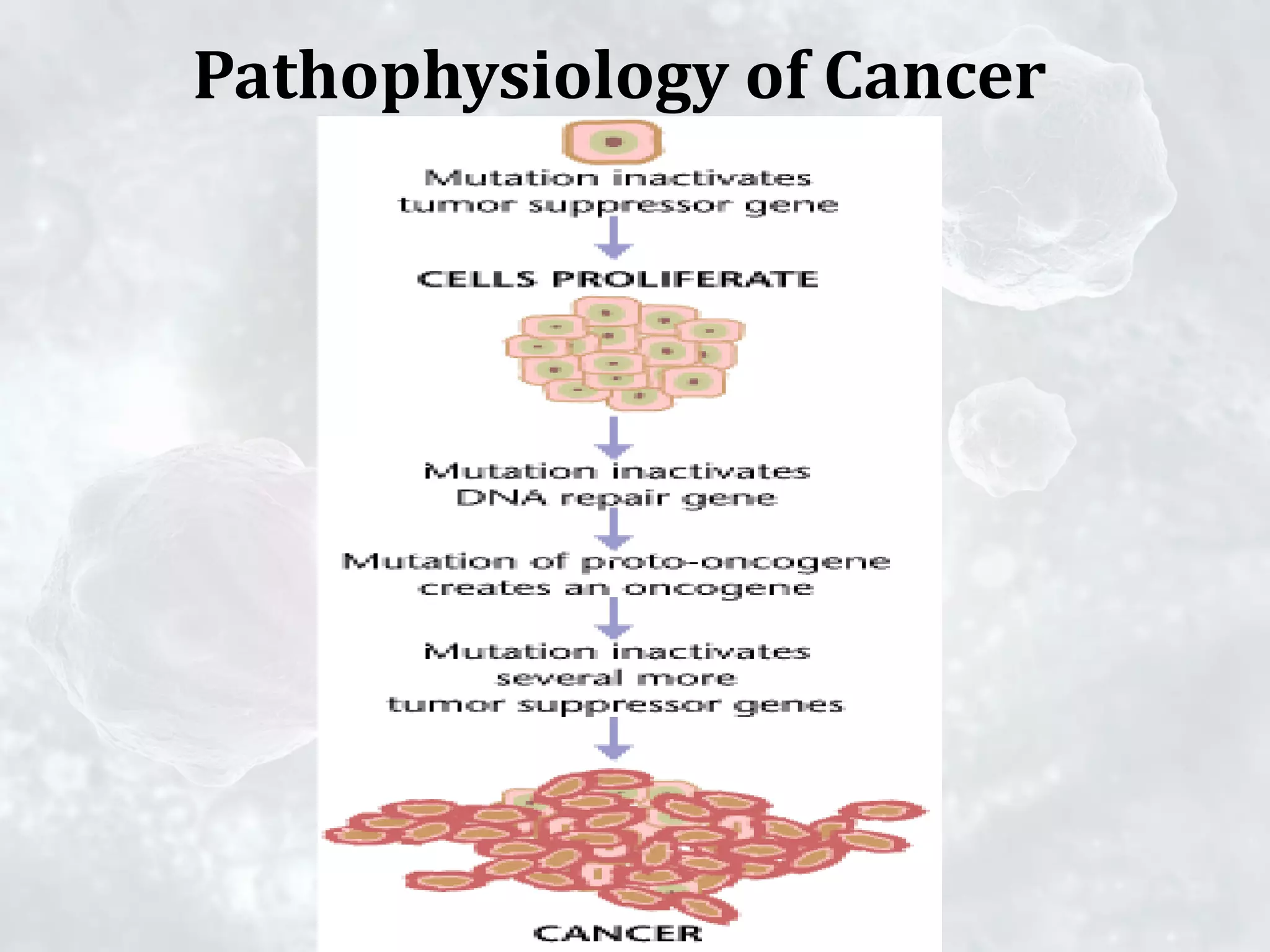


Malignant tumors are cancerous and can invade nearby tissues, spread to other parts of the body through the bloodstream, and form new tumors (metastasis). Benign tumors are not cancerous, do not invade tissues or spread, and can be surgically removed without threat to life. Cancer cells have characteristics like sustained growth signaling, evading growth suppression, resisting cell death, increased replication ability, inducing angiogenesis, and spreading to other areas (metastasis). These characteristics arise through genetic mutations that alter the functions of oncogenes and tumor suppressor genes.


