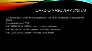
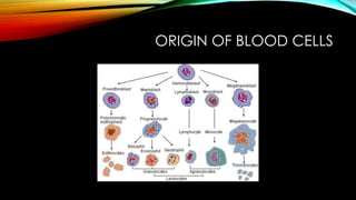
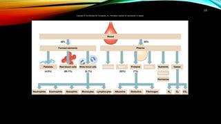
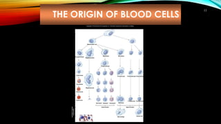
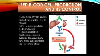
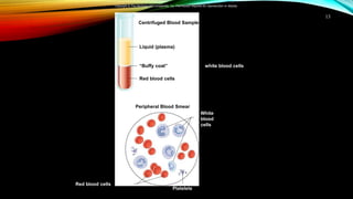
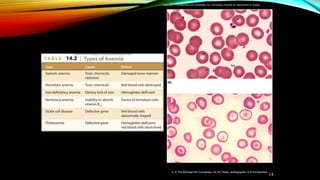
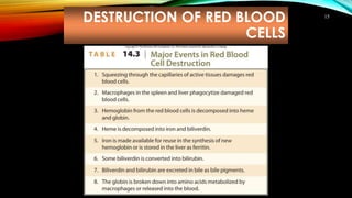
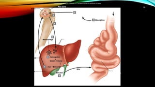
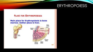
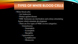
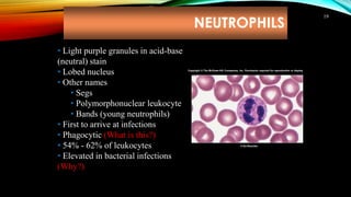
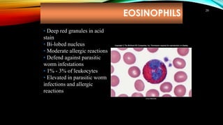
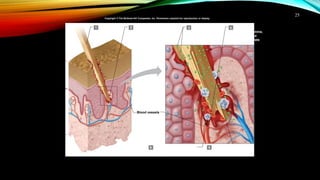
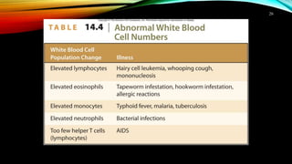
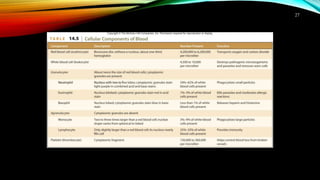
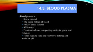
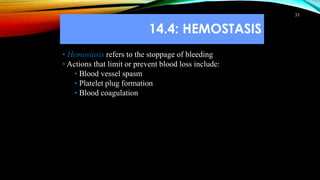
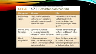
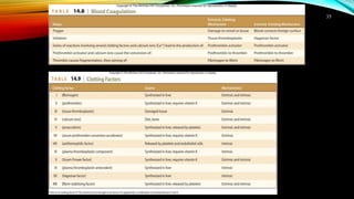
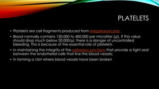
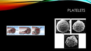
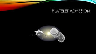
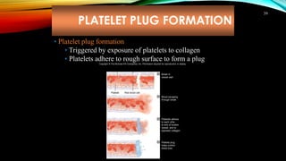
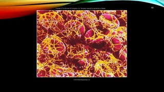
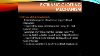
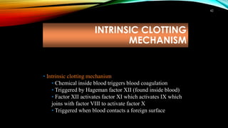
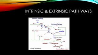
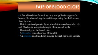
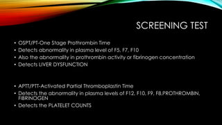
CARDIOVASULAR SYSTEM
Presented by
Dr. SINDHU. K,
MVSc SCHOLAR,
Dept of VPT,
COVAS,
POOKODE
CARDIO VASCULAR SYSTEM
CV physiology is study of the functions of the heart, the blood vessels and the
blood
3 main divisions of CVS
THE DISTRIBUTION SYSTEM - Heart, arteries, arterioles.
THE PERFUSION SYSTEM – arteries, arterioles, capillaries.
THE COLLECTING SYSTEM – venules, veins, heart.
MAJOR COMPONENTS OF THE
CVS
• RESISTANCE COMPONENT – the arterial system
• CAPACITANCE COMPONENT – the venous system
• CARDIAC OUTPUT
• PRE LOAD
• AFTER LOAD
• HYDROSTATIC PRESSURE
• CHRONOTROPIC EFFECT ( heart rate )
• IONOTROPIC EFFECT ( contractility )
IMPORTANCE OF CVS
1. The primary function of CVS is TRANSPORT ( metabolic substrates)
2. CONDUCTS chemical messengers ( harmones )
3. ELECTROLYTE BALANCE & tissue homogeneity
4. Maintains HEMOSTASIS
5. Helps to maintain COLLOIDAL OSMOTIC PRESSURE
WILLIAM HARVEY ( 1628 )
• Father of cardiovascular physiology
• Set forth the first proof that HEART PROPELS THE BLOOD THROUGH BLOOD
VESSELS IN A CIRCULATORY PATTERNS
• Before HARVEY’S proposal it was believed that blood flows in TIDAL FASHION
similar to respiratory system
• However the circularity of the cardiovascular system makes it difficult
• No clear ideas about tissue supply demand & supply of blood to pheriphery
PHYSICAL CHARACTERISTIC OF
CIRCULATION
• SYSTEMIC CIRCULATION
• Blood supplied to all parts of the body
• Also called GREATER / PERIPHERAL CIRCULATION
• PULMONARY CIRCULATION
• Blood transported to LUNGS only
PERFUSION PRESSURE >< TRANSMURAL PRESSURE
• PERFUSION PRESSURE
• Difference in the pressure between the two points in a blood vessel
• Causes the blood to flow through blood vessel
• TRANSMURAL PRESSURE
• Difference between the blood pressure inside a blood vessel & the fluid
pressure outside the vessel
• Causes blood to flow out of a vessel if you poked a hole in the vessel wall
MODE OF TRANSPORT OF BLOOD
FLOW
• BULK FLOW
• Rapid over long distances
• Transport requires ENERGY which is the HYDRO STATIC PRESSURE DIFFERENCE
• DIFFUSION
• Primary mechanism by which dissolved substances move across the wall of
blood vessels from the blood stream into the interstitial fluids or vice versa
• ENERGY source is CONCENTRATION GRADIENT
ORIGIN OF BLOOD CELLS
10
Blood
(4.8%) (95.1%) (0.1%)
Plasma
Hormones
MonocytesBasophilsEosinophilsNeutrophils
(54–62%) (1–3%) (<1%) (3–9%) (25–33%)
GlobulinsAlbumins
(92%) (7%)
N2 O2 CO2
Platelets Red blood cells Proteins Nutrients Gases
45% 55%
WastesWaterWhite blood cells Electrolytes
Vitamins
Lymphocytes Fibrinogen
Formed elements
Copyright © The McGraw-Hill Companies, Inc. Permission required for reproduction or display.
THE ORIGIN OF BLOOD CELLS
11
Copyright © The McGraw-Hill Companies, Inc. Permission required for reproduction or display.
(b)
Megakaryoblast
Myeloid stem cell
Megakaryocyte
Monocyte
Macrophage
T lymphocyte B lymphocyte
Plasma cell
Hematopoietic stem cell
Myeloblast
Progranulocyte
Erythroblast
Normoblast
Reticulocyte
Erythrocyte
Incirculatingblood
Neutrophil Basophil
Granulocytes
Eosinophil
Proerythroblast
Monoblast
Promonocyte Prolymphocyte Prolymphocyte
Inredbonemarrow
Agranulocytes
(a)
Lymphoid stem cell
Lymphoblast
B cell
precursor
Lymphoblast
T cell
precursor
Neutrophilic
myelocyte
Basophilic
myelocyte
Eosinophilic
myelocyte
Eosinophilic
band cell
Basophilic
band cell
Neutrophilic
band cell
Thrombocytes
(platelets)
Activatedintissues
(somecells)
RED BLOOD CELL PRODUCTION
AND ITS CONTROL
12
• Low blood oxygen causes
the kidneys and the liver to
release erythropoietin
(EPO) which stimulates
RBC production
• This is a negative
feedback mechanism
Within a few days many
new blood cells appear in
the circulating blood
Low blood oxygen
Liver Kidney
Erythropoietin
Red bone marrow
+
–
Bloodstream
Stimulation
Inhibition
Release into
bloodstream
Increased
oxygen-
carrying
capacity
Increased
number of
red blood
cells
Copyright © The McGraw-Hill Companies, Inc. Permission required for reproduction or display.
13
Copyright © The McGraw-Hill Companies, Inc. Permission required for reproduction or display.
Centrifuged Blood Sample
Peripheral Blood Smear
Liquid (plasma)
“Buffy coat” white blood cells and platelets)
Red blood cells
Red blood cells
Platelets
White
blood
cells
14
(b)
(a)
a: © The McGraw-Hill Companies, Inc./Al Telser, photographer :b © Ed Reschke
Copyright © The McGraw-Hill Companies, Inc. Permission required for reproduction or display.
DESTRUCTION OF RED BLOOD
CELLS
15
16
Bilirubin
Bone
Blood
Liver
Globin + Heme
3
2
1 Absorption
4
5
Macrophage
Hemoglobin
Iron + Biliverdin
8
6
7
Bile
Red bone
marrow
Red blood
cells produced
Red blood cells
circulate in
bloodstream for
about 120 days
Old red
blood cells
Blood transports
absorbed nutrients
Nutrients
from food
Vitamin B12
Folic acid
Iron
Small
intestine
Copyright © The McGraw-Hill Companies, Inc. Permission required for reproduction or display.
ERYTHROPOIESIS
TYPES OF WHITE BLOOD CELLS
18
• White blood cells:
• Are leukocytes
• Protect against disease
• WBC hormones are interleukins and colony-stimulating
factors which stimulate development
• There are five types of WBCs in two categories:
• Granulocytes
• Neutrophils
• Eosinophils
• Basophils
• Agranulocytes
• Lymphocytes
• Monocytes
NEUTROPHILS
19
• Light purple granules in acid-base
(neutral) stain
• Lobed nucleus
• Other names
• Segs
• Polymorphonuclear leukocyte
• Bands (young neutrophils)
• First to arrive at infections
• Phagocytic (What is this?)
• 54% - 62% of leukocytes
• Elevated in bacterial infections
(Why?)
© Ed Reschke
Copyright © The McGraw-Hill Companies, Inc. Permission required for reproduction or display.
EOSINOPHILS
20
• Deep red granules in acid
stain
• Bi-lobed nucleus
• Moderate allergic reactions
• Defend against parasitic
worm infestations
• 1% - 3% of leukocytes
• Elevated in parasitic worm
infections and allergic
reactions
© Ed Reschke
Copyright © The McGraw-Hill Companies, Inc. Permission required for reproduction or display.
BASOPHILS
21
• Deep blue granules in basic
stain
• Release histamine
•Release heparin Less than
1% of leukocytes
• Similar to Eosinophils in
size and shape of nuclei
© Ed Reschke
Copyright © The McGraw-Hill Companies, Inc. Permission required for reproduction or display.
MONOCYTES
22
• Largest of all blood cells
• Spherical, kidney-shaped,
oval or lobed nuclei
• May leave bloodstream to
become macrophages
• 3% - 9% of leukocytes
• Phagocytize bacteria, dead
cells, and other debris
© R. Kessel/Visuals Unlimited
Copyright © The McGraw-Hill Companies, Inc. Permission required for reproduction or display.
LYMPHOCYTES
23
• Slightly larger than RBC
• Large spherical nucleus surrounded
by thin rim of cytoplasm
• T cells and B cells
•Both important in immunity
• B cells produce antibodies
• 25% - 33% of leukocytes
© Ed Reschke
Copyright © The McGraw-Hill Companies, Inc. Permission required for reproduction or display.
FUNCTIONS OF WHITE BLOOD
CELLS
24
• WBCs protect against infection
• These leukocytes can squeeze between the cells of
a capillary wall and enter the tissue space outside the
blood vessel (called diapedesis)
Blood capillary
Leukocyte
Connective
tissue
Copyright © The McGraw-Hill Companies, Inc. Permission required for reproduction or display.
25
Epidermis
Dermis Blood vessels
1 Splinter
punctures
epidermis
5 6
2 3 4 Injured cells
release histamine,
causing blood
vessels to dilate
Bacteria
multiply
Bacteria are introduced
into the dermis
Neutrophils destroy
bacteria by phagocytosis
Neutrophils move through
blood vessel walls and
migrate toward bacteria
Copyright © The McGraw-Hill Companies, Inc. Permission required for reproduction or display.
26
27
14.3: BLOOD PLASMA
28
• Blood plasma is:
• Straw colored
• The liquid portion of blood
• 55% of blood volume
• 92% water
• Function includes transporting nutrients, gases, and
vitamins
• Helps regulate fluid and electrolyte balance and
maintain pH
PLASMA PROTEINS
29
• These are the most abundant dissolved substances
(solutes) in plasma
GASES AND NUTRIENTS
30
• The most important blood gases:
• Oxygen
• Carbon dioxide
• Plasma nutrients include:
• Amino acids
• Simple sugars
• Nucleotides
• Lipids
• Fats (triglycerides)
• Phospholipids
• Cholesterol
NONPROTEIN NITROGENOUS
SUBSTANCES
31
• These are molecules containing nitrogen but are not
proteins
• In plasma they include:
• Urea – product of protein catabolism; about 50% of
nonprotein nitrogenous substances
• Uric acid – product of nucleic acid catabolism
• Amino acids – product of protein catabolism
• Creatine – stores phosphates
•Creatinine – product of creatine metabolism
• BUN – blood urea nitrogen; indicates health of kidney
PLASMA ELECTROLYTES
32
• Plasma contains a variety of these ions called electrolytes
• They are absorbed from the intestine or released as by-
products of cellular metabolism
They include:
• Sodium (most abundant with chloride)
• Potassium
• Calcium
• Magnesium
• Chloride (most abundant with sodium)
• Bicarbonate
• Phosphate
• Sulfate
14.4: HEMOSTASIS
33
• Hemostasis refers to the stoppage of bleeding
• Actions that limit or prevent blood loss include:
• Blood vessel spasm
• Platelet plug formation
• Blood coagulation
34
35
PLATELETS
• Platelets are cell fragments produced from megakaryocytes.
• Blood normally contains 150,000 to 400,000 per microliter (µl). If this value
should drop much below 20,000/µl, there is a danger of uncontrolled
bleeding. This is because of the essential role of platelets
• in maintaining the integrity of the adherens junctions that provide a tight seal
between the endothelial cells that line the blood vessels;
• in forming a clot where blood vessels have been broken
PLATELETS
PLATELET ADHESION
PLATELET PLUG FORMATION
39
• Platelet plug formation
• Triggered by exposure of platelets to collagen
• Platelets adhere to rough surface to form a plug
Copyright © The McGraw-Hill Companies, Inc. Permission required for reproduction or display.
Endothelial lining Collagen fiber
Platelet Red blood cell
1
2
3
4
Break in
vessel wall
Blood escaping
through break
Platelet plug
helps control
blood loss
Platelets adhere
to each othe ,
to end of broken
vessel, and to
exposed collagen
40
© SPL/Photo Researchers, Inc.
Copyright © The McGraw-Hill Companies, Inc. Permission required for reproduction or display.
EXTRINSIC CLOTTING
MECHANISM
41
• Extrinsic clotting mechanism
• Chemical outside of blood vessel triggers blood
coagulation
• Triggered by tissue thromboplastin (factor III) (not
found in blood)
• A number of events occur that includes factor VII,
factor X, factor V, factor IV, and factor II (prothrombin)
• Triggered when blood contacts damaged blood vessel
walls or tissues
• This is an example of a positive feedback mechanism
INTRINSIC CLOTTING
MECHANISM
42
• Intrinsic clotting mechanism
• Chemical inside blood triggers blood coagulation
• Triggered by Hageman factor XII (found inside blood)
• Factor XII activates factor XI which activates IX which
joins with factor VIII to activate factor X
• Triggered when blood contacts a foreign surface
INTRINSIC & EXTRINSIC PATH WAYS
FATE OF BLOOD CLOTS
44
• After a blood clot forms it retracts and pulls the edges of a
broken blood vessel together while squeezing the fluid serum
from the clot
• Platelet-derived growth factor stimulates smooth muscle cells
and fibroblasts to repair damaged blood vessel walls
• Plasmin digests the blood clots
• A thrombus is an abnormal blood clot
• An embolus is a blood clot moving through the blood vessels
Antithrombin III
As its name suggests, this plasma protein (a serpin) inhibits the formation of
thrombin.
It does so by binding to and thus inactivating:
 prothrombin
 factor 9
 factor 10
Heparin is a mixture of polysaccharides that bind to antithrombin III, inducing
an allosteric change that greatly enhances its inhibition of thrombin synthesis.
Some surgical patients, especially those receiving hip or heart valve
replacements, and people at risk of ischemic stroke (clots in the brain), are
given heparin.
Protein C
With its many clot-promoting activities, it is probably no accident that thrombin sits
at the center of the control mechanism.
 Excess thrombin binds to cell-surface receptors called thrombomodulin.
 The resulting complex activates a plasma protein called Protein C and its
cofactor Protein S.
 Together these inhibit further thrombin formation
o directly — by inactivating Factor 5 and
o indirectly — by inactivating Factor 8.
Vitamin K
Vitamin K is a cofactor needed for the synthesis (in the liver) of
 factors 2 (prothrombin), 7, 9, and 10
 proteins C and S
So a deficiency of Vitamin K predisposes to bleeding.
Conversely, blocking the action of vitamin K helps to prevent inappropriate
clotting.
FIBRINOLYSIS
BLEEDING DISORDERS
• A deficiency of a clotting factor can lead to uncontrolled bleeding.
• The deficiency may arise because
• not enough of the factor is produced or
• a mvon Willebrand disease (the most common)
• hemophilia A for factor 8 deficiency
• hemophilia B for factor 9 deficiencyutant version of the factor fails to perform
properly.
• hemophilia C for factor 11 deficiency
SCREENING TEST
• OSPT/PT-One Stage Prothrombin Time
• Detects abnormality in plasma level of F5, F7, F10
• Also the abnormality in prothrombin activity or fibrinogen concentration
• Detects LIVER DYSFUNCTION
• APTT/PTT-Activated Partial Thromboplastin Time
• Detects the abnormality in plasma levels of F12, F10, F9, F8,PROTHROMBIN,
FIBRINOGEN
• Detects the PLATELET COUNTS
THANK YOU
• Physiology of cardiovascular system
• Origin of blood cells and circulation mechanisms
• Hemostasis
• Coagulation cascade
• Screening tests

CARDIOVASCULAR SYSTEM

  • 1.
    CARDIOVASULAR SYSTEM Presented by Dr.SINDHU. K, MVSc SCHOLAR, Dept of VPT, COVAS, POOKODE
  • 2.
    CARDIO VASCULAR SYSTEM CVphysiology is study of the functions of the heart, the blood vessels and the blood 3 main divisions of CVS THE DISTRIBUTION SYSTEM - Heart, arteries, arterioles. THE PERFUSION SYSTEM – arteries, arterioles, capillaries. THE COLLECTING SYSTEM – venules, veins, heart.
  • 3.
    MAJOR COMPONENTS OFTHE CVS • RESISTANCE COMPONENT – the arterial system • CAPACITANCE COMPONENT – the venous system • CARDIAC OUTPUT • PRE LOAD • AFTER LOAD • HYDROSTATIC PRESSURE • CHRONOTROPIC EFFECT ( heart rate ) • IONOTROPIC EFFECT ( contractility )
  • 4.
    IMPORTANCE OF CVS 1.The primary function of CVS is TRANSPORT ( metabolic substrates) 2. CONDUCTS chemical messengers ( harmones ) 3. ELECTROLYTE BALANCE & tissue homogeneity 4. Maintains HEMOSTASIS 5. Helps to maintain COLLOIDAL OSMOTIC PRESSURE
  • 5.
    WILLIAM HARVEY (1628 ) • Father of cardiovascular physiology • Set forth the first proof that HEART PROPELS THE BLOOD THROUGH BLOOD VESSELS IN A CIRCULATORY PATTERNS • Before HARVEY’S proposal it was believed that blood flows in TIDAL FASHION similar to respiratory system • However the circularity of the cardiovascular system makes it difficult • No clear ideas about tissue supply demand & supply of blood to pheriphery
  • 6.
    PHYSICAL CHARACTERISTIC OF CIRCULATION •SYSTEMIC CIRCULATION • Blood supplied to all parts of the body • Also called GREATER / PERIPHERAL CIRCULATION • PULMONARY CIRCULATION • Blood transported to LUNGS only
  • 7.
    PERFUSION PRESSURE ><TRANSMURAL PRESSURE • PERFUSION PRESSURE • Difference in the pressure between the two points in a blood vessel • Causes the blood to flow through blood vessel • TRANSMURAL PRESSURE • Difference between the blood pressure inside a blood vessel & the fluid pressure outside the vessel • Causes blood to flow out of a vessel if you poked a hole in the vessel wall
  • 8.
    MODE OF TRANSPORTOF BLOOD FLOW • BULK FLOW • Rapid over long distances • Transport requires ENERGY which is the HYDRO STATIC PRESSURE DIFFERENCE • DIFFUSION • Primary mechanism by which dissolved substances move across the wall of blood vessels from the blood stream into the interstitial fluids or vice versa • ENERGY source is CONCENTRATION GRADIENT
  • 9.
  • 10.
    10 Blood (4.8%) (95.1%) (0.1%) Plasma Hormones MonocytesBasophilsEosinophilsNeutrophils (54–62%)(1–3%) (<1%) (3–9%) (25–33%) GlobulinsAlbumins (92%) (7%) N2 O2 CO2 Platelets Red blood cells Proteins Nutrients Gases 45% 55% WastesWaterWhite blood cells Electrolytes Vitamins Lymphocytes Fibrinogen Formed elements Copyright © The McGraw-Hill Companies, Inc. Permission required for reproduction or display.
  • 11.
    THE ORIGIN OFBLOOD CELLS 11 Copyright © The McGraw-Hill Companies, Inc. Permission required for reproduction or display. (b) Megakaryoblast Myeloid stem cell Megakaryocyte Monocyte Macrophage T lymphocyte B lymphocyte Plasma cell Hematopoietic stem cell Myeloblast Progranulocyte Erythroblast Normoblast Reticulocyte Erythrocyte Incirculatingblood Neutrophil Basophil Granulocytes Eosinophil Proerythroblast Monoblast Promonocyte Prolymphocyte Prolymphocyte Inredbonemarrow Agranulocytes (a) Lymphoid stem cell Lymphoblast B cell precursor Lymphoblast T cell precursor Neutrophilic myelocyte Basophilic myelocyte Eosinophilic myelocyte Eosinophilic band cell Basophilic band cell Neutrophilic band cell Thrombocytes (platelets) Activatedintissues (somecells)
  • 12.
    RED BLOOD CELLPRODUCTION AND ITS CONTROL 12 • Low blood oxygen causes the kidneys and the liver to release erythropoietin (EPO) which stimulates RBC production • This is a negative feedback mechanism Within a few days many new blood cells appear in the circulating blood Low blood oxygen Liver Kidney Erythropoietin Red bone marrow + – Bloodstream Stimulation Inhibition Release into bloodstream Increased oxygen- carrying capacity Increased number of red blood cells Copyright © The McGraw-Hill Companies, Inc. Permission required for reproduction or display.
  • 13.
    13 Copyright © TheMcGraw-Hill Companies, Inc. Permission required for reproduction or display. Centrifuged Blood Sample Peripheral Blood Smear Liquid (plasma) “Buffy coat” white blood cells and platelets) Red blood cells Red blood cells Platelets White blood cells
  • 14.
    14 (b) (a) a: © TheMcGraw-Hill Companies, Inc./Al Telser, photographer :b © Ed Reschke Copyright © The McGraw-Hill Companies, Inc. Permission required for reproduction or display.
  • 15.
    DESTRUCTION OF REDBLOOD CELLS 15
  • 16.
    16 Bilirubin Bone Blood Liver Globin + Heme 3 2 1Absorption 4 5 Macrophage Hemoglobin Iron + Biliverdin 8 6 7 Bile Red bone marrow Red blood cells produced Red blood cells circulate in bloodstream for about 120 days Old red blood cells Blood transports absorbed nutrients Nutrients from food Vitamin B12 Folic acid Iron Small intestine Copyright © The McGraw-Hill Companies, Inc. Permission required for reproduction or display.
  • 17.
  • 18.
    TYPES OF WHITEBLOOD CELLS 18 • White blood cells: • Are leukocytes • Protect against disease • WBC hormones are interleukins and colony-stimulating factors which stimulate development • There are five types of WBCs in two categories: • Granulocytes • Neutrophils • Eosinophils • Basophils • Agranulocytes • Lymphocytes • Monocytes
  • 19.
    NEUTROPHILS 19 • Light purplegranules in acid-base (neutral) stain • Lobed nucleus • Other names • Segs • Polymorphonuclear leukocyte • Bands (young neutrophils) • First to arrive at infections • Phagocytic (What is this?) • 54% - 62% of leukocytes • Elevated in bacterial infections (Why?) © Ed Reschke Copyright © The McGraw-Hill Companies, Inc. Permission required for reproduction or display.
  • 20.
    EOSINOPHILS 20 • Deep redgranules in acid stain • Bi-lobed nucleus • Moderate allergic reactions • Defend against parasitic worm infestations • 1% - 3% of leukocytes • Elevated in parasitic worm infections and allergic reactions © Ed Reschke Copyright © The McGraw-Hill Companies, Inc. Permission required for reproduction or display.
  • 21.
    BASOPHILS 21 • Deep bluegranules in basic stain • Release histamine •Release heparin Less than 1% of leukocytes • Similar to Eosinophils in size and shape of nuclei © Ed Reschke Copyright © The McGraw-Hill Companies, Inc. Permission required for reproduction or display.
  • 22.
    MONOCYTES 22 • Largest ofall blood cells • Spherical, kidney-shaped, oval or lobed nuclei • May leave bloodstream to become macrophages • 3% - 9% of leukocytes • Phagocytize bacteria, dead cells, and other debris © R. Kessel/Visuals Unlimited Copyright © The McGraw-Hill Companies, Inc. Permission required for reproduction or display.
  • 23.
    LYMPHOCYTES 23 • Slightly largerthan RBC • Large spherical nucleus surrounded by thin rim of cytoplasm • T cells and B cells •Both important in immunity • B cells produce antibodies • 25% - 33% of leukocytes © Ed Reschke Copyright © The McGraw-Hill Companies, Inc. Permission required for reproduction or display.
  • 24.
    FUNCTIONS OF WHITEBLOOD CELLS 24 • WBCs protect against infection • These leukocytes can squeeze between the cells of a capillary wall and enter the tissue space outside the blood vessel (called diapedesis) Blood capillary Leukocyte Connective tissue Copyright © The McGraw-Hill Companies, Inc. Permission required for reproduction or display.
  • 25.
    25 Epidermis Dermis Blood vessels 1Splinter punctures epidermis 5 6 2 3 4 Injured cells release histamine, causing blood vessels to dilate Bacteria multiply Bacteria are introduced into the dermis Neutrophils destroy bacteria by phagocytosis Neutrophils move through blood vessel walls and migrate toward bacteria Copyright © The McGraw-Hill Companies, Inc. Permission required for reproduction or display.
  • 26.
  • 27.
  • 28.
    14.3: BLOOD PLASMA 28 •Blood plasma is: • Straw colored • The liquid portion of blood • 55% of blood volume • 92% water • Function includes transporting nutrients, gases, and vitamins • Helps regulate fluid and electrolyte balance and maintain pH
  • 29.
    PLASMA PROTEINS 29 • Theseare the most abundant dissolved substances (solutes) in plasma
  • 30.
    GASES AND NUTRIENTS 30 •The most important blood gases: • Oxygen • Carbon dioxide • Plasma nutrients include: • Amino acids • Simple sugars • Nucleotides • Lipids • Fats (triglycerides) • Phospholipids • Cholesterol
  • 31.
    NONPROTEIN NITROGENOUS SUBSTANCES 31 • Theseare molecules containing nitrogen but are not proteins • In plasma they include: • Urea – product of protein catabolism; about 50% of nonprotein nitrogenous substances • Uric acid – product of nucleic acid catabolism • Amino acids – product of protein catabolism • Creatine – stores phosphates •Creatinine – product of creatine metabolism • BUN – blood urea nitrogen; indicates health of kidney
  • 32.
    PLASMA ELECTROLYTES 32 • Plasmacontains a variety of these ions called electrolytes • They are absorbed from the intestine or released as by- products of cellular metabolism They include: • Sodium (most abundant with chloride) • Potassium • Calcium • Magnesium • Chloride (most abundant with sodium) • Bicarbonate • Phosphate • Sulfate
  • 33.
    14.4: HEMOSTASIS 33 • Hemostasisrefers to the stoppage of bleeding • Actions that limit or prevent blood loss include: • Blood vessel spasm • Platelet plug formation • Blood coagulation
  • 34.
  • 35.
  • 36.
    PLATELETS • Platelets arecell fragments produced from megakaryocytes. • Blood normally contains 150,000 to 400,000 per microliter (µl). If this value should drop much below 20,000/µl, there is a danger of uncontrolled bleeding. This is because of the essential role of platelets • in maintaining the integrity of the adherens junctions that provide a tight seal between the endothelial cells that line the blood vessels; • in forming a clot where blood vessels have been broken
  • 37.
  • 38.
  • 39.
    PLATELET PLUG FORMATION 39 •Platelet plug formation • Triggered by exposure of platelets to collagen • Platelets adhere to rough surface to form a plug Copyright © The McGraw-Hill Companies, Inc. Permission required for reproduction or display. Endothelial lining Collagen fiber Platelet Red blood cell 1 2 3 4 Break in vessel wall Blood escaping through break Platelet plug helps control blood loss Platelets adhere to each othe , to end of broken vessel, and to exposed collagen
  • 40.
    40 © SPL/Photo Researchers,Inc. Copyright © The McGraw-Hill Companies, Inc. Permission required for reproduction or display.
  • 41.
    EXTRINSIC CLOTTING MECHANISM 41 • Extrinsicclotting mechanism • Chemical outside of blood vessel triggers blood coagulation • Triggered by tissue thromboplastin (factor III) (not found in blood) • A number of events occur that includes factor VII, factor X, factor V, factor IV, and factor II (prothrombin) • Triggered when blood contacts damaged blood vessel walls or tissues • This is an example of a positive feedback mechanism
  • 42.
    INTRINSIC CLOTTING MECHANISM 42 • Intrinsicclotting mechanism • Chemical inside blood triggers blood coagulation • Triggered by Hageman factor XII (found inside blood) • Factor XII activates factor XI which activates IX which joins with factor VIII to activate factor X • Triggered when blood contacts a foreign surface
  • 43.
  • 44.
    FATE OF BLOODCLOTS 44 • After a blood clot forms it retracts and pulls the edges of a broken blood vessel together while squeezing the fluid serum from the clot • Platelet-derived growth factor stimulates smooth muscle cells and fibroblasts to repair damaged blood vessel walls • Plasmin digests the blood clots • A thrombus is an abnormal blood clot • An embolus is a blood clot moving through the blood vessels
  • 45.
    Antithrombin III As itsname suggests, this plasma protein (a serpin) inhibits the formation of thrombin. It does so by binding to and thus inactivating:  prothrombin  factor 9  factor 10 Heparin is a mixture of polysaccharides that bind to antithrombin III, inducing an allosteric change that greatly enhances its inhibition of thrombin synthesis. Some surgical patients, especially those receiving hip or heart valve replacements, and people at risk of ischemic stroke (clots in the brain), are given heparin.
  • 46.
    Protein C With itsmany clot-promoting activities, it is probably no accident that thrombin sits at the center of the control mechanism.  Excess thrombin binds to cell-surface receptors called thrombomodulin.  The resulting complex activates a plasma protein called Protein C and its cofactor Protein S.  Together these inhibit further thrombin formation o directly — by inactivating Factor 5 and o indirectly — by inactivating Factor 8. Vitamin K Vitamin K is a cofactor needed for the synthesis (in the liver) of  factors 2 (prothrombin), 7, 9, and 10  proteins C and S So a deficiency of Vitamin K predisposes to bleeding. Conversely, blocking the action of vitamin K helps to prevent inappropriate clotting.
  • 47.
  • 48.
    BLEEDING DISORDERS • Adeficiency of a clotting factor can lead to uncontrolled bleeding. • The deficiency may arise because • not enough of the factor is produced or • a mvon Willebrand disease (the most common) • hemophilia A for factor 8 deficiency • hemophilia B for factor 9 deficiencyutant version of the factor fails to perform properly. • hemophilia C for factor 11 deficiency
  • 49.
    SCREENING TEST • OSPT/PT-OneStage Prothrombin Time • Detects abnormality in plasma level of F5, F7, F10 • Also the abnormality in prothrombin activity or fibrinogen concentration • Detects LIVER DYSFUNCTION • APTT/PTT-Activated Partial Thromboplastin Time • Detects the abnormality in plasma levels of F12, F10, F9, F8,PROTHROMBIN, FIBRINOGEN • Detects the PLATELET COUNTS
  • 50.
    THANK YOU • Physiologyof cardiovascular system • Origin of blood cells and circulation mechanisms • Hemostasis • Coagulation cascade • Screening tests