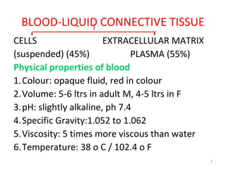
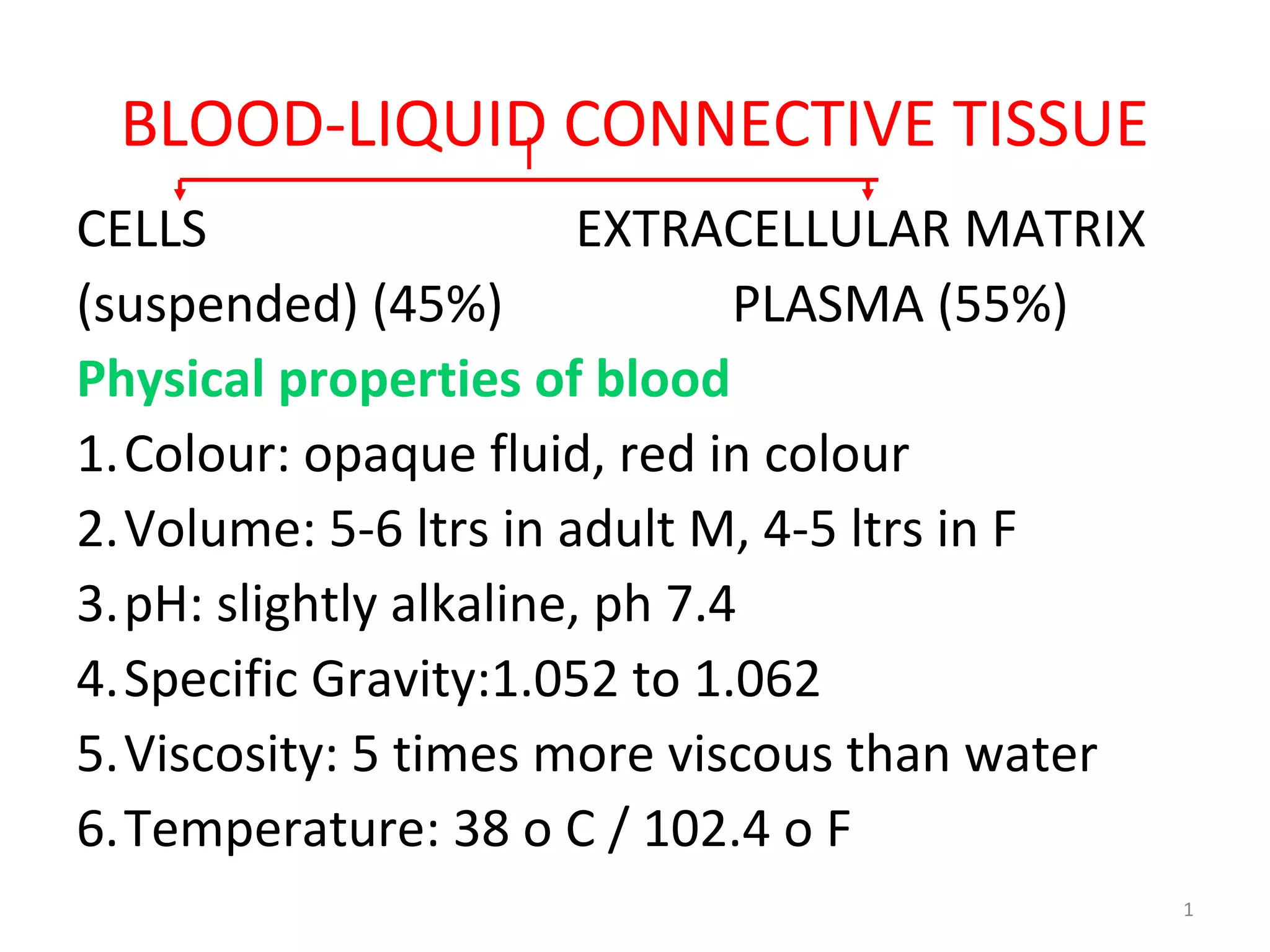
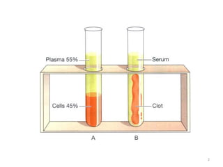
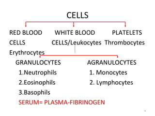
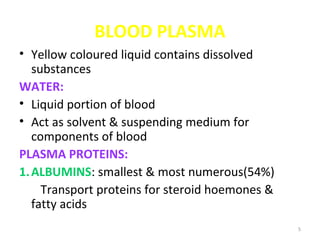
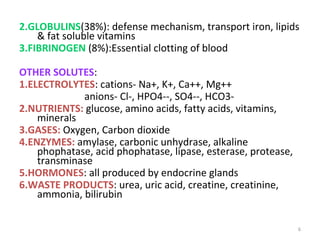
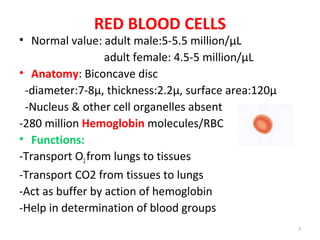
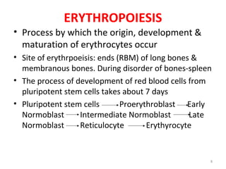
1
BLOOD-LIQUID CONNECTIVE TISSUE
CELLS EXTRACELLULAR MATRIX
(suspended) (45%) PLASMA (55%)
Physical properties of blood
1.Colour: opaque fluid, red in colour
2.Volume: 5-6 ltrs in adult M, 4-5 ltrs in F
3.pH: slightly alkaline, ph 7.4
4.Specific Gravity:1.052 to 1.062
5.Viscosity: 5 times more viscous than water
6.Temperature: 38 o C / 102.4 o F
2
3
PLASMA
WATER 91-92% SOLUTES 8-9%
PROTEINS OTHER SOLUTES
Albumin Electrolytes
Globulin Nutrients
Fibrinogen Gases
Enzymes
Hormones
Waste products
4
CELLS
RED BLOOD WHITE BLOOD PLATELETS
CELLS CELLS/Leukocytes Thrombocytes
Erythrocytes
GRANULOCYTES AGRANULOCYTES
1.Neutrophils 1. Monocytes
2.Eosinophils 2. Lymphocytes
3.Basophils
SERUM= PLASMA-FIBRINOGEN
5
BLOOD PLASMA
• Yellow coloured liquid contains dissolved
substances
WATER:
• Liquid portion of blood
• Act as solvent & suspending medium for
components of blood
PLASMA PROTEINS:
1.ALBUMINS: smallest & most numerous(54%)
Transport proteins for steroid hoemones &
fatty acids
6
2.GLOBULINS(38%): defense mechanism, transport iron, lipids
& fat soluble vitamins
3.FIBRINOGEN (8%):Essential clotting of blood
OTHER SOLUTES:
1.ELECTROLYTES: cations- Na+, K+, Ca++, Mg++
anions- Cl-, HPO4--, SO4--, HCO3-
2.NUTRIENTS: glucose, amino acids, fatty acids, vitamins,
minerals
3.GASES: Oxygen, Carbon dioxide
4.ENZYMES: amylase, carbonic unhydrase, alkaline
phophatase, acid phophatase, lipase, esterase, protease,
transminase
5.HORMONES: all produced by endocrine glands
6.WASTE PRODUCTS: urea, uric acid, creatine, creatinine,
ammonia, bilirubin
7
RED BLOOD CELLS
• Normal value: adult male:5-5.5 million/µL
adult female: 4.5-5 million/µL
• Anatomy: Biconcave disc
-diameter:7-8µ, thickness:2.2µ, surface area:120µ
-Nucleus & other cell organelles absent
-280 million Hemoglobin molecules/RBC
• Functions:
-Transport O2from lungs to tissues
-Transport CO2 from tissues to lungs
-Act as buffer by action of hemoglobin
-Help in determination of blood groups
8
ERYTHROPOIESIS
• Process by which the origin, development &
maturation of erythrocytes occur
• Site of erythrpoeisis: ends (RBM) of long bones &
membranous bones. During disorder of bones-spleen
• The process of development of red blood cells from
pluripotent stem cells takes about 7 days
• Pluripotent stem cells Proerythroblast Early
Normoblast Intermediate Normoblast Late
Normoblast Reticulocyte Erythyrocyte
9
1. PROERYTHROBLAST (Megaloblast)
• First cell derived from Stem Cell
• Size of cell is large:Dia-20-25µ
• Nucleus is large, occupies the cell
• Does not contain hemoglobin
• It multiplies several times and forms next
stage
2. EARLY NORMOBLAST
• Slightly smaller , diameter of about 15µ
• Nucleoli disappear
• Condensation of chromatin network
10
3.INTERMEDIATE NORMOBLAST:
• Small than previous cell, dia of about 10-12µ
• Nucleus still present
• Further condensation of chromatin
• Hemoglobin starts appearing
4.LATE NORMOBLAST:
• Further reduction in cell size, dia 8-10 µ
• Ink spot nucleus
• Increase hemoglobin quantity
• Disintegration of nucleus & disappears
11
5.RETICULOCYTE:
• Slightly larger than matured RBC
• Cytoplasm contains reticulum formed by
remnants of disintegrated organelles
• Below 1% of RBC
• Cells enter capillaries
6.MATURED ERYTHROCYTE:
• Reticulum starts disappearing
• Biconcave & smaller in size-7.2µ
• With hemoglobin & without nucleus
12
WHITE BLOOD CELLS/LEUKOCYTES
• Normal value:4000-11000/mm3
• WBCs have nucleus and of various sizes
• Account for about 1% of the blood volume
WHITE BLOOD CELLS/LEUKOCYTES
GRANULOCYTES AGRANULOCYTES
1.Neutrophils 1. Monocytes
2.Eosinophils 2. Lymphocytes
3.Basophils
13
GRANULOCYTES
• Neutrophils: 10-12µ diameter, nucleus has 2-
5 lobes connected by thin strands of
chromatin, cytoplasm has very fine violet
granules
• Function-Phagocytosis: destruction of bacteria
• Eosinophils:10-12µ diameter, nucleus has 2
lobes, large red-orange granules, almost cover
nucleus
• Function-phagocytize antigen-antibody complex,
parasitic invasion; overcomes effects of histamine
involved in inflammation during allergic reactions
14
•Basophils:8-10µ in diameter, nucleus has 2
lobes, deep blue granules
• Function-liberate heparin, histamine &
sertotonin at inflammation site in allergic
reactions, that intensify overall inflammatory
response
15
AGRANULOCYTES
• Lymphocytes: Small 6-9µ in diameter, large
10-14µ in diameter, nucleus is round
or slightly indented, cytoplasm forms
a rim around the nucleus, sky
blue colour. Larger the cell, more
cytoplasm is visible
• Function: These develop into plasma cells
which secret antibodies
• Monocytes:12-20µ diameter, nucleus is
kidney shaped, cytoplasm is blue-grey
• Function: Phagocytosis
16
SIGNIFICANCE OF LEUKOCYTE COUNT
TYPE OF
WBC
NORMAL
RANGE (%)
HIGH COUNT
INDICATIONS
LOW COUNT
INDICATIONS
Neutrophil 50 - 70 Bacterial infection,
Burns, Stress,
Inflammation
Radiation exposure,
Drug toxicity, Vit B12
deficiency, SLE
Eosinophil 1 - 6 Allergic reactions,
Parasitic infections,
Autoimmune disease
Drug toxicity, Stress
Basophil Less than 1 Allergic reactions,
Leukemia,
Hypothyroidism
Pregnancy, Stress,
Hyperthyroidism
Monocytes 2 - 10 Viral, Fungal infections,
TB, Chronic diseases
Bone marrow
suppression,
treatment with
cortisol
Lymphocytes 20 - 30 Viral infections,
Leukemia
Prolonged illness,
immunosuppression
17
PLATELETS/THROMBOCYTES
• Normal Value:1.5-4.0 lacks/µL.
• Disc shaped, 2-4µ in diameter, has many
vesicles but no nucleus
• Function: help stop blood loss from damaged
vessels by forming platelet plug. Platelet
release chemicals that provide vascular spasm
and blood clotting.
18
Functions of Blood
• Transportation
Blood transports oxygen from lungs to
cells of body and co2 from body cells to
lungs
Carries nutrients from GIT to body cells
Transport enzymes and hormones to
other body cells
Transports waste products to various
organs for elimination from body
19
Regulation
•Blood helps regulate pH through
use of buffers
•Helps adjust body temperature
through heat absorbing and
cooling properties of water in
plasma
•Blood regulates water balance
between intracellular & extra-
cellular fluid
20
Protection
•Blood protects excessive loss of
blood by formation of clot after
injury
•WBC protect against disease by
carrying phagocytosis
•Several types of blood proteins,
interferons help protect against
disease various ways
21
BLOOD GROUPS
• The surfaces of erythrocytes contain a
genetically determined collections of antigens.
These are composed of glycoproteins &
glycolipids.
• Based on the presence or absence of various
antigens, blood is categorized into different
blood groups.
• Within a blood group there may be two or
more different blood types.
• Two blood group systems:-ABO and Rh
22
ABO Blood Group System
Blood
Type
Type A Type B Type AB
(universal
recipient)
Type O
(universal
donor)
Antigen
on
surface of
RBC
A-
antigen
B-antigen Both A &
B-
antigen
Neither A
nor B-
antigen
Antibody
in Plasma
Anti-B
antibody
Anti-A
antibody
Neither
antibody
Both anti-
A, anti-B
23
Rh Blood Group
• Antigen Rh was
discovered in the
blood of Rhesus
Monkey
Blood
Type
Rh +
(positive)
Rh –
(negative)
Antigen Rh
antigen
No Rh
antigen
Antibody No
antibody
No
antibody
24
BLOOD CLOTTING
• Clotting or process of gel formation is a series of
chemical reaction that ends in formation of fibrin
threads.
• Clotting factors: Calcium ions, several inactive
enzymes and various molecules associated with
platelets or released by damaged tissues
• Clotting is a complex cascade of enzymatic reactions
in which each clotting factor activates next factor in
a fixed sequence. Finally, large quantity of product
(Fibrin) is formed.
25
Clotting- 3 Stages
1. Formation of Prothrombinase, either by
extrinsic pathway or intrinsic pathway
Next 2 stages are same-Common pathway
2. Conversion of Prothrombin into Thrombin
3. Conversion of soluble fibrinogen into
insoluble fibrin
Fibrin + Cells Clot
26
Extrinsic Pathway
• Surface of damaged cells release a tissue protein called Tissue
Factor (TF) i.e. Thromboplastin in blood (outside the blood)
• In the presence of Ca++
, TF begins a sequence of reactions that
activates clotting factor X. This factor combines with factor V
in presence of Ca++
forms active enzyme Prothrombinase
Intrinsic Pathway
• Damaged blood vessels activates clotting factor XII, which
begins a sequence of reactions that activates factor X
• Factor X combines with factor V and forms enzyme
Prothrombinase
27
Common Pathway
• Prothrombinase and Ca++
catalyze
conversion of prothrombin into thrombin
• In last stage, thrombin in presence of Ca+
+, converts soluble fibrinogen into loose
fibrin threads, fibrin
• Thrombin also activates factor XIII which
strengthens and stabilizes fibrin threads
into clot.
Blood Transfusion
• Indications:
• Transfusion of whole blood from one
individual to another is indicated for two main
reasons: firstly, when the volume of blood
within the circulation system of the patient is
less than that required to sustain life and,
secondly, when the red blood cells are
deficient either in quantity or quality.
28
Indications
• Anaemia
• Major Surgical Operation
• Accidents resulting in considerable blood loss
• Cancer patients requiring therapy
• Women in childbirth and newborn babies in
certain cases
• Patients of hereditary disorders like
Haemophilia and Thalassaemia
• Severe burn victims
29
30
BLOOD DISORDERS
• Anemia-a condition in which oxygen carrying
capacity of blood is reduced
• Classification of Anemia
1. Impaired erythrocyte production
– Iron deficiency
– Megaloblastic
– Hypoplastic
2. Increased erythrocyte loss
– Hemolytic anemia
– Normocytic anemia
31
• Polycythemia-abnormally large number of
erythrocytes in the blood
• Leukopenia- In this condition, total blood leukocyte
count is less than 4000/mm3
• Granulocytopenia-abnormal reduction in numbers of
circulating granulocytes
• Leukocytosis-In this condition, total blood leukocyte
count more than 11000/mm3
• Leukemia-Uncontrolled increase in production of
leukocytes/ or their precursors
• Thrombocytopenia-In this condition blood platelet
count is below 150000/mm3

05 blood liquid connective tissue

  • 1.
    1 BLOOD-LIQUID CONNECTIVE TISSUE CELLSEXTRACELLULAR MATRIX (suspended) (45%) PLASMA (55%) Physical properties of blood 1.Colour: opaque fluid, red in colour 2.Volume: 5-6 ltrs in adult M, 4-5 ltrs in F 3.pH: slightly alkaline, ph 7.4 4.Specific Gravity:1.052 to 1.062 5.Viscosity: 5 times more viscous than water 6.Temperature: 38 o C / 102.4 o F
  • 2.
  • 3.
    3 PLASMA WATER 91-92% SOLUTES8-9% PROTEINS OTHER SOLUTES Albumin Electrolytes Globulin Nutrients Fibrinogen Gases Enzymes Hormones Waste products
  • 4.
    4 CELLS RED BLOOD WHITEBLOOD PLATELETS CELLS CELLS/Leukocytes Thrombocytes Erythrocytes GRANULOCYTES AGRANULOCYTES 1.Neutrophils 1. Monocytes 2.Eosinophils 2. Lymphocytes 3.Basophils SERUM= PLASMA-FIBRINOGEN
  • 5.
    5 BLOOD PLASMA • Yellowcoloured liquid contains dissolved substances WATER: • Liquid portion of blood • Act as solvent & suspending medium for components of blood PLASMA PROTEINS: 1.ALBUMINS: smallest & most numerous(54%) Transport proteins for steroid hoemones & fatty acids
  • 6.
    6 2.GLOBULINS(38%): defense mechanism,transport iron, lipids & fat soluble vitamins 3.FIBRINOGEN (8%):Essential clotting of blood OTHER SOLUTES: 1.ELECTROLYTES: cations- Na+, K+, Ca++, Mg++ anions- Cl-, HPO4--, SO4--, HCO3- 2.NUTRIENTS: glucose, amino acids, fatty acids, vitamins, minerals 3.GASES: Oxygen, Carbon dioxide 4.ENZYMES: amylase, carbonic unhydrase, alkaline phophatase, acid phophatase, lipase, esterase, protease, transminase 5.HORMONES: all produced by endocrine glands 6.WASTE PRODUCTS: urea, uric acid, creatine, creatinine, ammonia, bilirubin
  • 7.
    7 RED BLOOD CELLS •Normal value: adult male:5-5.5 million/µL adult female: 4.5-5 million/µL • Anatomy: Biconcave disc -diameter:7-8µ, thickness:2.2µ, surface area:120µ -Nucleus & other cell organelles absent -280 million Hemoglobin molecules/RBC • Functions: -Transport O2from lungs to tissues -Transport CO2 from tissues to lungs -Act as buffer by action of hemoglobin -Help in determination of blood groups
  • 8.
    8 ERYTHROPOIESIS • Process bywhich the origin, development & maturation of erythrocytes occur • Site of erythrpoeisis: ends (RBM) of long bones & membranous bones. During disorder of bones-spleen • The process of development of red blood cells from pluripotent stem cells takes about 7 days • Pluripotent stem cells Proerythroblast Early Normoblast Intermediate Normoblast Late Normoblast Reticulocyte Erythyrocyte
  • 9.
    9 1. PROERYTHROBLAST (Megaloblast) •First cell derived from Stem Cell • Size of cell is large:Dia-20-25µ • Nucleus is large, occupies the cell • Does not contain hemoglobin • It multiplies several times and forms next stage 2. EARLY NORMOBLAST • Slightly smaller , diameter of about 15µ • Nucleoli disappear • Condensation of chromatin network
  • 10.
    10 3.INTERMEDIATE NORMOBLAST: • Smallthan previous cell, dia of about 10-12µ • Nucleus still present • Further condensation of chromatin • Hemoglobin starts appearing 4.LATE NORMOBLAST: • Further reduction in cell size, dia 8-10 µ • Ink spot nucleus • Increase hemoglobin quantity • Disintegration of nucleus & disappears
  • 11.
    11 5.RETICULOCYTE: • Slightly largerthan matured RBC • Cytoplasm contains reticulum formed by remnants of disintegrated organelles • Below 1% of RBC • Cells enter capillaries 6.MATURED ERYTHROCYTE: • Reticulum starts disappearing • Biconcave & smaller in size-7.2µ • With hemoglobin & without nucleus
  • 12.
    12 WHITE BLOOD CELLS/LEUKOCYTES •Normal value:4000-11000/mm3 • WBCs have nucleus and of various sizes • Account for about 1% of the blood volume WHITE BLOOD CELLS/LEUKOCYTES GRANULOCYTES AGRANULOCYTES 1.Neutrophils 1. Monocytes 2.Eosinophils 2. Lymphocytes 3.Basophils
  • 13.
    13 GRANULOCYTES • Neutrophils: 10-12µdiameter, nucleus has 2- 5 lobes connected by thin strands of chromatin, cytoplasm has very fine violet granules • Function-Phagocytosis: destruction of bacteria • Eosinophils:10-12µ diameter, nucleus has 2 lobes, large red-orange granules, almost cover nucleus • Function-phagocytize antigen-antibody complex, parasitic invasion; overcomes effects of histamine involved in inflammation during allergic reactions
  • 14.
    14 •Basophils:8-10µ in diameter,nucleus has 2 lobes, deep blue granules • Function-liberate heparin, histamine & sertotonin at inflammation site in allergic reactions, that intensify overall inflammatory response
  • 15.
    15 AGRANULOCYTES • Lymphocytes: Small6-9µ in diameter, large 10-14µ in diameter, nucleus is round or slightly indented, cytoplasm forms a rim around the nucleus, sky blue colour. Larger the cell, more cytoplasm is visible • Function: These develop into plasma cells which secret antibodies • Monocytes:12-20µ diameter, nucleus is kidney shaped, cytoplasm is blue-grey • Function: Phagocytosis
  • 16.
    16 SIGNIFICANCE OF LEUKOCYTECOUNT TYPE OF WBC NORMAL RANGE (%) HIGH COUNT INDICATIONS LOW COUNT INDICATIONS Neutrophil 50 - 70 Bacterial infection, Burns, Stress, Inflammation Radiation exposure, Drug toxicity, Vit B12 deficiency, SLE Eosinophil 1 - 6 Allergic reactions, Parasitic infections, Autoimmune disease Drug toxicity, Stress Basophil Less than 1 Allergic reactions, Leukemia, Hypothyroidism Pregnancy, Stress, Hyperthyroidism Monocytes 2 - 10 Viral, Fungal infections, TB, Chronic diseases Bone marrow suppression, treatment with cortisol Lymphocytes 20 - 30 Viral infections, Leukemia Prolonged illness, immunosuppression
  • 17.
    17 PLATELETS/THROMBOCYTES • Normal Value:1.5-4.0lacks/µL. • Disc shaped, 2-4µ in diameter, has many vesicles but no nucleus • Function: help stop blood loss from damaged vessels by forming platelet plug. Platelet release chemicals that provide vascular spasm and blood clotting.
  • 18.
    18 Functions of Blood •Transportation Blood transports oxygen from lungs to cells of body and co2 from body cells to lungs Carries nutrients from GIT to body cells Transport enzymes and hormones to other body cells Transports waste products to various organs for elimination from body
  • 19.
    19 Regulation •Blood helps regulatepH through use of buffers •Helps adjust body temperature through heat absorbing and cooling properties of water in plasma •Blood regulates water balance between intracellular & extra- cellular fluid
  • 20.
    20 Protection •Blood protects excessiveloss of blood by formation of clot after injury •WBC protect against disease by carrying phagocytosis •Several types of blood proteins, interferons help protect against disease various ways
  • 21.
    21 BLOOD GROUPS • Thesurfaces of erythrocytes contain a genetically determined collections of antigens. These are composed of glycoproteins & glycolipids. • Based on the presence or absence of various antigens, blood is categorized into different blood groups. • Within a blood group there may be two or more different blood types. • Two blood group systems:-ABO and Rh
  • 22.
    22 ABO Blood GroupSystem Blood Type Type A Type B Type AB (universal recipient) Type O (universal donor) Antigen on surface of RBC A- antigen B-antigen Both A & B- antigen Neither A nor B- antigen Antibody in Plasma Anti-B antibody Anti-A antibody Neither antibody Both anti- A, anti-B
  • 23.
    23 Rh Blood Group •Antigen Rh was discovered in the blood of Rhesus Monkey Blood Type Rh + (positive) Rh – (negative) Antigen Rh antigen No Rh antigen Antibody No antibody No antibody
  • 24.
    24 BLOOD CLOTTING • Clottingor process of gel formation is a series of chemical reaction that ends in formation of fibrin threads. • Clotting factors: Calcium ions, several inactive enzymes and various molecules associated with platelets or released by damaged tissues • Clotting is a complex cascade of enzymatic reactions in which each clotting factor activates next factor in a fixed sequence. Finally, large quantity of product (Fibrin) is formed.
  • 25.
    25 Clotting- 3 Stages 1.Formation of Prothrombinase, either by extrinsic pathway or intrinsic pathway Next 2 stages are same-Common pathway 2. Conversion of Prothrombin into Thrombin 3. Conversion of soluble fibrinogen into insoluble fibrin Fibrin + Cells Clot
  • 26.
    26 Extrinsic Pathway • Surfaceof damaged cells release a tissue protein called Tissue Factor (TF) i.e. Thromboplastin in blood (outside the blood) • In the presence of Ca++ , TF begins a sequence of reactions that activates clotting factor X. This factor combines with factor V in presence of Ca++ forms active enzyme Prothrombinase Intrinsic Pathway • Damaged blood vessels activates clotting factor XII, which begins a sequence of reactions that activates factor X • Factor X combines with factor V and forms enzyme Prothrombinase
  • 27.
    27 Common Pathway • Prothrombinaseand Ca++ catalyze conversion of prothrombin into thrombin • In last stage, thrombin in presence of Ca+ +, converts soluble fibrinogen into loose fibrin threads, fibrin • Thrombin also activates factor XIII which strengthens and stabilizes fibrin threads into clot.
  • 28.
    Blood Transfusion • Indications: •Transfusion of whole blood from one individual to another is indicated for two main reasons: firstly, when the volume of blood within the circulation system of the patient is less than that required to sustain life and, secondly, when the red blood cells are deficient either in quantity or quality. 28
  • 29.
    Indications • Anaemia • MajorSurgical Operation • Accidents resulting in considerable blood loss • Cancer patients requiring therapy • Women in childbirth and newborn babies in certain cases • Patients of hereditary disorders like Haemophilia and Thalassaemia • Severe burn victims 29
  • 30.
    30 BLOOD DISORDERS • Anemia-acondition in which oxygen carrying capacity of blood is reduced • Classification of Anemia 1. Impaired erythrocyte production – Iron deficiency – Megaloblastic – Hypoplastic 2. Increased erythrocyte loss – Hemolytic anemia – Normocytic anemia
  • 31.
    31 • Polycythemia-abnormally largenumber of erythrocytes in the blood • Leukopenia- In this condition, total blood leukocyte count is less than 4000/mm3 • Granulocytopenia-abnormal reduction in numbers of circulating granulocytes • Leukocytosis-In this condition, total blood leukocyte count more than 11000/mm3 • Leukemia-Uncontrolled increase in production of leukocytes/ or their precursors • Thrombocytopenia-In this condition blood platelet count is below 150000/mm3