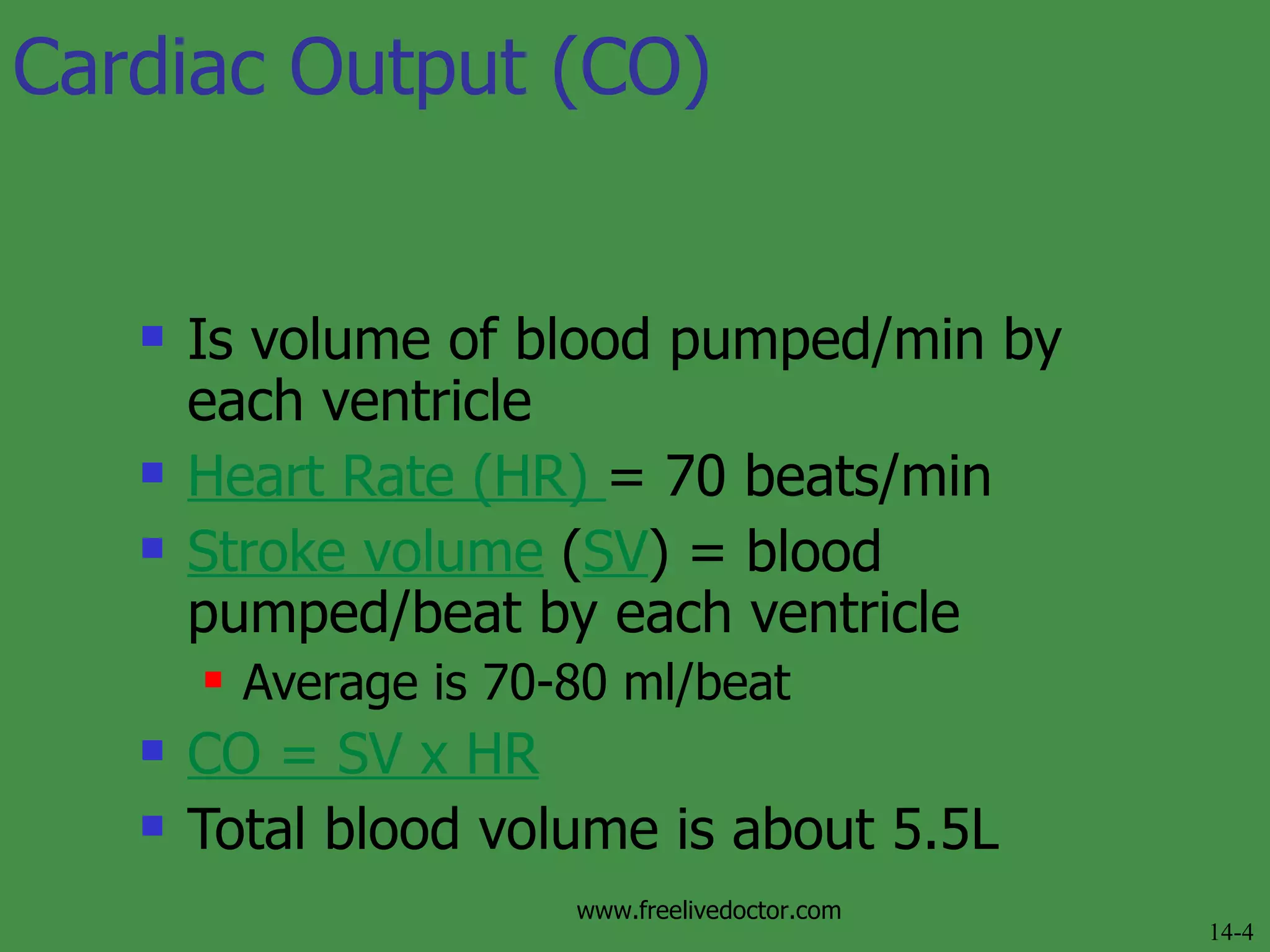
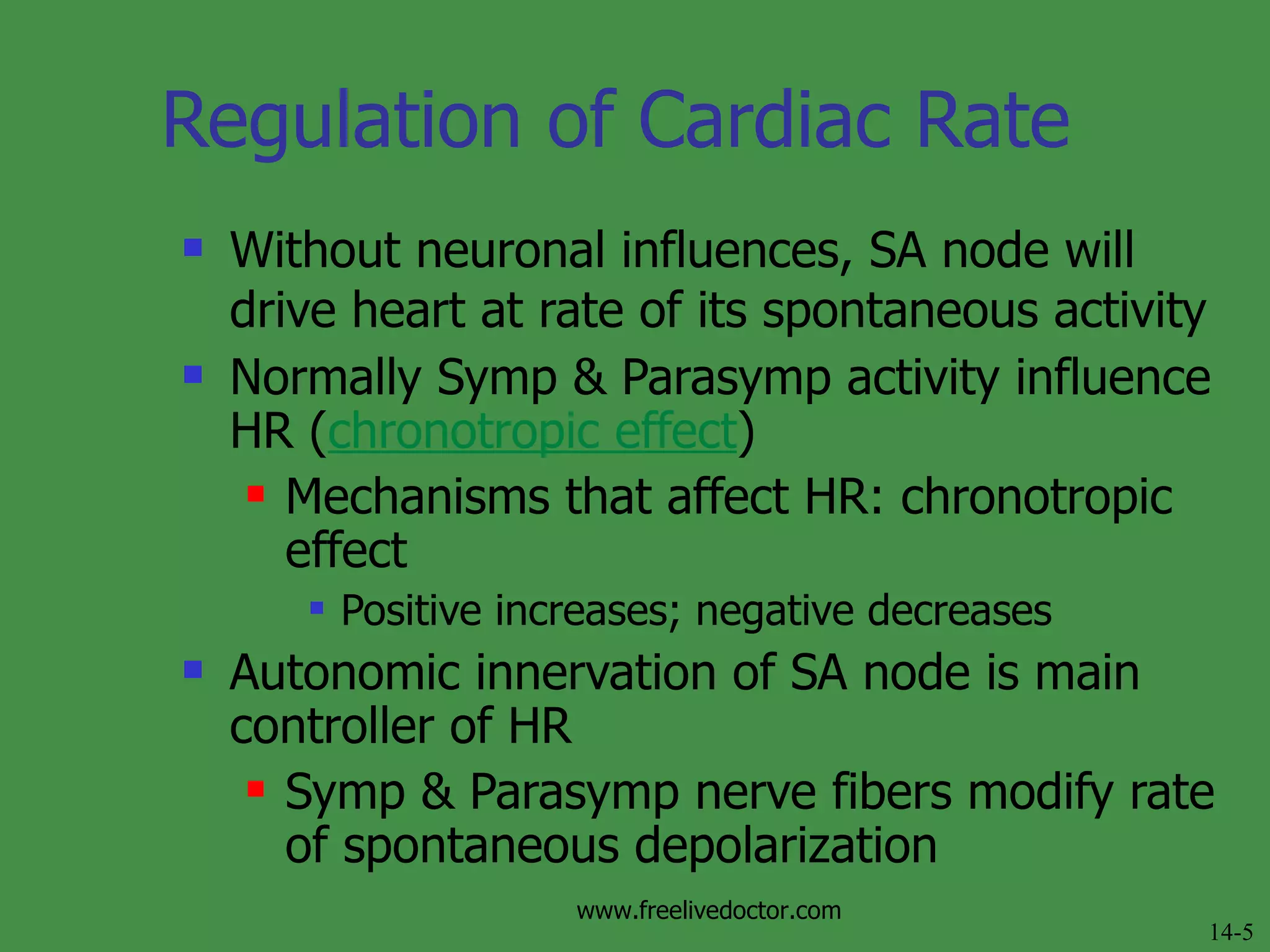
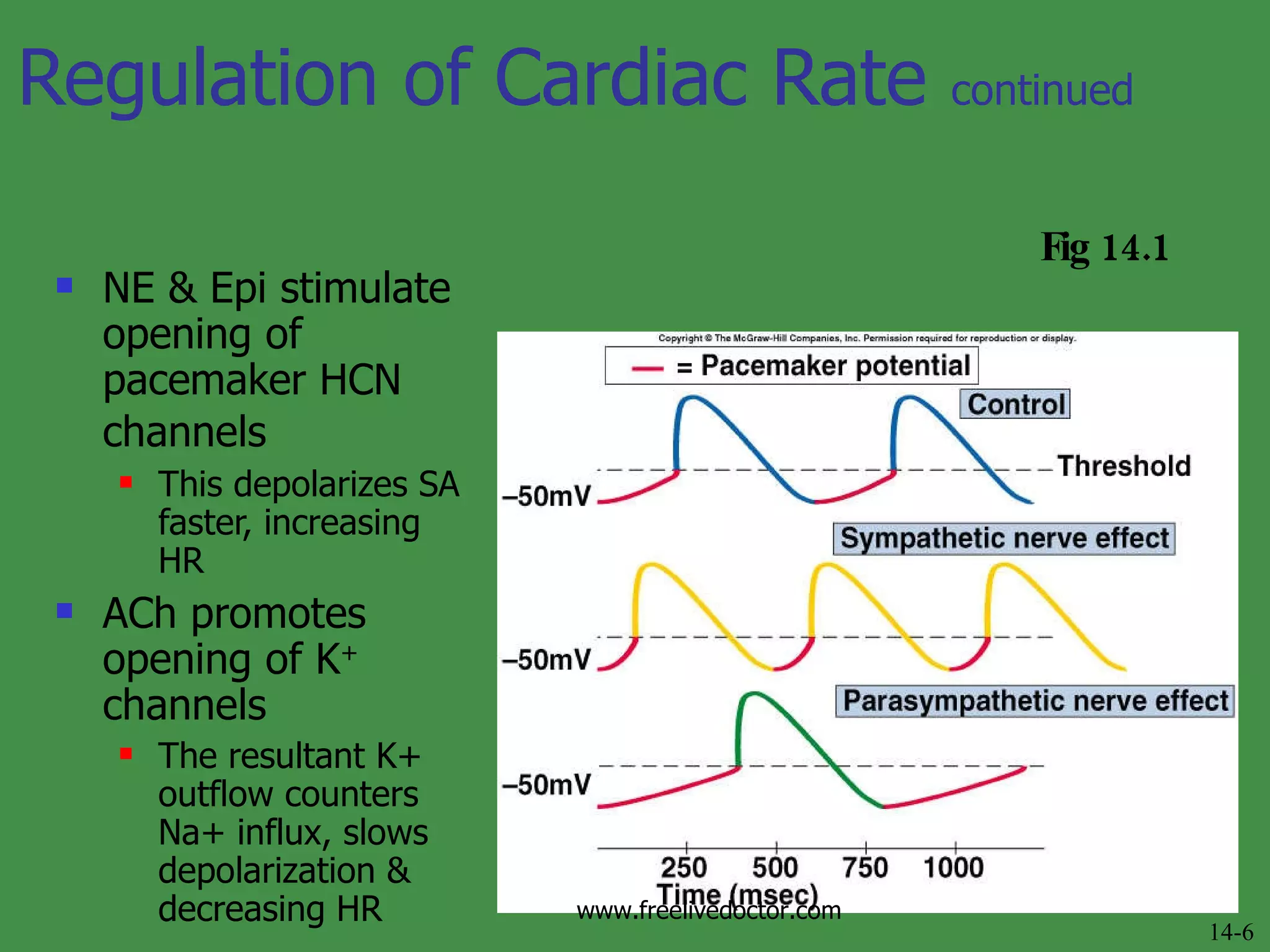
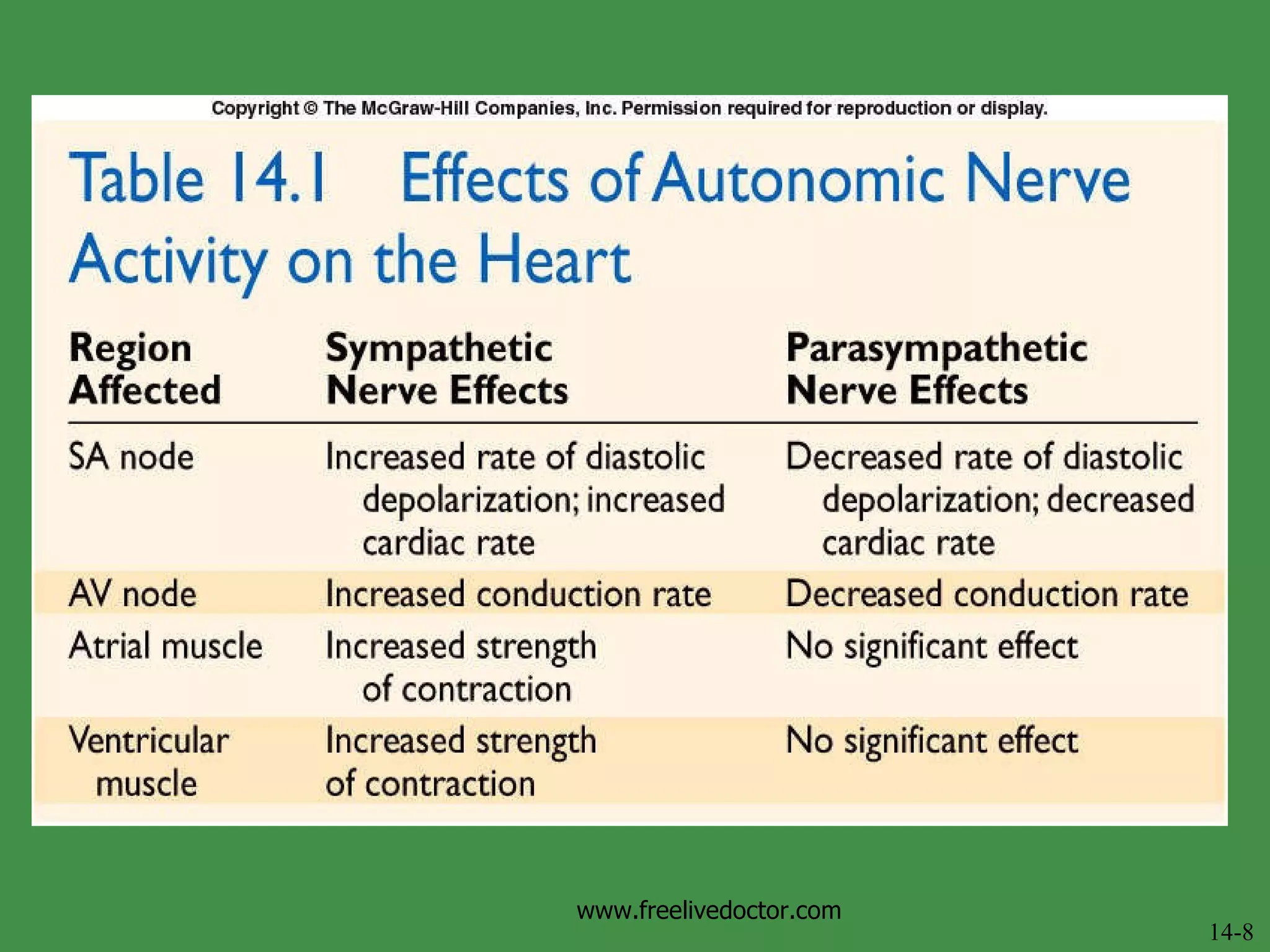
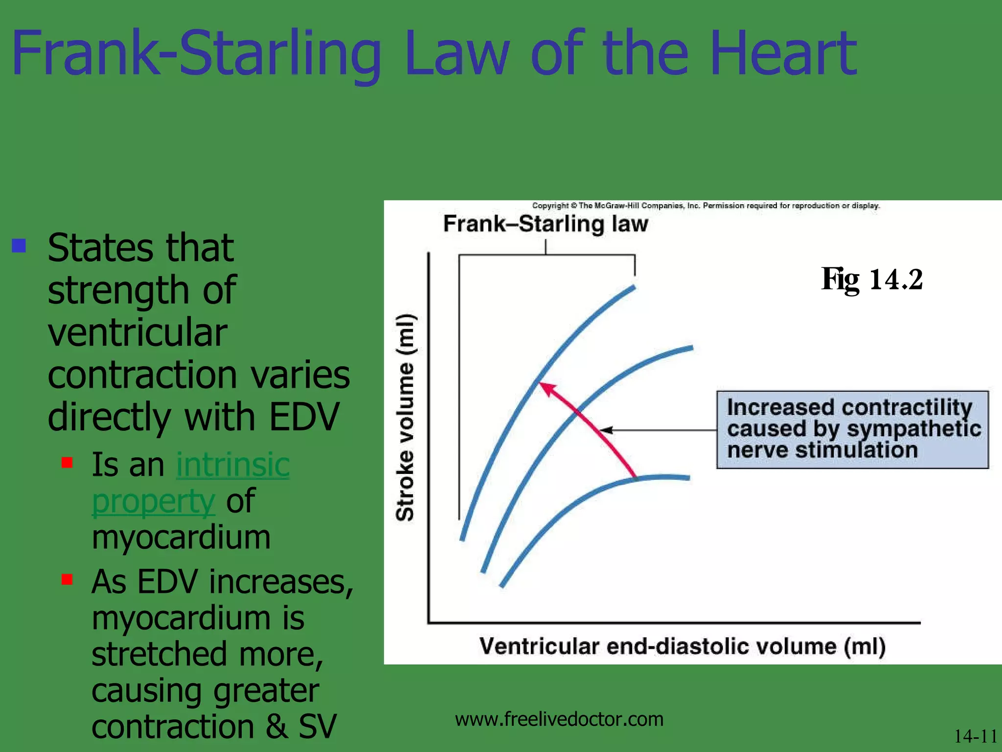
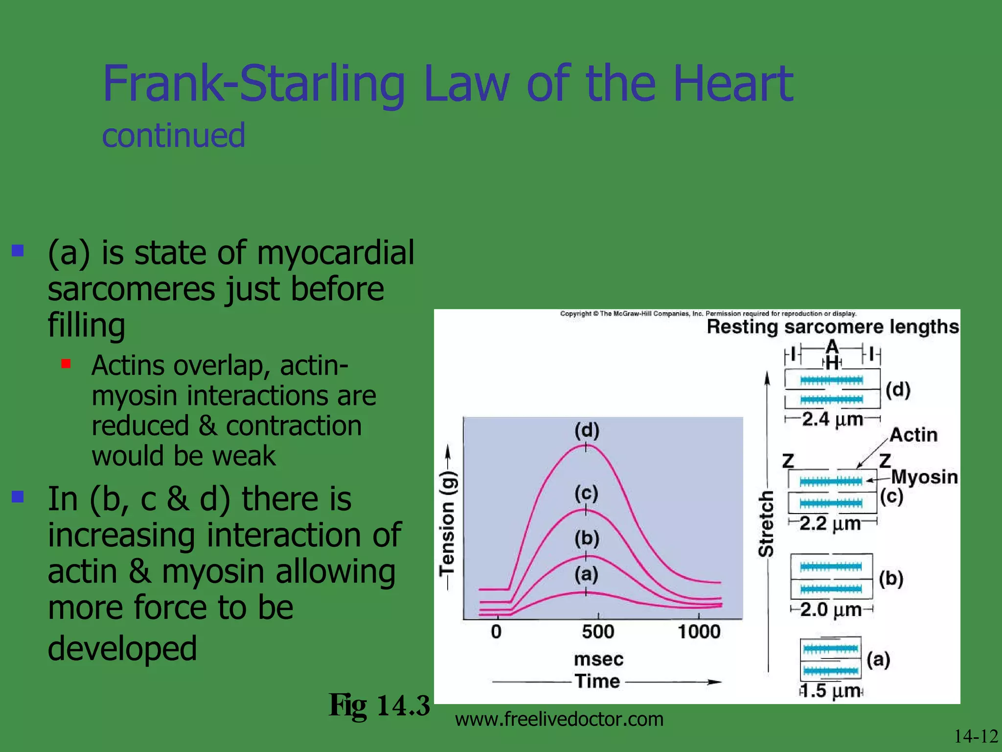
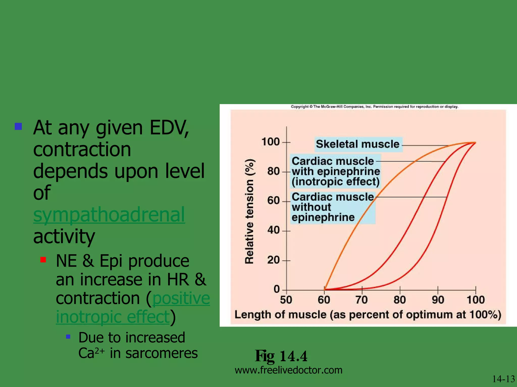
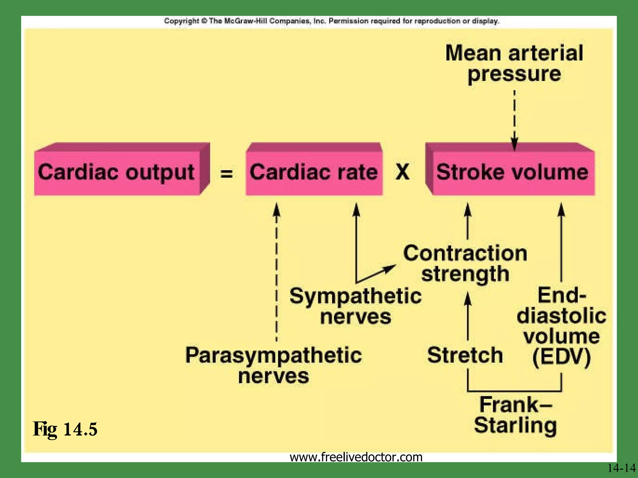
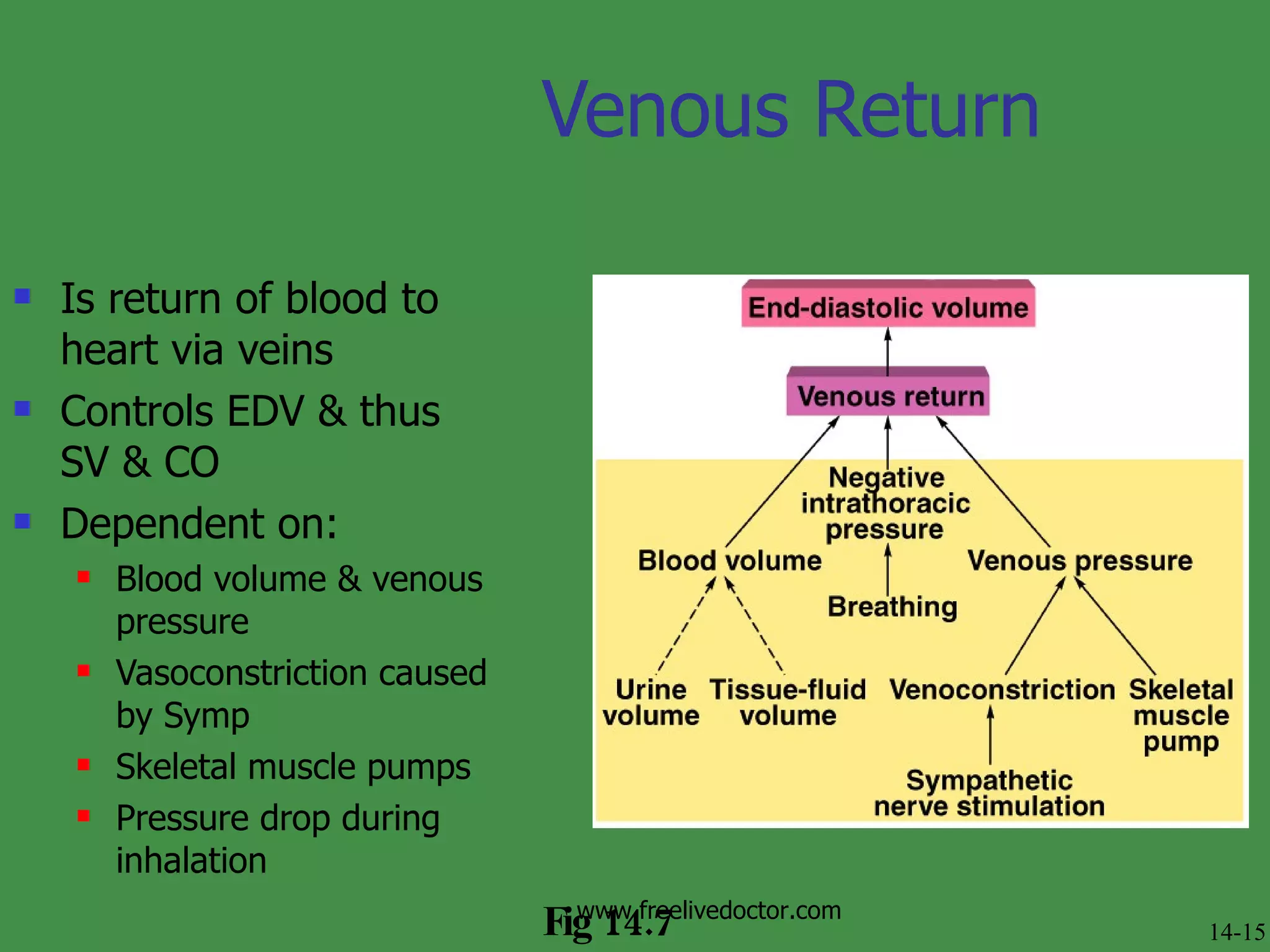
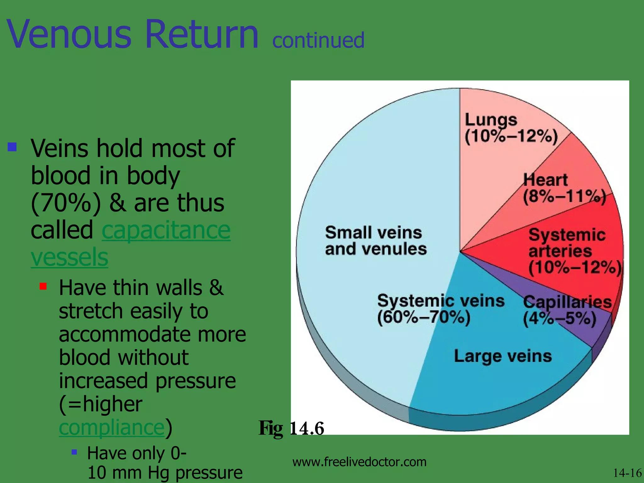
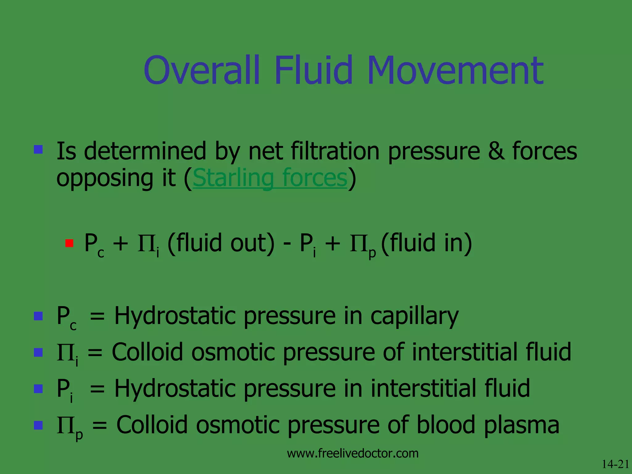
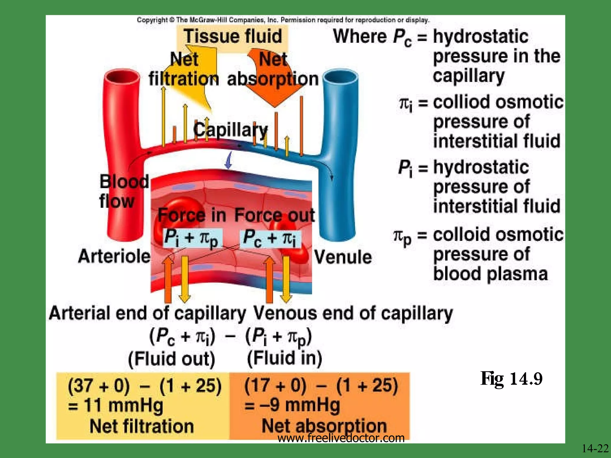
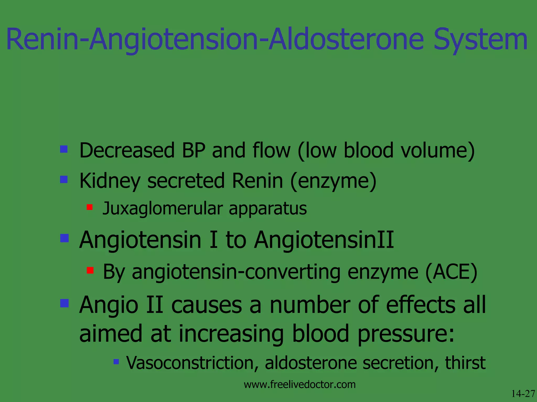
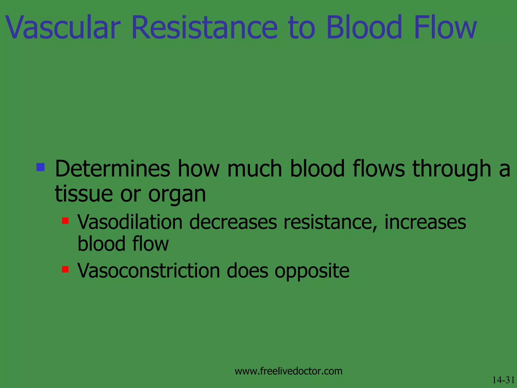
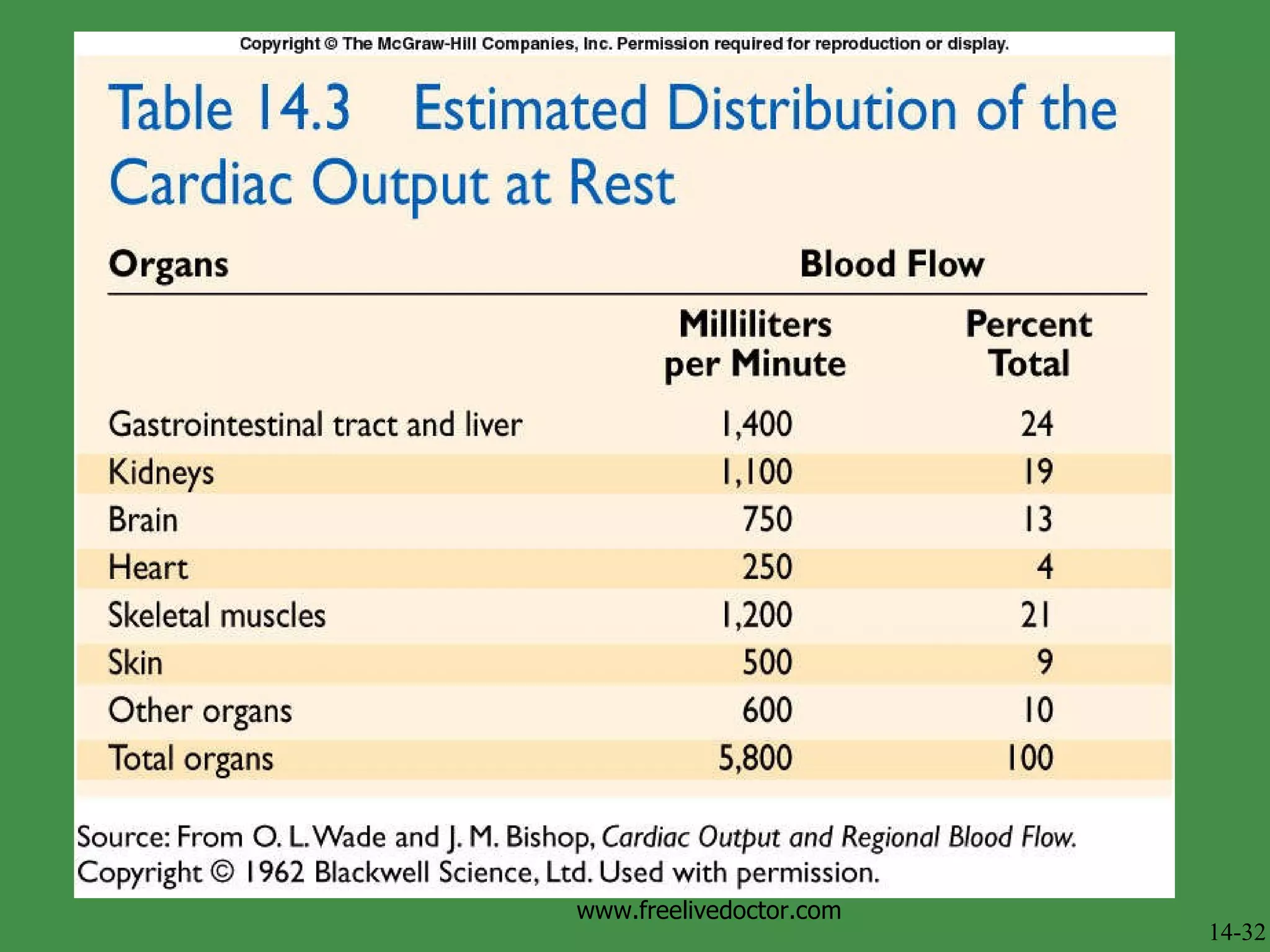
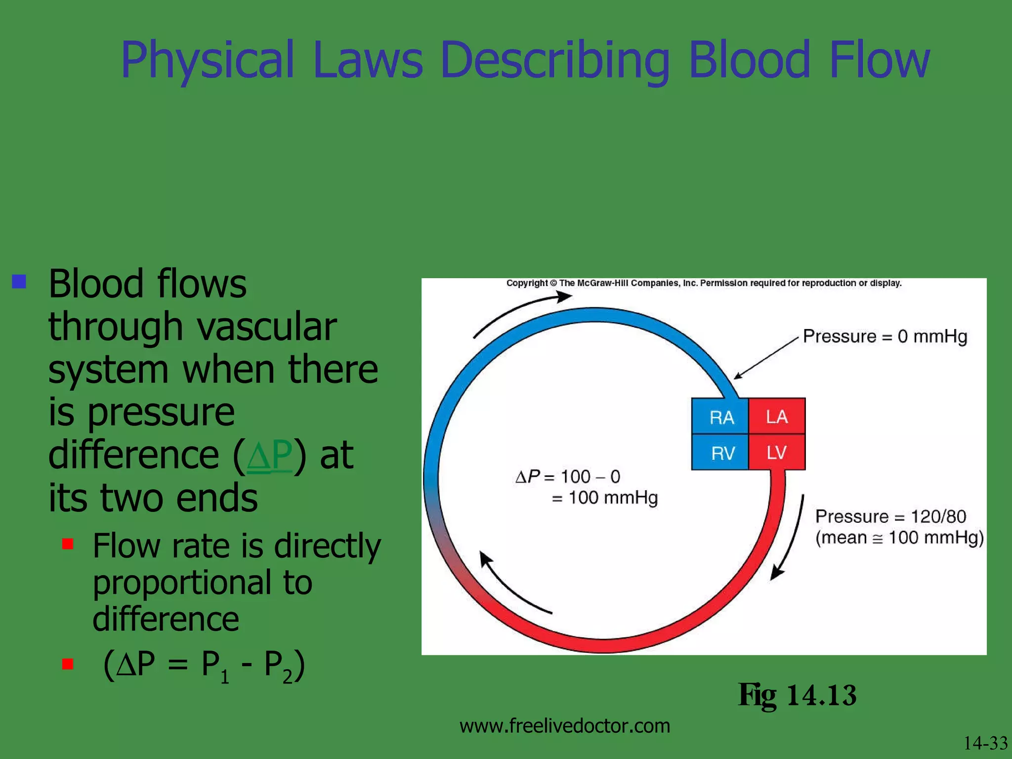
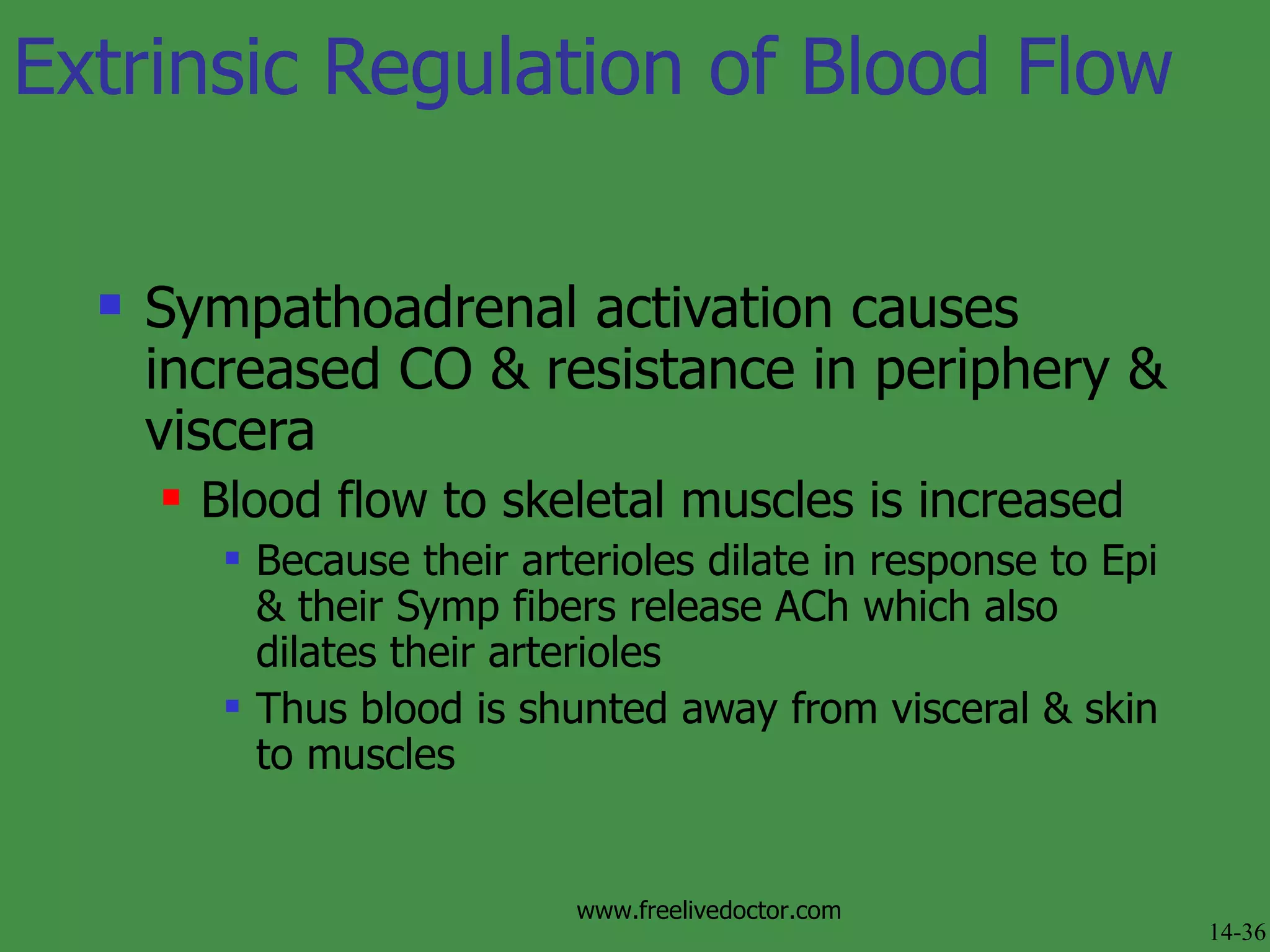
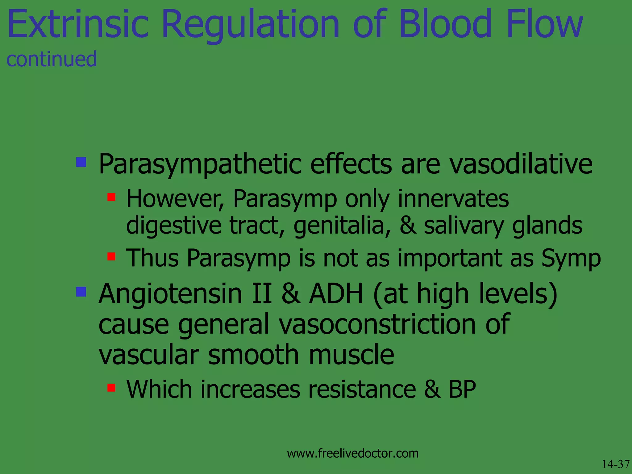
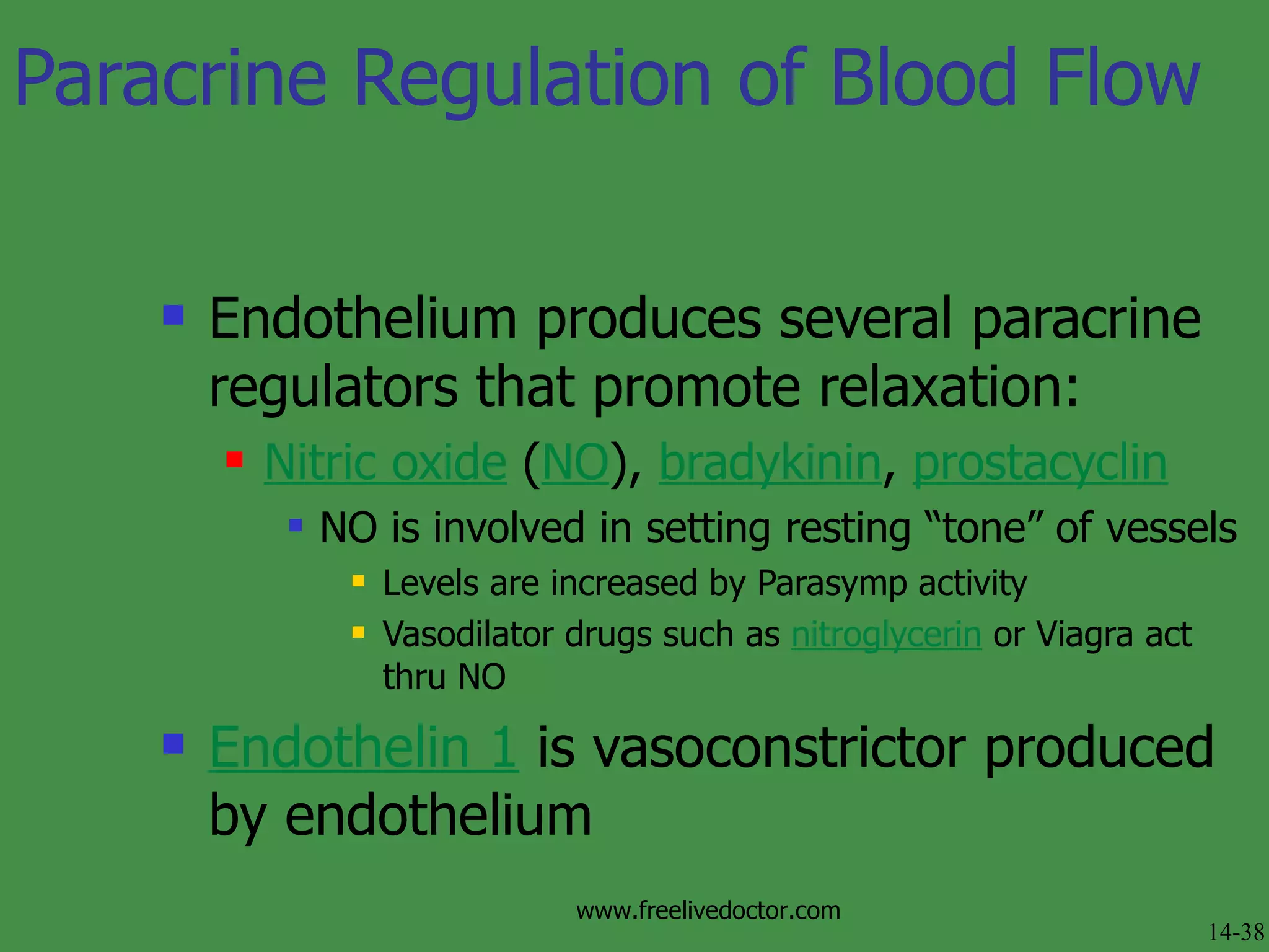
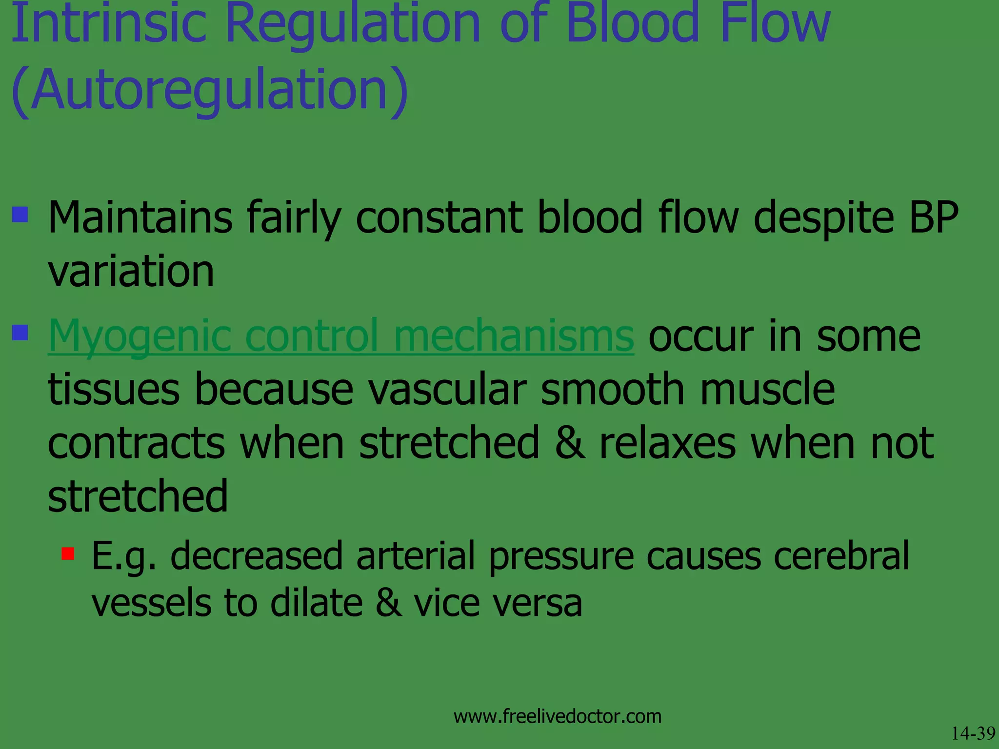
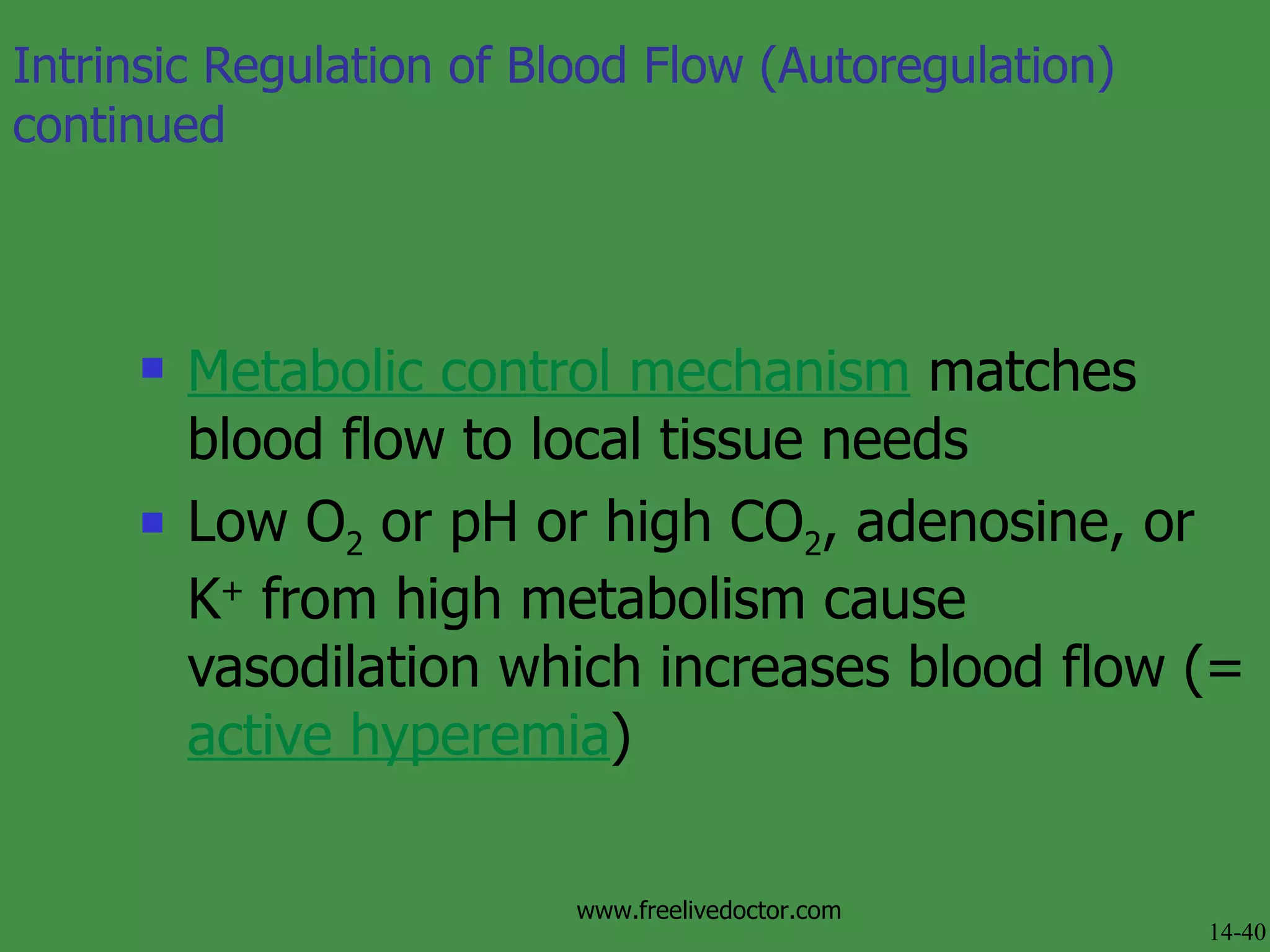
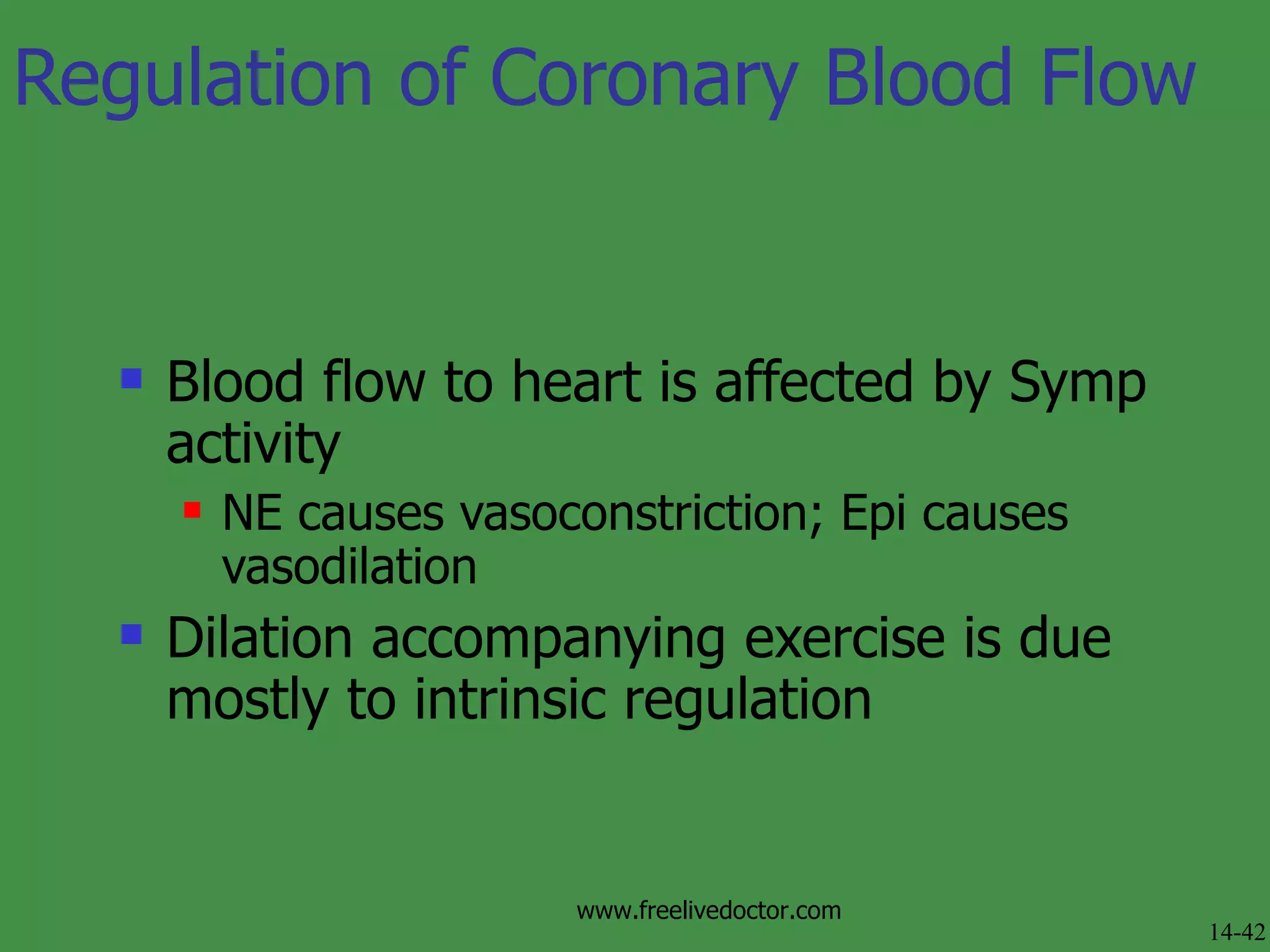
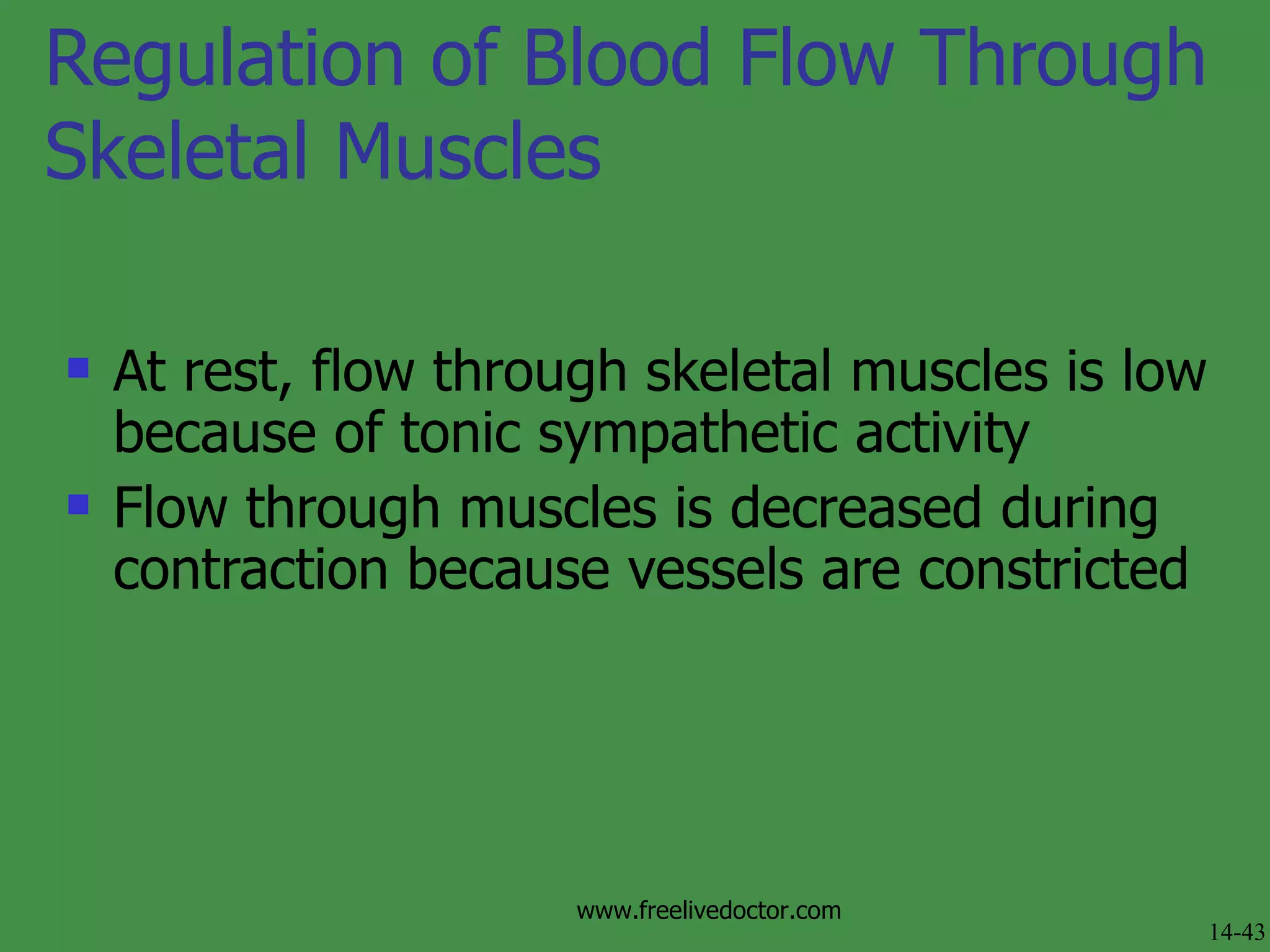
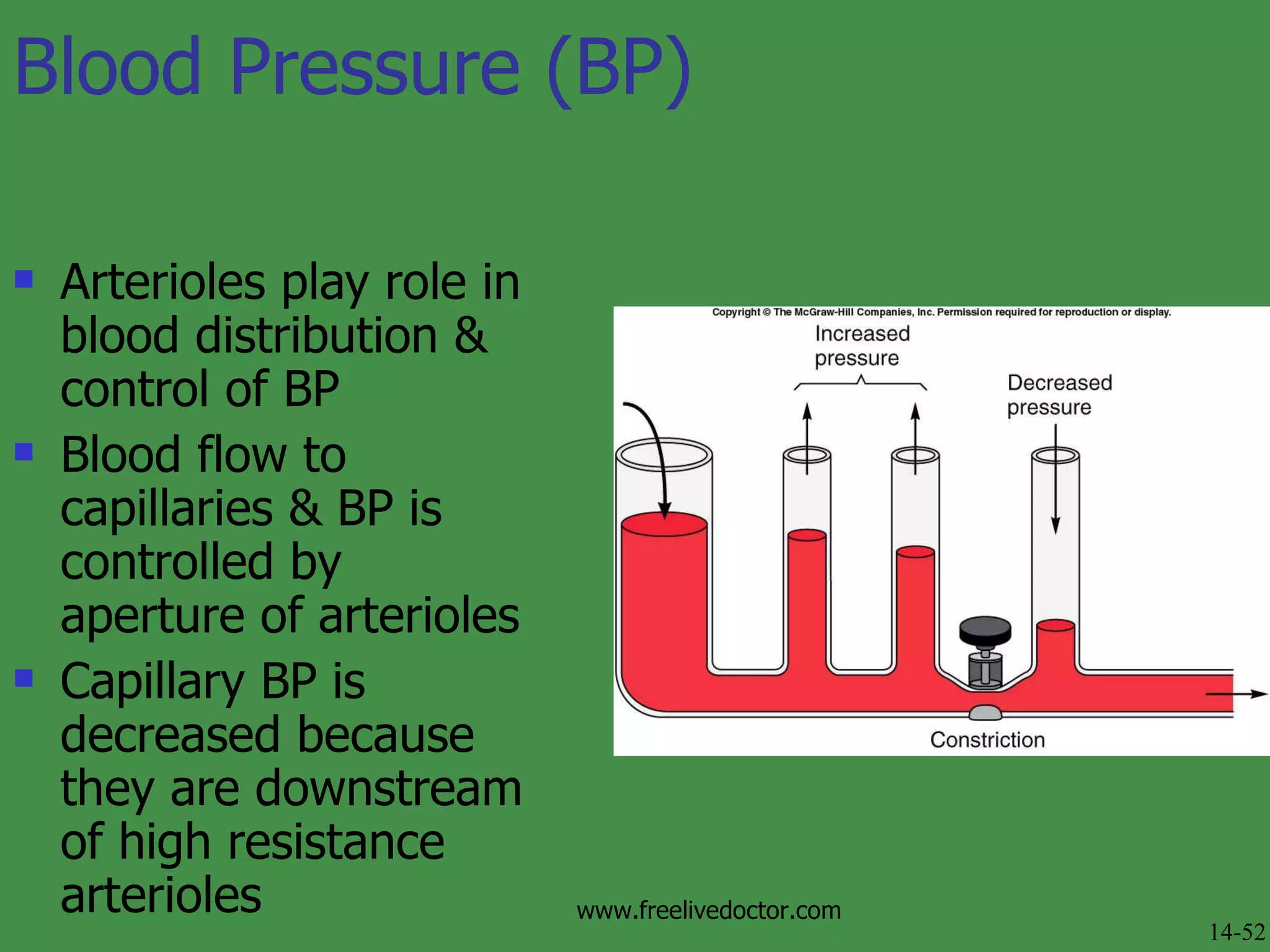
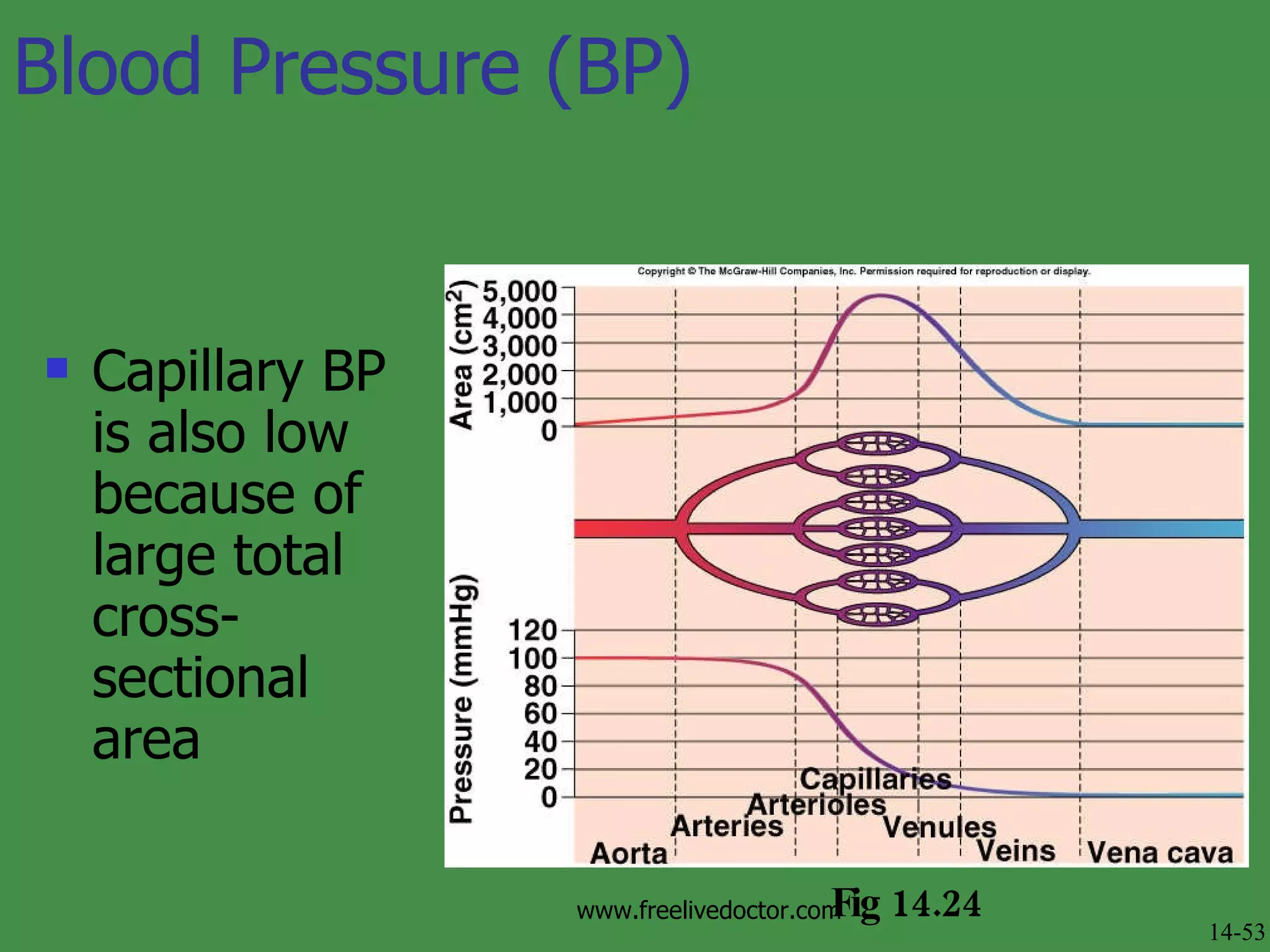
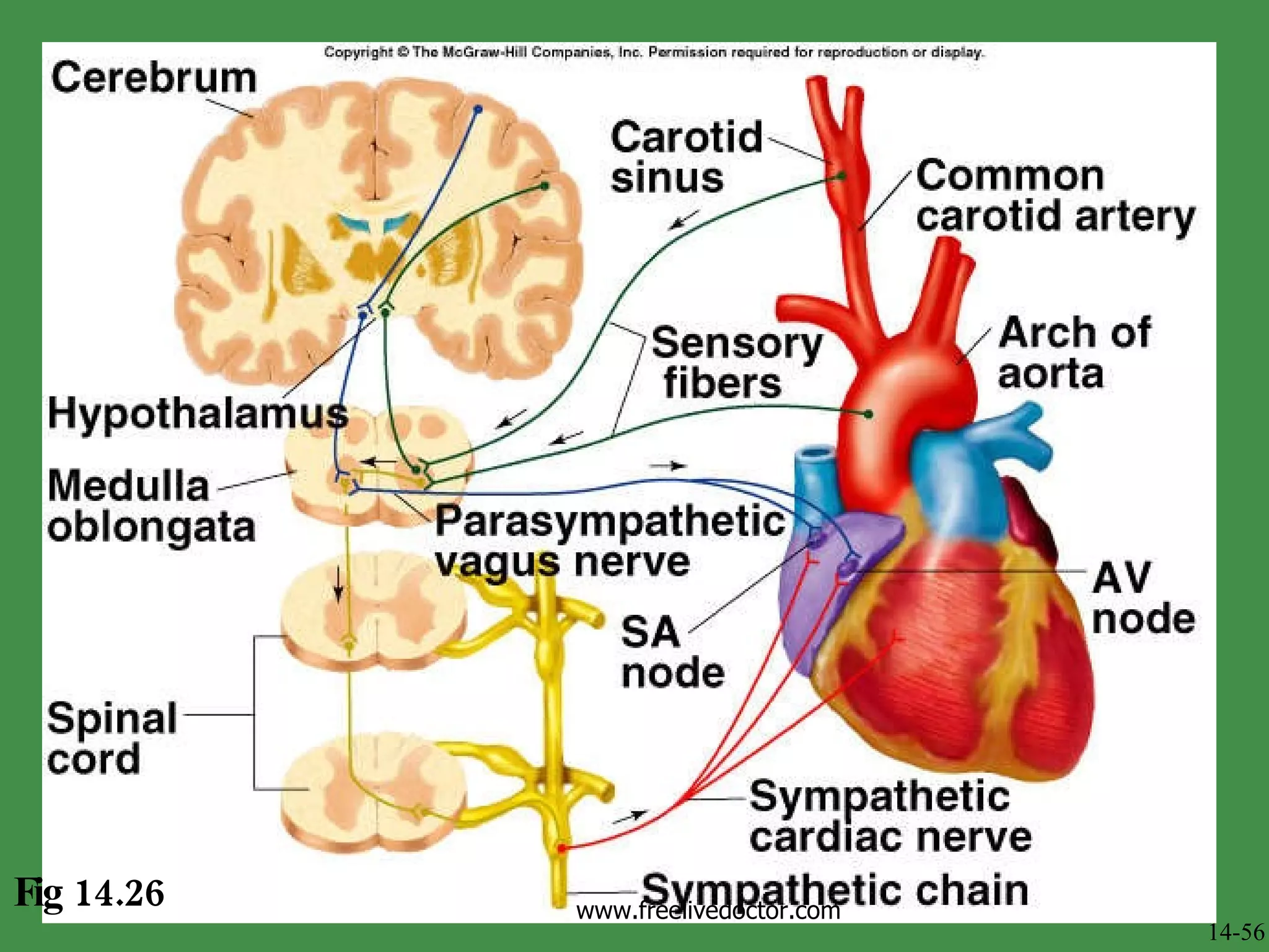
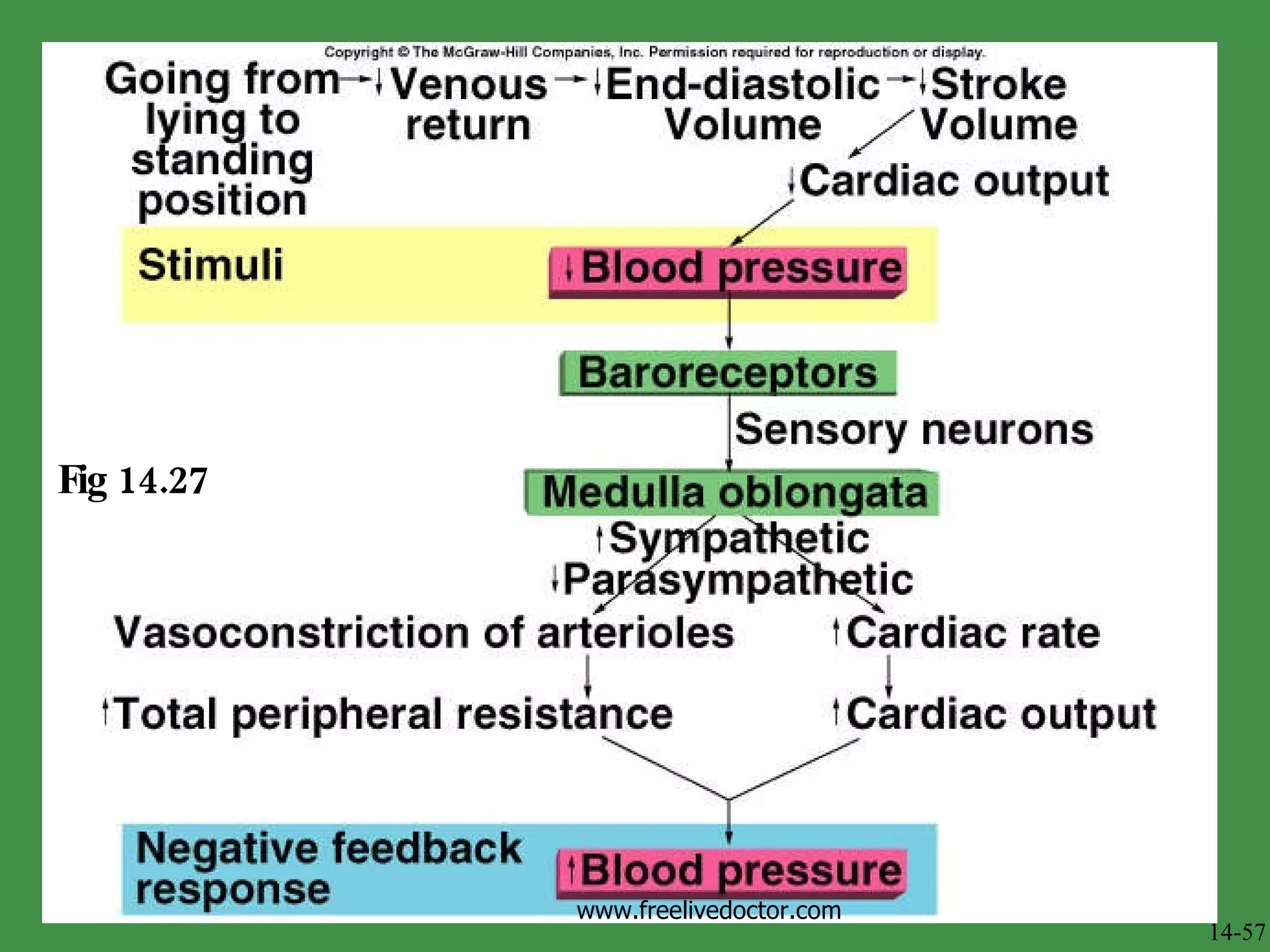
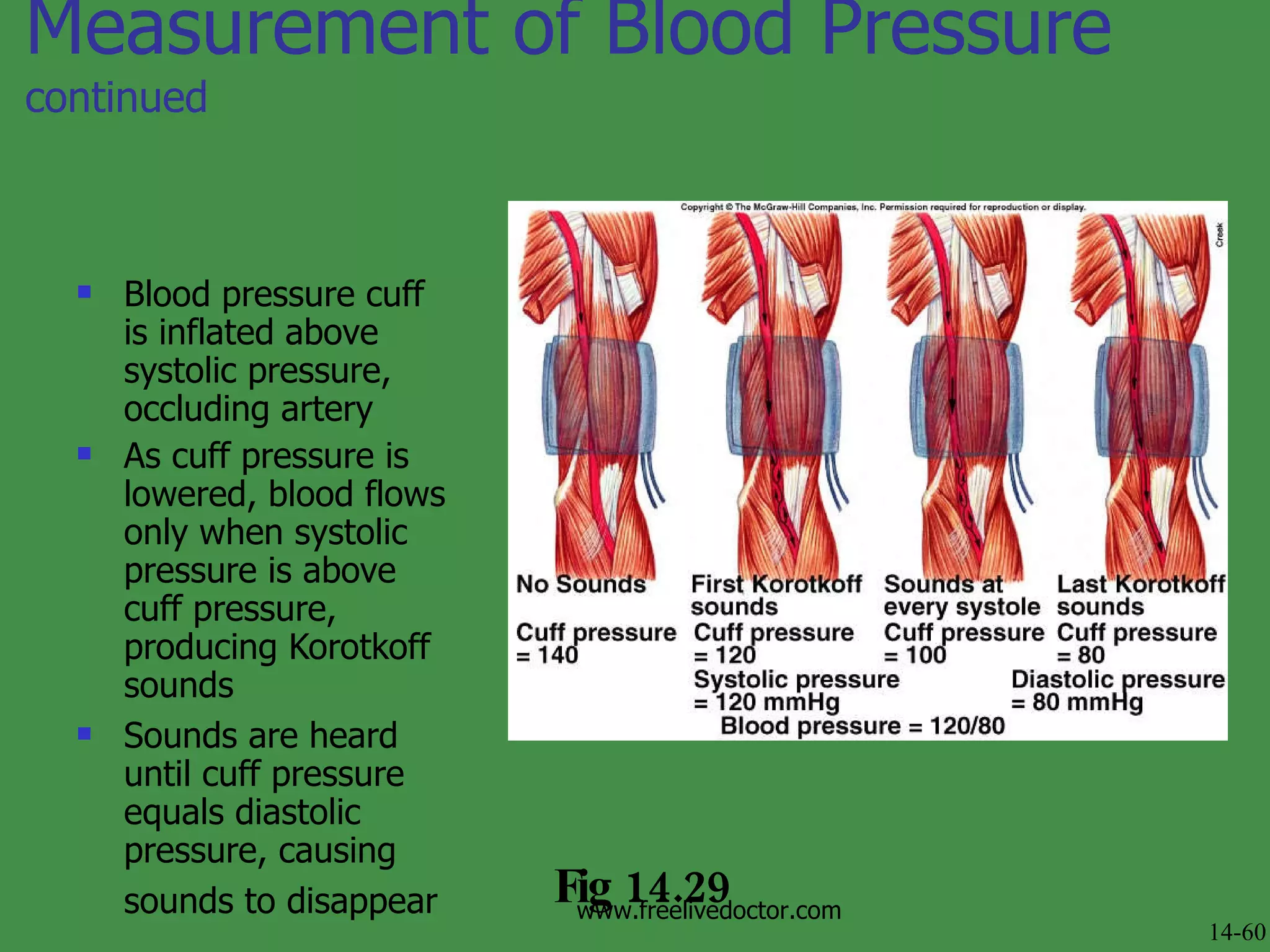
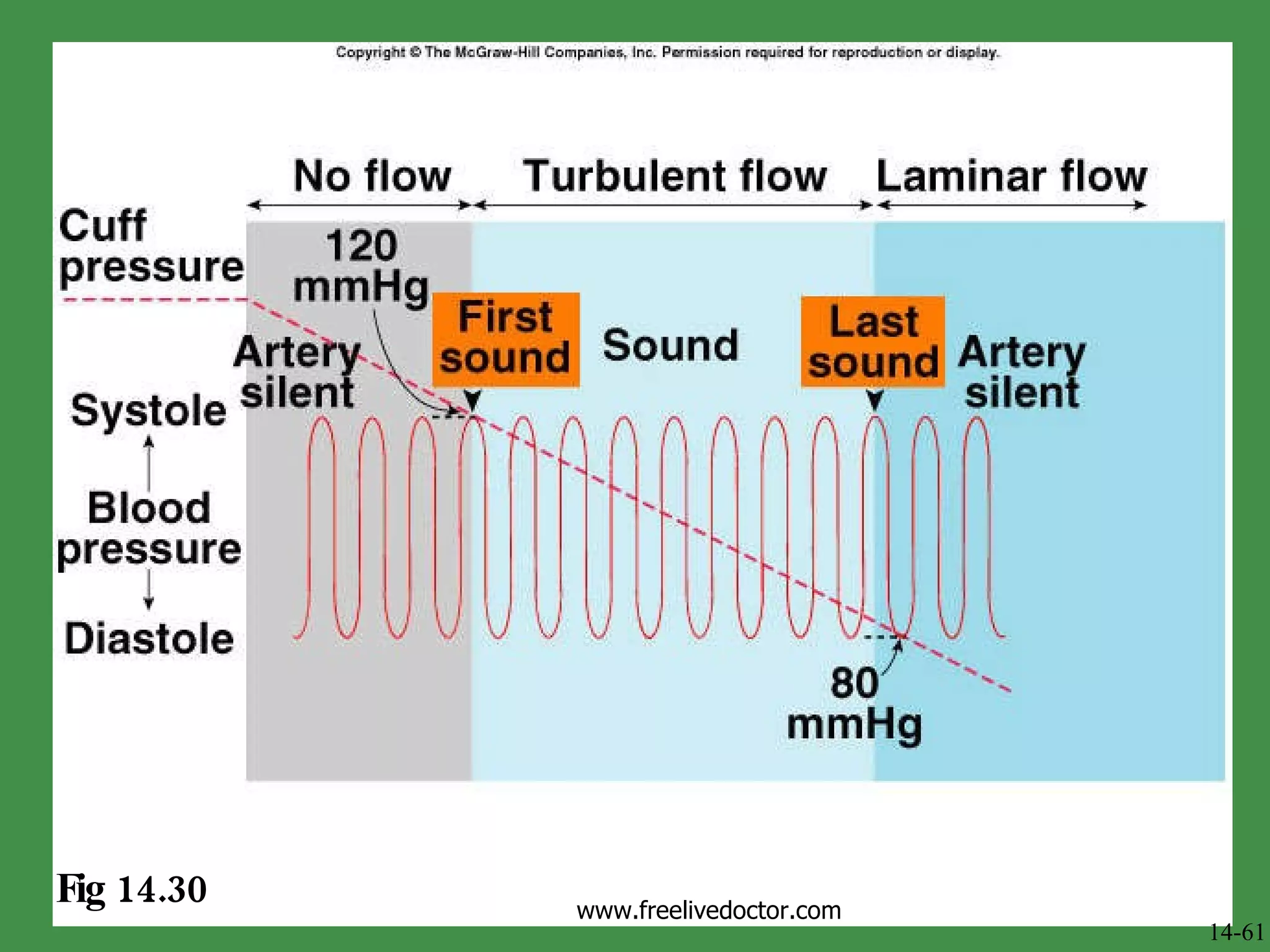
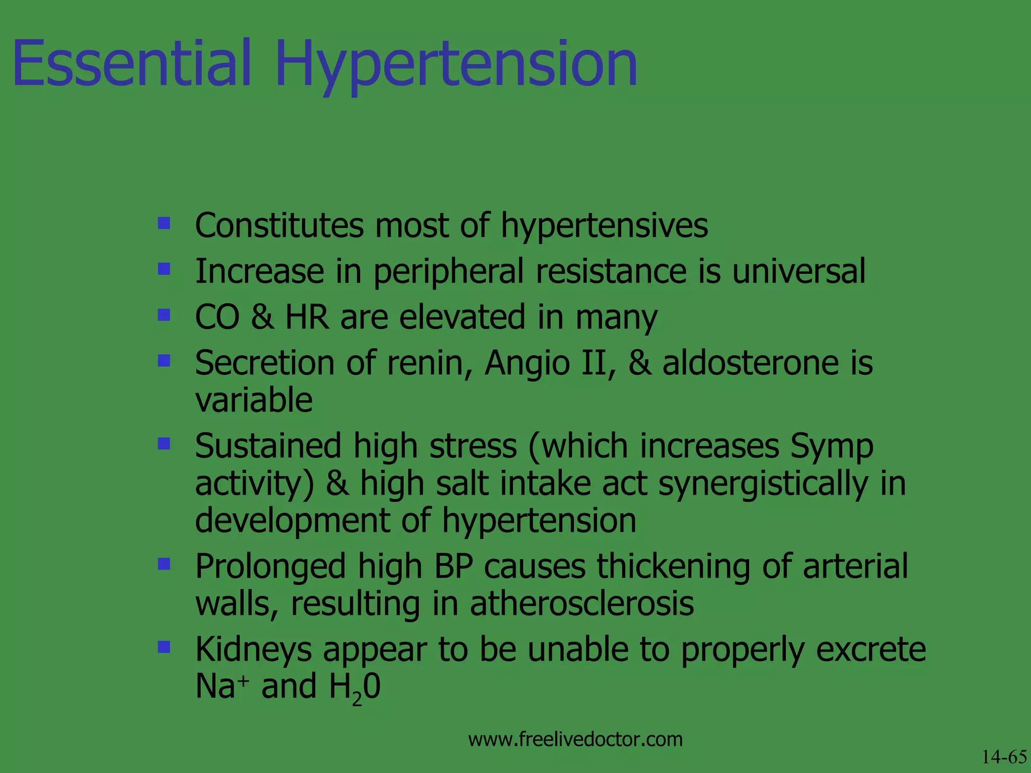
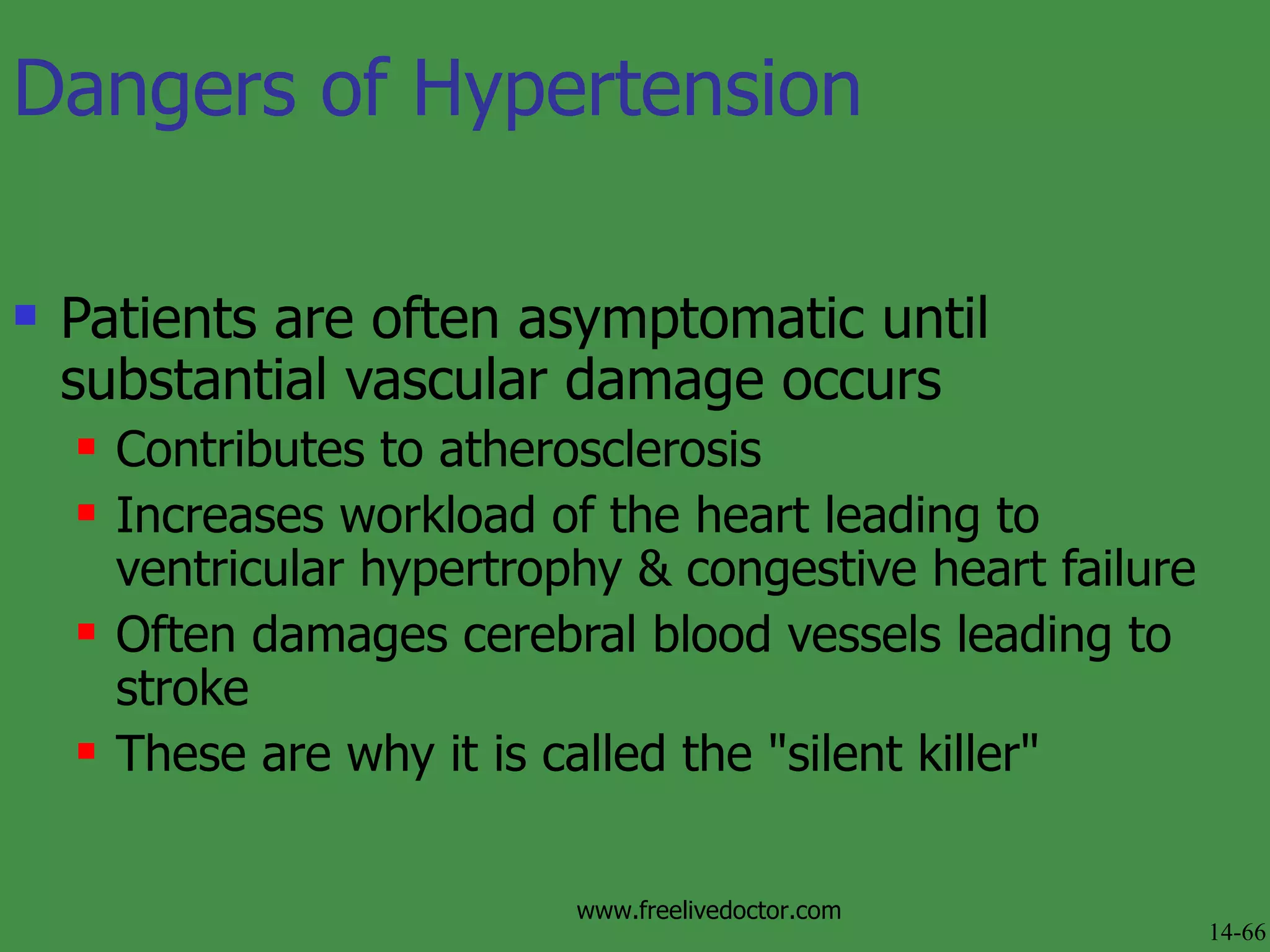
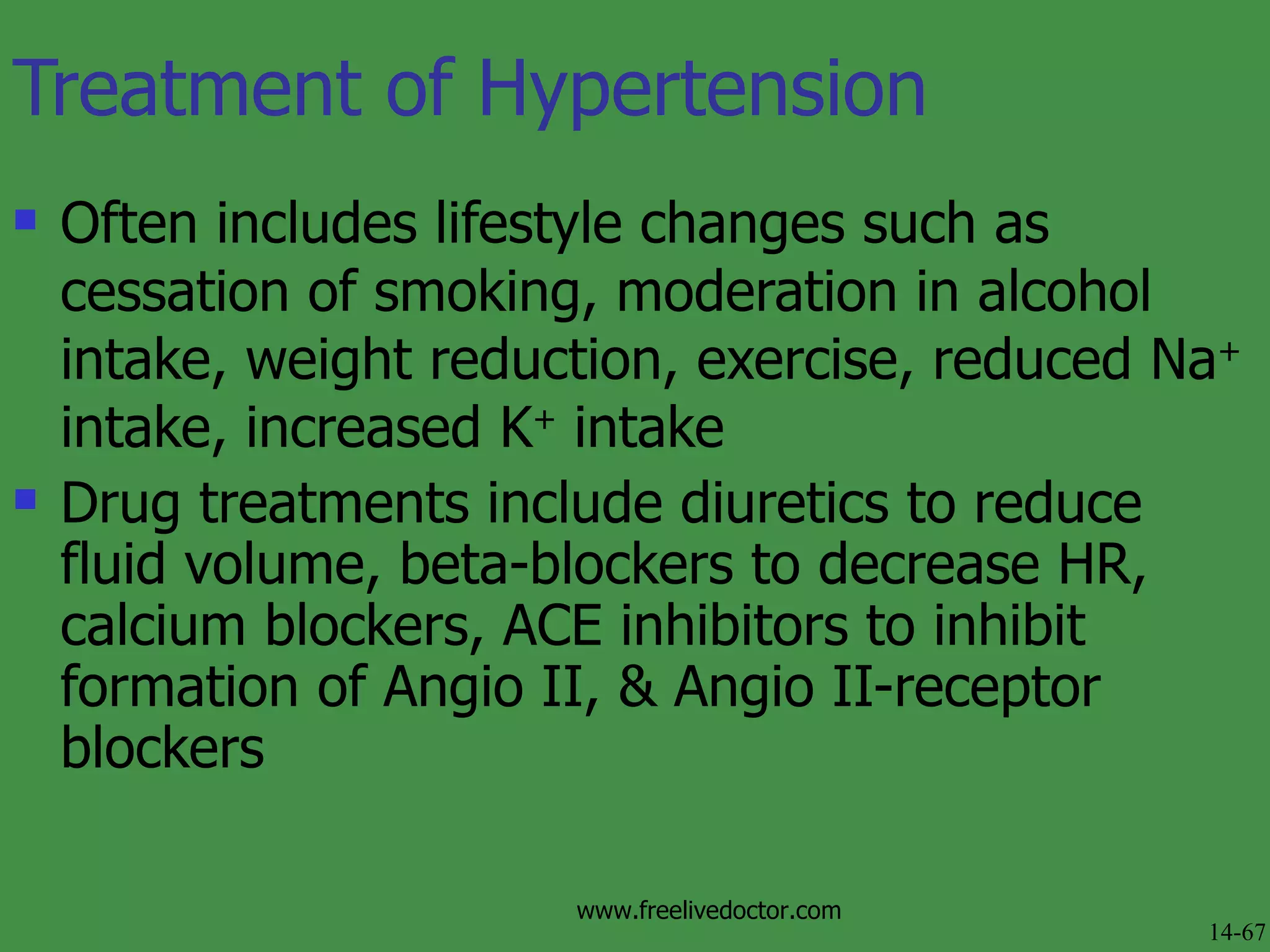
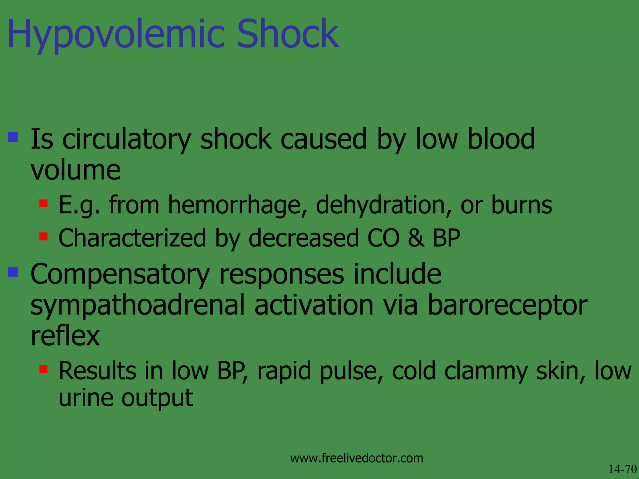
Cardiac Output, Blood Flow, and Blood Pressure 14-1 www.freelivedoctor.com
Outline Cardiac Output Blood & Body Fluid Volumes Factors Affecting Blood Flow Blood Pressure Hypertension Circulatory Shock 14-2 www.freelivedoctor.com
Cardiac Output  14-3 www.freelivedoctor.com
Is volume of blood pumped/min by each ventricle Heart Rate (HR)  = 70 beats/min Stroke volume  ( SV ) = blood pumped/beat by each ventricle Average is 70-80 ml/beat CO = SV x HR Total blood volume is about 5.5L Cardiac Output (CO) 14-4 www.freelivedoctor.com
Regulation of Cardiac Rate Without neuronal influences, SA node will drive heart at rate of its spontaneous activity   Normally Symp & Parasymp activity influence HR ( chronotropic effect ) Mechanisms that affect HR: chronotropic effect Positive increases; negative decreases Autonomic innervation of SA node is main controller of HR Symp & Parasymp nerve fibers modify rate of spontaneous depolarization  14-5 www.freelivedoctor.com
Regulation of Cardiac Rate  continued NE & Epi stimulate opening of pacemaker HCN channels   This depolarizes SA faster, increasing HR ACh promotes opening of K +  channels The resultant K+ outflow counters Na+ influx, slows depolarization & decreasing HR Fig 14.1 14-6 www.freelivedoctor.com
Vagus nerve: Decrease activity :  increases heart rate Increased activity: slows heart Cardiac control center  of medulla coordinates activity of autonomic innervation Sympathetic endings in atria & ventricles can stimulate increased strength of contraction Regulation of Cardiac Rate  continued 14-7 www.freelivedoctor.com
14-8 www.freelivedoctor.com
Stroke Volume Is determined by 3 variables: End diastolic volume  ( EDV ) = volume of blood in ventricles at end of diastole Total peripheral resistance  ( TPR ) = impedance to blood flow in arteries Contractility  = strength of ventricular contraction 14-9 www.freelivedoctor.com
EDV is workload ( preload ) on heart prior to contraction SV is directly proportional to preload & contractility Strength of contraction varies directly with EDV Total peripheral resistance =  afterload  which impedes ejection from ventricle SV is inversely proportional to TPR Ejection fraction  is SV/ EDV ( ~80ml/130ml=62%) Normally is 60%; useful clinical diagnostic tool Regulation of Stroke Volume 14-10 www.freelivedoctor.com
Frank-Starling Law of the Heart States that strength of ventricular contraction varies directly with EDV Is an  intrinsic   property  of myocardium As EDV increases, myocardium is stretched more, causing greater contraction & SV Fig 14.2 14-11 www.freelivedoctor.com
Frank-Starling Law of the Heart   continued (a) is state of myocardial sarcomeres just before filling Actins overlap, actin-myosin interactions are reduced & contraction would be weak In (b, c & d) there is increasing interaction of actin & myosin allowing more force to be developed   Fig 14.3 14-12 www.freelivedoctor.com
At any given EDV, contraction depends upon level of  sympathoadrenal  activity NE & Epi produce an increase in HR & contraction ( positive inotropic effect )  Due to increased Ca 2+  in sarcomeres Fig 14.4 14-13 www.freelivedoctor.com
Extrinsic Control of Contractility Parasympathetic stimulation Negative chronotropic effect Through innervation of the SA node and myocardial cell Slower heart rate means increased EDV Increases SV through Frank-Starling law www.freelivedoctor.com
Fig 14.5 14-14 www.freelivedoctor.com
Venous Return Is return of blood to heart via veins Controls EDV & thus SV & CO Dependent on: Blood volume & venous pressure Vasoconstriction caused by Symp Skeletal muscle pumps Pressure drop during inhalation Fig 14.7 14-15 www.freelivedoctor.com
Venous Return  continued Veins hold most of blood in body (70%) & are thus called  capacitance vessels   Have thin walls & stretch easily to accommodate more blood without increased pressure (=higher  compliance ) Have only 0- 10 mm Hg pressure Fig 14.6 14-16 www.freelivedoctor.com
Blood & Body Fluid Volumes 14-17 www.freelivedoctor.com
Blood Volume Constitutes small fraction of total body fluid 2/3 of body H 2 0 is inside cells ( intracellular compartment )  1/3 total body H 2 0 is in  extracellular compartment 80% of this is interstitial fluid; 20% is blood plasma Fig 14.8 14-18 www.freelivedoctor.com
Exchange of Fluid between Capillaries & Tissues Distribution of ECF between blood & interstitial compartments is in state of dynamic equilibrium Movement out of capillaries is driven by hydrostatic pressure exerted against capillary wall Promotes formation of tissue fluid  Net filtration pressure = hydrostatic pressure in capillary (17-37 mm Hg) - hydrostatic pressure of ECF (1 mm Hg) 14-19 www.freelivedoctor.com
Exchange of Fluid between Capillaries & Tissues Movement also affected by  colloid osmotic pressure = osmotic pressure exerted by proteins in fluid Difference between osmotic pressures in & outside of capillaries ( oncotic pressure ) affects fluid movement Plasma osmotic pressure = 25 mm Hg; interstitial osmotic pressure = 0 mm Hg 14-20 www.freelivedoctor.com
Overall Fluid Movement Is determined by net filtration pressure & forces opposing it ( Starling forces ) P c  +   i  (fluid out) - P i  +   p  (fluid in) P c  = Hydrostatic pressure in capillary  i  = Colloid osmotic pressure of interstitial fluid P i   = Hydrostatic pressure in interstitial fluid  p  = Colloid osmotic pressure of blood plasma 14-21 www.freelivedoctor.com
Fig 14.9 14-22 www.freelivedoctor.com
Edema Normally filtration, osmotic reuptake, & lymphatic drainage maintain proper ECF levels Edema  is excessive accumulation of ECF resulting from: High blood pressure Venous obstruction Leakage of plasma proteins into ECF Myxedema  (excess production of glycoproteins in extracellular matrix) from hypothyroidism Low plasma protein levels resulting from liver disease Obstruction of lymphatic drainage 14-23 www.freelivedoctor.com
Regulation of Blood Volume by Kidney Urine formation begins with filtration of plasma in  glomerulus Filtrate passes through & is modified by nephron Volume of urine excreted can be varied by changes in  reabsorption  of filtrate Adjusted according to needs of body by action of hormones 14-24 www.freelivedoctor.com
ADH (vasopressin) ADH  released by Post Pit when  osmoreceptors  detect high osmolality From excess salt intake or dehydration Causes thirst  Stimulates H 2 0 reabsorption from urine ADH release inhibited by low osmolality   Fig 14.11 14-25 www.freelivedoctor.com
Aldosterone Is steroid hormone secreted by adrenal cortex Helps maintain blood volume & pressure through reabsorption & retention of salt & water Release stimulated by salt deprivation, low blood volume, & pressure 14-26 www.freelivedoctor.com
Renin-Angiotension-Aldosterone System Decreased BP and flow (low blood volume) Kidney secreted Renin (enzyme) Juxaglomerular apparatus Angiotensin I to AngiotensinII By angiotensin-converting enzyme (ACE) Angio II causes a number of effects all aimed at increasing blood pressure: Vasoconstriction, aldosterone secretion, thirst 14-27 www.freelivedoctor.com
Fig 14.12 shows when & how Angio II is produced, & its effects   Angiotensin II 14-28 www.freelivedoctor.com
Atrial Natriuretic Peptide (ANP) Expanded blood volume is detected by stretch receptors in left atrium & causes release of ANP Inhibits aldosterone, promoting salt & water excretion to lower blood volume Promotes vasodilation 14-29 www.freelivedoctor.com
Factors Affecting Blood Flow 14-30 www.freelivedoctor.com
Vascular Resistance to Blood Flow Determines how much blood flows through a tissue or organ Vasodilation decreases resistance, increases blood flow Vasoconstriction does opposite 14-31 www.freelivedoctor.com
14-32 www.freelivedoctor.com
Physical Laws Describing Blood Flow Blood flows through vascular system when there is pressure difference (  P ) at its two ends  Flow rate is directly proportional to difference (  P = P 1  - P 2 ) Fig 14.13 14-33 www.freelivedoctor.com
Physical Laws Describing Blood Flow Flow rate is inversely proportional to resistance Flow =  P/R Resistance is directly proportional to length of vessel (L) & viscosity of blood (  ) Inversely proportional to 4th power of radius So diameter of vessel is very important for resistance Poiseuille's Law  describes factors affecting blood flow Blood flow =    Pr 4 (  )    L(8) 14-34 www.freelivedoctor.com
Fig 14.14.  Relationship between blood flow,  radius & resistance 14-35 www.freelivedoctor.com
Sympathoadrenal activation causes increased CO & resistance in periphery & viscera Blood flow to skeletal muscles is increased Because their arterioles dilate in response to Epi & their Symp fibers release ACh which also dilates their arterioles  Thus blood is shunted away from visceral & skin to muscles Extrinsic Regulation of Blood Flow 14-36 www.freelivedoctor.com
Parasympathetic effects are vasodilative However, Parasymp only innervates digestive tract, genitalia, & salivary glands Thus Parasymp is not as important as Symp Angiotensin II & ADH (at high levels) cause general vasoconstriction of vascular smooth muscle Which increases resistance & BP Extrinsic Regulation of Blood Flow  continued 14-37 www.freelivedoctor.com
Paracrine Regulation of Blood Flow Endothelium produces several paracrine regulators that promote relaxation: Nitric oxide  ( NO ),  bradykinin ,  prostacyclin NO is involved in setting resting “tone” of vessels Levels are increased by Parasymp activity Vasodilator drugs such as  nitroglycerin  or Viagra act thru NO Endothelin 1  is vasoconstrictor produced by endothelium 14-38 www.freelivedoctor.com
Intrinsic Regulation of Blood Flow (Autoregulation) Maintains fairly constant blood flow despite BP variation Myogenic control mechanisms  occur in some tissues because vascular smooth muscle contracts when stretched & relaxes when not stretched E.g. decreased arterial pressure causes cerebral vessels to dilate & vice versa 14-39 www.freelivedoctor.com
Metabolic control mechanism  matches blood flow to local tissue needs Low O 2  or pH or high CO 2 , adenosine, or K +  from high metabolism cause vasodilation which increases blood flow (=  active hyperemia ) Intrinsic Regulation of Blood Flow (Autoregulation) continued 14-40 www.freelivedoctor.com
Aerobic Requirements of the Heart Heart (& brain) must receive adequate blood supply at all times Heart is most aerobic tissue--each myocardial cell is within 10 m of capillary Contains lots of mitochondria & aerobic enzymes During systole coronary vessels are occluded Heart gets around this by having lots of  myoglobin Myoglobin is an 0 2  storage molecule that releases 0 2  to heart during systole 14-41 www.freelivedoctor.com
Regulation of Coronary Blood Flow Blood flow to heart is affected by Symp activity NE causes vasoconstriction; Epi causes vasodilation Dilation accompanying exercise is due mostly to intrinsic regulation 14-42 www.freelivedoctor.com
Regulation of Blood Flow Through Skeletal Muscles At rest, flow through skeletal muscles is low because of tonic sympathetic activity Flow through muscles is decreased during contraction because vessels are constricted 14-43 www.freelivedoctor.com
Circulatory Changes During Exercise At beginning of exercise, Symp activity causes vasodilation via Epi & local ACh release Blood flow is shunted from periphery & visceral to active skeletal muscles Blood flow to brain stays same As exercise continues, intrinsic regulation is major vasodilator Symp effects cause SV & CO to increase HR & ejection fraction increases vascular resistance 14-44 www.freelivedoctor.com
Fig 14.19 14-45 www.freelivedoctor.com
Fig 14.20 14-46 www.freelivedoctor.com
Cerebral Circulation Gets about 15% of total resting CO  Held constant (750ml/min) over varying conditions Because loss of consciousness occurs after few secs of interrupted flow Is not normally influenced by sympathetic activity 14-47 www.freelivedoctor.com
Cerebral Circulation Is regulated almost exclusively by intrinsic mechanisms When BP increases, cerebral arterioles constrict; when BP decreases, arterioles dilate (= myogenic regulation )  Arterioles dilate & constrict in response to changes in C0 2  levels Arterioles are very sensitive to increases in local neural activity (= metabolic regulation )  Areas of brain with high metabolic activity receive most blood 14-48 www.freelivedoctor.com
Fig 14.21 14-49 www.freelivedoctor.com
Cutaneous Blood Flow Skin serves as a heat exchanger for  thermoregulation Skin blood flow is adjusted to keep deep-body at 37 o C By arterial dilation or constriction & activity of  arteriovenous anastomoses  which control blood flow through surface capillaries Symp activity closes surface beds during cold & fight-or-flight, & opens them in heat & exercise Fig 14.22 14-50 www.freelivedoctor.com
Blood Pressure 14-51 www.freelivedoctor.com
Blood Pressure (BP) Arterioles play role in blood distribution & control of BP Blood flow to capillaries & BP is controlled by aperture of arterioles  Capillary BP is decreased because they are downstream of high resistance arterioles Fig 14.23  14-52 www.freelivedoctor.com
Blood Pressure (BP) Capillary BP is also low because of large total cross-sectional area Fig 14.24  14-53 www.freelivedoctor.com
Blood Pressure (BP) Is controlled mainly by  HR ,  SV , &  peripheral resistance An increase in any of these can result in increased BP Sympathoadrenal activity raises BP via arteriole vasoconstriction & by increased CO Kidney plays role in BP by regulating blood volume & thus stroke volume 14-54 www.freelivedoctor.com
Baroreceptor Reflex Is activated by changes in BP Which is detected by  baroreceptors  (stretch receptors) located in aortic arch & carotid sinuses Increase in BP causes walls of these regions to stretch, increasing frequency of APs Baroreceptors send APs to  vasomotor  &  cardiac   control centers  in medulla  Is most sensitive to decrease & sudden changes in BP  14-55 www.freelivedoctor.com
Fig 14.26 14-56 www.freelivedoctor.com
Fig 14.27 14-57 www.freelivedoctor.com
Atrial Stretch Receptors Are activated by increased venous return & act to reduce BP Stimulate reflex  tachycardia  (slow HR) Inhibit ADH release & promote secretion of ANP 14-58 www.freelivedoctor.com
Measurement of Blood Pressure Is via  auscultation  (to examine by listening) No sound is heard during  laminar flow  (normal, quiet, smooth blood flow) Korotkoff sounds  can be heard when  sphygmomanometer  cuff pressure is greater than diastolic but lower than systolic pressure Cuff constricts artery creating turbulent flow & noise as blood passes constriction during systole & is blocked during diastole 1st Korotkoff sound is heard at pressure that blood is 1st able to pass thru cuff; last occurs when can no long hear systole because cuff pressure = diastolic pressure 14-59 www.freelivedoctor.com
Measurement of Blood Pressure  continued Blood pressure cuff is inflated above systolic pressure, occluding artery  As cuff pressure is lowered, blood flows only when systolic pressure is above cuff pressure, producing Korotkoff sounds  Sounds are heard until cuff pressure equals diastolic pressure, causing sounds to disappear   Fig 14.29 14-60 www.freelivedoctor.com
Fig 14.30 14-61 www.freelivedoctor.com
Pulse Pressure Pulse pressure  = (systolic pressure) – (diastolic pressure) Mean arterial pressure  ( MAP ) represents average arterial pressure during cardiac cycle Has to be approximated because period of diastole is longer than period of systole MAP  = diastolic pressure + 1/3 pulse pressure 14-62 www.freelivedoctor.com
Hypertension 14-63 www.freelivedoctor.com
Hypertension  Is blood pressure in excess of normal range for age & gender (> 140/90 mmHg) Afflicts about 20 % of adults Primary  or  essential   hypertension  is caused by complex & poorly understood processes Secondary hypertension  is caused by known disease processes 14-64 www.freelivedoctor.com
Essential Hypertension Constitutes most of hypertensives Increase in peripheral resistance is universal CO & HR are elevated in many Secretion of renin, Angio II, & aldosterone is variable Sustained high stress (which increases Symp activity) & high salt intake act synergistically in development of hypertension Prolonged high BP causes thickening of arterial walls, resulting in atherosclerosis Kidneys appear to be unable to properly excrete Na +  and H 2 0 14-65 www.freelivedoctor.com
Dangers of Hypertension Patients are often asymptomatic until substantial vascular damage occurs  Contributes to atherosclerosis Increases workload of the heart leading to ventricular hypertrophy & congestive heart failure Often damages cerebral blood vessels leading to stroke These are why it is called the "silent killer" 14-66 www.freelivedoctor.com
Treatment of Hypertension Often includes lifestyle changes such as cessation of smoking, moderation in alcohol intake, weight reduction, exercise, reduced Na +  intake, increased K +  intake Drug treatments include diuretics to reduce fluid volume, beta-blockers to decrease HR, calcium blockers, ACE inhibitors to inhibit formation of Angio II, & Angio II-receptor blockers 14-67 www.freelivedoctor.com
Circulatory Shock 14-68 www.freelivedoctor.com
Circulatory Shock Occurs when there is inadequate blood flow to, &/or O 2  usage by, tissues  Cardiovascular system undergoes compensatory changes Sometimes shock becomes irreversible & death ensues  14-69 www.freelivedoctor.com
Hypovolemic Shock Is circulatory shock caused by low blood volume E.g. from hemorrhage, dehydration, or burns Characterized by decreased CO & BP Compensatory responses include sympathoadrenal activation via baroreceptor reflex Results in low BP, rapid pulse, cold clammy skin, low urine output 14-70 www.freelivedoctor.com
Refers to dangerously low blood pressure resulting from  sepsis  (infection) Mortality rate is high (50-70%) Often occurs as a result of  endotoxin  release from bacteria Endotoxin induces NO production causing vasodilation & resultant low BP Effective treatment includes drugs that inhibit production of NO Septic Shock 14-71 www.freelivedoctor.com
Other Causes of Circulatory Shock  Severe allergic reaction can cause a rapid fall in BP called  anaphylactic shock Due to generalized release of histamine causing vasodilation Rapid fall in BP called  neurogenic   shock  can result from decrease in Symp tone following spinal cord damage or anesthesia Cardiogenic   shock  is common following cardiac failure resulting from infarction that causes significant myocardial loss 14-72 www.freelivedoctor.com
Congestive Heart Failure Occurs when CO is insufficient to maintain blood flow required by body Caused by MI (most common), congenital defects, hypertension, aortic valve stenosis, disturbances in electrolyte levels Compensatory responses are similar to those of hypovolemic shock Treated with digitalis, vasodilators, & diuretics 14-73 www.freelivedoctor.com

Physiology Of Circulation

  • 1.
    Cardiac Output, BloodFlow, and Blood Pressure 14-1 www.freelivedoctor.com
  • 2.
    Outline Cardiac OutputBlood & Body Fluid Volumes Factors Affecting Blood Flow Blood Pressure Hypertension Circulatory Shock 14-2 www.freelivedoctor.com
  • 3.
    Cardiac Output 14-3 www.freelivedoctor.com
  • 4.
    Is volume ofblood pumped/min by each ventricle Heart Rate (HR) = 70 beats/min Stroke volume ( SV ) = blood pumped/beat by each ventricle Average is 70-80 ml/beat CO = SV x HR Total blood volume is about 5.5L Cardiac Output (CO) 14-4 www.freelivedoctor.com
  • 5.
    Regulation of CardiacRate Without neuronal influences, SA node will drive heart at rate of its spontaneous activity Normally Symp & Parasymp activity influence HR ( chronotropic effect ) Mechanisms that affect HR: chronotropic effect Positive increases; negative decreases Autonomic innervation of SA node is main controller of HR Symp & Parasymp nerve fibers modify rate of spontaneous depolarization 14-5 www.freelivedoctor.com
  • 6.
    Regulation of CardiacRate continued NE & Epi stimulate opening of pacemaker HCN channels This depolarizes SA faster, increasing HR ACh promotes opening of K + channels The resultant K+ outflow counters Na+ influx, slows depolarization & decreasing HR Fig 14.1 14-6 www.freelivedoctor.com
  • 7.
    Vagus nerve: Decreaseactivity : increases heart rate Increased activity: slows heart Cardiac control center of medulla coordinates activity of autonomic innervation Sympathetic endings in atria & ventricles can stimulate increased strength of contraction Regulation of Cardiac Rate continued 14-7 www.freelivedoctor.com
  • 8.
  • 9.
    Stroke Volume Isdetermined by 3 variables: End diastolic volume ( EDV ) = volume of blood in ventricles at end of diastole Total peripheral resistance ( TPR ) = impedance to blood flow in arteries Contractility = strength of ventricular contraction 14-9 www.freelivedoctor.com
  • 10.
    EDV is workload( preload ) on heart prior to contraction SV is directly proportional to preload & contractility Strength of contraction varies directly with EDV Total peripheral resistance = afterload which impedes ejection from ventricle SV is inversely proportional to TPR Ejection fraction is SV/ EDV ( ~80ml/130ml=62%) Normally is 60%; useful clinical diagnostic tool Regulation of Stroke Volume 14-10 www.freelivedoctor.com
  • 11.
    Frank-Starling Law ofthe Heart States that strength of ventricular contraction varies directly with EDV Is an intrinsic property of myocardium As EDV increases, myocardium is stretched more, causing greater contraction & SV Fig 14.2 14-11 www.freelivedoctor.com
  • 12.
    Frank-Starling Law ofthe Heart continued (a) is state of myocardial sarcomeres just before filling Actins overlap, actin-myosin interactions are reduced & contraction would be weak In (b, c & d) there is increasing interaction of actin & myosin allowing more force to be developed Fig 14.3 14-12 www.freelivedoctor.com
  • 13.
    At any givenEDV, contraction depends upon level of sympathoadrenal activity NE & Epi produce an increase in HR & contraction ( positive inotropic effect ) Due to increased Ca 2+ in sarcomeres Fig 14.4 14-13 www.freelivedoctor.com
  • 14.
    Extrinsic Control ofContractility Parasympathetic stimulation Negative chronotropic effect Through innervation of the SA node and myocardial cell Slower heart rate means increased EDV Increases SV through Frank-Starling law www.freelivedoctor.com
  • 15.
    Fig 14.5 14-14www.freelivedoctor.com
  • 16.
    Venous Return Isreturn of blood to heart via veins Controls EDV & thus SV & CO Dependent on: Blood volume & venous pressure Vasoconstriction caused by Symp Skeletal muscle pumps Pressure drop during inhalation Fig 14.7 14-15 www.freelivedoctor.com
  • 17.
    Venous Return continued Veins hold most of blood in body (70%) & are thus called capacitance vessels Have thin walls & stretch easily to accommodate more blood without increased pressure (=higher compliance ) Have only 0- 10 mm Hg pressure Fig 14.6 14-16 www.freelivedoctor.com
  • 18.
    Blood & BodyFluid Volumes 14-17 www.freelivedoctor.com
  • 19.
    Blood Volume Constitutessmall fraction of total body fluid 2/3 of body H 2 0 is inside cells ( intracellular compartment ) 1/3 total body H 2 0 is in extracellular compartment 80% of this is interstitial fluid; 20% is blood plasma Fig 14.8 14-18 www.freelivedoctor.com
  • 20.
    Exchange of Fluidbetween Capillaries & Tissues Distribution of ECF between blood & interstitial compartments is in state of dynamic equilibrium Movement out of capillaries is driven by hydrostatic pressure exerted against capillary wall Promotes formation of tissue fluid Net filtration pressure = hydrostatic pressure in capillary (17-37 mm Hg) - hydrostatic pressure of ECF (1 mm Hg) 14-19 www.freelivedoctor.com
  • 21.
    Exchange of Fluidbetween Capillaries & Tissues Movement also affected by colloid osmotic pressure = osmotic pressure exerted by proteins in fluid Difference between osmotic pressures in & outside of capillaries ( oncotic pressure ) affects fluid movement Plasma osmotic pressure = 25 mm Hg; interstitial osmotic pressure = 0 mm Hg 14-20 www.freelivedoctor.com
  • 22.
    Overall Fluid MovementIs determined by net filtration pressure & forces opposing it ( Starling forces ) P c +  i (fluid out) - P i +  p (fluid in) P c = Hydrostatic pressure in capillary  i = Colloid osmotic pressure of interstitial fluid P i = Hydrostatic pressure in interstitial fluid  p = Colloid osmotic pressure of blood plasma 14-21 www.freelivedoctor.com
  • 23.
    Fig 14.9 14-22www.freelivedoctor.com
  • 24.
    Edema Normally filtration,osmotic reuptake, & lymphatic drainage maintain proper ECF levels Edema is excessive accumulation of ECF resulting from: High blood pressure Venous obstruction Leakage of plasma proteins into ECF Myxedema (excess production of glycoproteins in extracellular matrix) from hypothyroidism Low plasma protein levels resulting from liver disease Obstruction of lymphatic drainage 14-23 www.freelivedoctor.com
  • 25.
    Regulation of BloodVolume by Kidney Urine formation begins with filtration of plasma in glomerulus Filtrate passes through & is modified by nephron Volume of urine excreted can be varied by changes in reabsorption of filtrate Adjusted according to needs of body by action of hormones 14-24 www.freelivedoctor.com
  • 26.
    ADH (vasopressin) ADH released by Post Pit when osmoreceptors detect high osmolality From excess salt intake or dehydration Causes thirst Stimulates H 2 0 reabsorption from urine ADH release inhibited by low osmolality Fig 14.11 14-25 www.freelivedoctor.com
  • 27.
    Aldosterone Is steroidhormone secreted by adrenal cortex Helps maintain blood volume & pressure through reabsorption & retention of salt & water Release stimulated by salt deprivation, low blood volume, & pressure 14-26 www.freelivedoctor.com
  • 28.
    Renin-Angiotension-Aldosterone System DecreasedBP and flow (low blood volume) Kidney secreted Renin (enzyme) Juxaglomerular apparatus Angiotensin I to AngiotensinII By angiotensin-converting enzyme (ACE) Angio II causes a number of effects all aimed at increasing blood pressure: Vasoconstriction, aldosterone secretion, thirst 14-27 www.freelivedoctor.com
  • 29.
    Fig 14.12 showswhen & how Angio II is produced, & its effects Angiotensin II 14-28 www.freelivedoctor.com
  • 30.
    Atrial Natriuretic Peptide(ANP) Expanded blood volume is detected by stretch receptors in left atrium & causes release of ANP Inhibits aldosterone, promoting salt & water excretion to lower blood volume Promotes vasodilation 14-29 www.freelivedoctor.com
  • 31.
    Factors Affecting BloodFlow 14-30 www.freelivedoctor.com
  • 32.
    Vascular Resistance toBlood Flow Determines how much blood flows through a tissue or organ Vasodilation decreases resistance, increases blood flow Vasoconstriction does opposite 14-31 www.freelivedoctor.com
  • 33.
  • 34.
    Physical Laws DescribingBlood Flow Blood flows through vascular system when there is pressure difference (  P ) at its two ends Flow rate is directly proportional to difference (  P = P 1 - P 2 ) Fig 14.13 14-33 www.freelivedoctor.com
  • 35.
    Physical Laws DescribingBlood Flow Flow rate is inversely proportional to resistance Flow =  P/R Resistance is directly proportional to length of vessel (L) & viscosity of blood (  ) Inversely proportional to 4th power of radius So diameter of vessel is very important for resistance Poiseuille's Law describes factors affecting blood flow Blood flow =  Pr 4 (  )  L(8) 14-34 www.freelivedoctor.com
  • 36.
    Fig 14.14. Relationship between blood flow, radius & resistance 14-35 www.freelivedoctor.com
  • 37.
    Sympathoadrenal activation causesincreased CO & resistance in periphery & viscera Blood flow to skeletal muscles is increased Because their arterioles dilate in response to Epi & their Symp fibers release ACh which also dilates their arterioles Thus blood is shunted away from visceral & skin to muscles Extrinsic Regulation of Blood Flow 14-36 www.freelivedoctor.com
  • 38.
    Parasympathetic effects arevasodilative However, Parasymp only innervates digestive tract, genitalia, & salivary glands Thus Parasymp is not as important as Symp Angiotensin II & ADH (at high levels) cause general vasoconstriction of vascular smooth muscle Which increases resistance & BP Extrinsic Regulation of Blood Flow continued 14-37 www.freelivedoctor.com
  • 39.
    Paracrine Regulation ofBlood Flow Endothelium produces several paracrine regulators that promote relaxation: Nitric oxide ( NO ), bradykinin , prostacyclin NO is involved in setting resting “tone” of vessels Levels are increased by Parasymp activity Vasodilator drugs such as nitroglycerin or Viagra act thru NO Endothelin 1 is vasoconstrictor produced by endothelium 14-38 www.freelivedoctor.com
  • 40.
    Intrinsic Regulation ofBlood Flow (Autoregulation) Maintains fairly constant blood flow despite BP variation Myogenic control mechanisms occur in some tissues because vascular smooth muscle contracts when stretched & relaxes when not stretched E.g. decreased arterial pressure causes cerebral vessels to dilate & vice versa 14-39 www.freelivedoctor.com
  • 41.
    Metabolic control mechanism matches blood flow to local tissue needs Low O 2 or pH or high CO 2 , adenosine, or K + from high metabolism cause vasodilation which increases blood flow (= active hyperemia ) Intrinsic Regulation of Blood Flow (Autoregulation) continued 14-40 www.freelivedoctor.com
  • 42.
    Aerobic Requirements ofthe Heart Heart (& brain) must receive adequate blood supply at all times Heart is most aerobic tissue--each myocardial cell is within 10 m of capillary Contains lots of mitochondria & aerobic enzymes During systole coronary vessels are occluded Heart gets around this by having lots of myoglobin Myoglobin is an 0 2 storage molecule that releases 0 2 to heart during systole 14-41 www.freelivedoctor.com
  • 43.
    Regulation of CoronaryBlood Flow Blood flow to heart is affected by Symp activity NE causes vasoconstriction; Epi causes vasodilation Dilation accompanying exercise is due mostly to intrinsic regulation 14-42 www.freelivedoctor.com
  • 44.
    Regulation of BloodFlow Through Skeletal Muscles At rest, flow through skeletal muscles is low because of tonic sympathetic activity Flow through muscles is decreased during contraction because vessels are constricted 14-43 www.freelivedoctor.com
  • 45.
    Circulatory Changes DuringExercise At beginning of exercise, Symp activity causes vasodilation via Epi & local ACh release Blood flow is shunted from periphery & visceral to active skeletal muscles Blood flow to brain stays same As exercise continues, intrinsic regulation is major vasodilator Symp effects cause SV & CO to increase HR & ejection fraction increases vascular resistance 14-44 www.freelivedoctor.com
  • 46.
    Fig 14.19 14-45www.freelivedoctor.com
  • 47.
    Fig 14.20 14-46www.freelivedoctor.com
  • 48.
    Cerebral Circulation Getsabout 15% of total resting CO Held constant (750ml/min) over varying conditions Because loss of consciousness occurs after few secs of interrupted flow Is not normally influenced by sympathetic activity 14-47 www.freelivedoctor.com
  • 49.
    Cerebral Circulation Isregulated almost exclusively by intrinsic mechanisms When BP increases, cerebral arterioles constrict; when BP decreases, arterioles dilate (= myogenic regulation ) Arterioles dilate & constrict in response to changes in C0 2 levels Arterioles are very sensitive to increases in local neural activity (= metabolic regulation ) Areas of brain with high metabolic activity receive most blood 14-48 www.freelivedoctor.com
  • 50.
    Fig 14.21 14-49www.freelivedoctor.com
  • 51.
    Cutaneous Blood FlowSkin serves as a heat exchanger for thermoregulation Skin blood flow is adjusted to keep deep-body at 37 o C By arterial dilation or constriction & activity of arteriovenous anastomoses which control blood flow through surface capillaries Symp activity closes surface beds during cold & fight-or-flight, & opens them in heat & exercise Fig 14.22 14-50 www.freelivedoctor.com
  • 52.
    Blood Pressure 14-51www.freelivedoctor.com
  • 53.
    Blood Pressure (BP)Arterioles play role in blood distribution & control of BP Blood flow to capillaries & BP is controlled by aperture of arterioles Capillary BP is decreased because they are downstream of high resistance arterioles Fig 14.23 14-52 www.freelivedoctor.com
  • 54.
    Blood Pressure (BP)Capillary BP is also low because of large total cross-sectional area Fig 14.24 14-53 www.freelivedoctor.com
  • 55.
    Blood Pressure (BP)Is controlled mainly by HR , SV , & peripheral resistance An increase in any of these can result in increased BP Sympathoadrenal activity raises BP via arteriole vasoconstriction & by increased CO Kidney plays role in BP by regulating blood volume & thus stroke volume 14-54 www.freelivedoctor.com
  • 56.
    Baroreceptor Reflex Isactivated by changes in BP Which is detected by baroreceptors (stretch receptors) located in aortic arch & carotid sinuses Increase in BP causes walls of these regions to stretch, increasing frequency of APs Baroreceptors send APs to vasomotor & cardiac control centers in medulla Is most sensitive to decrease & sudden changes in BP 14-55 www.freelivedoctor.com
  • 57.
    Fig 14.26 14-56www.freelivedoctor.com
  • 58.
    Fig 14.27 14-57www.freelivedoctor.com
  • 59.
    Atrial Stretch ReceptorsAre activated by increased venous return & act to reduce BP Stimulate reflex tachycardia (slow HR) Inhibit ADH release & promote secretion of ANP 14-58 www.freelivedoctor.com
  • 60.
    Measurement of BloodPressure Is via auscultation (to examine by listening) No sound is heard during laminar flow (normal, quiet, smooth blood flow) Korotkoff sounds can be heard when sphygmomanometer cuff pressure is greater than diastolic but lower than systolic pressure Cuff constricts artery creating turbulent flow & noise as blood passes constriction during systole & is blocked during diastole 1st Korotkoff sound is heard at pressure that blood is 1st able to pass thru cuff; last occurs when can no long hear systole because cuff pressure = diastolic pressure 14-59 www.freelivedoctor.com
  • 61.
    Measurement of BloodPressure continued Blood pressure cuff is inflated above systolic pressure, occluding artery As cuff pressure is lowered, blood flows only when systolic pressure is above cuff pressure, producing Korotkoff sounds Sounds are heard until cuff pressure equals diastolic pressure, causing sounds to disappear Fig 14.29 14-60 www.freelivedoctor.com
  • 62.
    Fig 14.30 14-61www.freelivedoctor.com
  • 63.
    Pulse Pressure Pulsepressure = (systolic pressure) – (diastolic pressure) Mean arterial pressure ( MAP ) represents average arterial pressure during cardiac cycle Has to be approximated because period of diastole is longer than period of systole MAP = diastolic pressure + 1/3 pulse pressure 14-62 www.freelivedoctor.com
  • 64.
  • 65.
    Hypertension Isblood pressure in excess of normal range for age & gender (> 140/90 mmHg) Afflicts about 20 % of adults Primary or essential hypertension is caused by complex & poorly understood processes Secondary hypertension is caused by known disease processes 14-64 www.freelivedoctor.com
  • 66.
    Essential Hypertension Constitutesmost of hypertensives Increase in peripheral resistance is universal CO & HR are elevated in many Secretion of renin, Angio II, & aldosterone is variable Sustained high stress (which increases Symp activity) & high salt intake act synergistically in development of hypertension Prolonged high BP causes thickening of arterial walls, resulting in atherosclerosis Kidneys appear to be unable to properly excrete Na + and H 2 0 14-65 www.freelivedoctor.com
  • 67.
    Dangers of HypertensionPatients are often asymptomatic until substantial vascular damage occurs Contributes to atherosclerosis Increases workload of the heart leading to ventricular hypertrophy & congestive heart failure Often damages cerebral blood vessels leading to stroke These are why it is called the "silent killer" 14-66 www.freelivedoctor.com
  • 68.
    Treatment of HypertensionOften includes lifestyle changes such as cessation of smoking, moderation in alcohol intake, weight reduction, exercise, reduced Na + intake, increased K + intake Drug treatments include diuretics to reduce fluid volume, beta-blockers to decrease HR, calcium blockers, ACE inhibitors to inhibit formation of Angio II, & Angio II-receptor blockers 14-67 www.freelivedoctor.com
  • 69.
    Circulatory Shock 14-68www.freelivedoctor.com
  • 70.
    Circulatory Shock Occurswhen there is inadequate blood flow to, &/or O 2 usage by, tissues Cardiovascular system undergoes compensatory changes Sometimes shock becomes irreversible & death ensues 14-69 www.freelivedoctor.com
  • 71.
    Hypovolemic Shock Iscirculatory shock caused by low blood volume E.g. from hemorrhage, dehydration, or burns Characterized by decreased CO & BP Compensatory responses include sympathoadrenal activation via baroreceptor reflex Results in low BP, rapid pulse, cold clammy skin, low urine output 14-70 www.freelivedoctor.com
  • 72.
    Refers to dangerouslylow blood pressure resulting from sepsis (infection) Mortality rate is high (50-70%) Often occurs as a result of endotoxin release from bacteria Endotoxin induces NO production causing vasodilation & resultant low BP Effective treatment includes drugs that inhibit production of NO Septic Shock 14-71 www.freelivedoctor.com
  • 73.
    Other Causes ofCirculatory Shock Severe allergic reaction can cause a rapid fall in BP called anaphylactic shock Due to generalized release of histamine causing vasodilation Rapid fall in BP called neurogenic shock can result from decrease in Symp tone following spinal cord damage or anesthesia Cardiogenic shock is common following cardiac failure resulting from infarction that causes significant myocardial loss 14-72 www.freelivedoctor.com
  • 74.
    Congestive Heart FailureOccurs when CO is insufficient to maintain blood flow required by body Caused by MI (most common), congenital defects, hypertension, aortic valve stenosis, disturbances in electrolyte levels Compensatory responses are similar to those of hypovolemic shock Treated with digitalis, vasodilators, & diuretics 14-73 www.freelivedoctor.com