BLOOD
REPRESENTEDBY
MDMEHBUBALAM
Free Educational YouTube Channel Md Nursing Education
CONTENTINDETAIL
• Definition of blood and
haematology
• Functions and properties of
blood
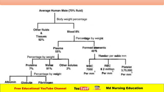
• Blood plasma and formed
elements
• Haemopoietin
• Red blood cells and
erythropoiesis
• White blood cells and platelets
• Mechanism of homeostasis
• Pathway of blood clotting
• Blood groups
Free Educational YouTube Channel Md Nursing Education
INTRODUCTIONOFBLOOD
• Blood is a specialized fluid connective tissue which
contains of plasma and corpuscles (RBC, WBC and
platelets).
• The liquid component of blood is called plasma. It a
mixture of water, sugar, fat, protein and salt.
• Blood is the primary transport medium that is
responsible for continuously supply oxygen and
nutrients to the active cells of the body.
Free Educational YouTube Channel Md Nursing Education
DEFINITIONOFBLOODANDHEMATOLOGY
• Blood:-A liquid connective tissue composed of
plasma and corpuscles.
• Haematology:-The branch of science that
deals with the study of blood, tissue and blood
disorders.
Free Educational YouTube Channel Md Nursing Education
PROPERTIESOFBLOOD
• Colour – Scarlet red
• Volume – 5-6 litres
• Reaction and pH – lightly alkaline and ph-7.4
• Specific gravity – 1.052-1.061
• Viscosity – 5 time more viscous than water.
Free Educational YouTube Channel Md Nursing Education
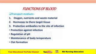
FUNCTIONSOFBLOOD
Transport medium:-
1. Oxygen, nutrients and waste material
2. Hormones to there target tissue
3. Protective antibodies to the site of infection
• Protection against infection
• Regulation of pH
• Maintenance of body temperature
• Clot formation
Free Educational YouTube Channel Md Nursing Education
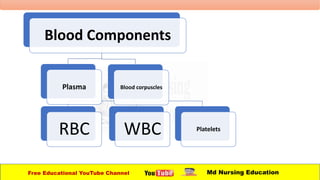
Blood Components
Plasma Blood corpuscles
RBC WBC Platelets
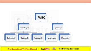
WBC
Granulocytes
Eosinophils Basophils Neutrophils
Agranulocytes
Lymphocytes Monocytes
Free Educational YouTube Channel Md Nursing Education
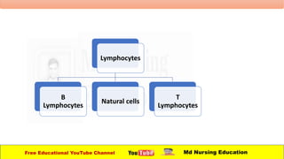
Lymphocytes
B
Lymphocytes
Natural cells
T
Lymphocytes
Free Educational YouTube Channel Md Nursing Education
REDBLOODCELLS
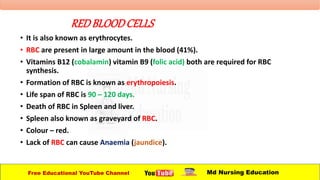
• It is also known as erythrocytes.
• RBC are present in large amount in the blood (41%).
• Vitamins B12 (cobalamin) vitamin B9 (folic acid) both are required for RBC
synthesis.
• Formation of RBC is known as erythropoiesis.
• Life span of RBC is 90 – 120 days.
• Death of RBC in Spleen and liver.
• Spleen also known as graveyard of RBC.
• Colour – red.
• Lack of RBC can cause Anaemia (jaundice).
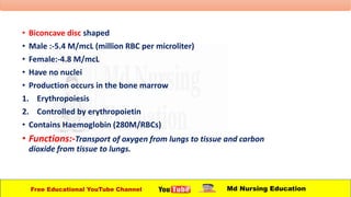
• Biconcave disc shaped
• Male :-5.4 M/mcL (million RBC per microliter)
• Female:-4.8 M/mcL
• Have no nuclei
• Production occurs in the bone marrow
1. Erythropoiesis
2. Controlled by erythropoietin
• Contains Haemoglobin (280M/RBCs)
• Functions:-Transport of oxygen from lungs to tissue and carbon
dioxide from tissue to lungs.
Free Educational YouTube Channel Md Nursing Education
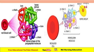
HAEMOGLOBIN
• Consists of:
- 4 globin molecules:
colourless protein (96%).
- 4 heme molecules (4%):
Transport of oxygen
iron is required for oxygen
transport
Normal values:
• Female :11.6 to 15gm/dl
• Male: 13.2 to 16.6gm/dl
• Infant:-14 to 20 gm/dl
Free Educational YouTube Channel Md Nursing Education
FORMATIONANDDESTRUCTIONOF RBC
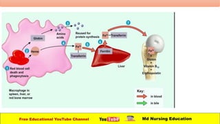
• After 120 days RBC will ruptured or damaged.
• Without a nucleus and other organelles RBCs can’t
synthesize new components to replace damaged once.
• Ruptured RBCs removed from circulation and
destroyed by macrophages in the spleen and liver, and
breakdown products are recycled.
• Globin is breakdown into amino acids which can be
reused to synthesize other proteins.
Free Educational YouTube Channel Md Nursing Education
FORMATIONANDDESTRUCTIONOF RBC
• Iron is removed in the form of Fe3, which associates with
the plasma protein transferrin.
• In muscles fibres liver cells and macrophages of the spleen
and liver Fe3 detaches from transferrin and attaches to an
iron storage protein called ferritin.
• Upon release from a storage site or absorption from the
gastrointestinal tract, Fe3 reattaches to transferrin.
• The fe3-transferrin complex is then carried to red bone
marrow, where RBC precursor cells use it in Haemoglobin
synthesis.
• Erythropoiesis in red bone marrow results in the
production of red blood cells, which enter the circulation.
Free Educational YouTube Channel Md Nursing Education
FORMATIONANDDISTRIBUTIONOFRBCs
• When iron is removed from heme , the non iron
protein of heme is converted to biliverdin, a green
pigment and then into bilirubin a yellow orange
pigment.
• Bilirubin enters the blood and is transported to the
liver.
• Within the liver bilirubin is released by liver cells into
bile which pass into the small intestine and then into
the large intestine.
• In the large intestine
Free Educational YouTube Channel Md Nursing Education
BONEMARROW
• It the cellulovascular tissue occupying the Medullary cavities and
the cancellous space of the bone.
• Active bone marrow in the adult is estimated at from 3.5-6% of
body weight.
• The volume of bone marrow is 70 ml at birth and 4000 ml in the
adult.
• Red bone marrow (active) and yellow bone marrow (inactive).
• In adults red bone marrow is present in the axial skeleton (skull,
pelvis, ribs, sternum, vertebrae, and proximal ends of femur and
humerus.
• In newborn hematopoiesis is going on out side the bone marrow,
i.e., in the liver, spleen and lymph nodes, it is called extra
Medullary Hematopoiesis.
Free Educational YouTube Channel Md Nursing Education
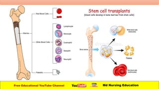
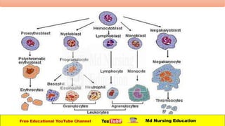
HEMATOPOIESIS
• The process of formation of blood cells, i.e. RBC, WBC
and platelets is called hematopoiesis and the sites
where it occurs are known as hematopoietic tissue or
organ (bone marrow, liver and spleen).
• The cells responsible for hematopoiesis are first seen in
yolk sac of embryo in third week of embryonic
development and these cells are known as
hematopoietic stem cells.
Free Educational YouTube Channel Md Nursing Education
HEMATOPOIESIS
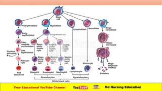
• Hematopoiesis is completed through five process:
1.Erythropoiesis
2.Lymphopoiesis
3.Granulopoiesis
4.Monopolises
5.Thrombopoiesis
Free Educational YouTube Channel Md Nursing Education
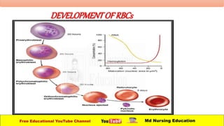
ERYTHROPOIESIS
• It is a process by which erythrocytes are producing.
• The process is triggered by erythropoietin, which is a
kidney hormone and is released during hypoxia.
• Erythroblast present in cytoplasm has a large nucleus
with blue cytoplasm and differentiates to become
progressively smaller size.
Free Educational YouTube Channel Md Nursing Education
DEVELOPMENTOFRBCs
Free Educational YouTube Channel Md Nursing Education
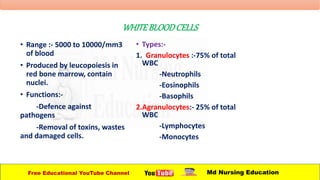
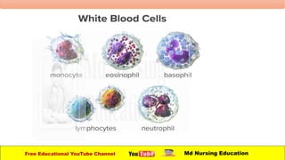
WHITEBLOODCELLS
• Range :- 5000 to 10000/mm3
of blood
• Produced by leucopoiesis in
red bone marrow, contain
nuclei.
• Functions:-
-Defence against
pathogens
-Removal of toxins, wastes
and damaged cells.
• Types:-
1. Granulocytes :-75% of total
WBC
-Neutrophils
-Eosinophils
-Basophils
2.Agranulocytes:- 25% of total
WBC
-Lymphocytes
-Monocytes
Free Educational YouTube Channel Md Nursing Education
Leucocytosisand leukopenia
• Leucocytosis:-High white blood cells count is called
leucocytosis.
• Low white blood cells count is called as leukopenia.
Free Educational YouTube Channel Md Nursing Education
Neutrophils
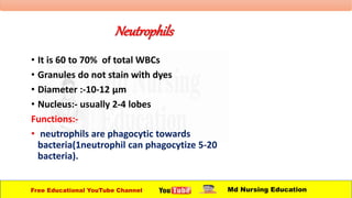
• It is 60 to 70% of total WBCs
• Granules do not stain with dyes
• Diameter :-10-12 µm
• Nucleus:- usually 2-4 lobes
Functions:-
• neutrophils are phagocytic towards
bacteria(1neutrophil can phagocytize 5-20
bacteria).
Free Educational YouTube Channel Md Nursing Education
EOSINOPHILS
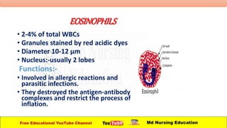
• 2-4% of total WBCs
• Granules stained by red acidic dyes
• Diameter 10-12 µm
• Nucleus:-usually 2 lobes
Functions:-
• Involved in allergic reactions and
parasitic infections.
• They destroyed the antigen-antibody
complexes and restrict the process of
inflation.
Free Educational YouTube Channel Md Nursing Education
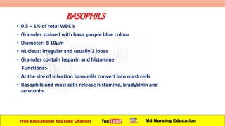
BASOPHILS
• 0.5 – 1% of total WBC’s
• Granules stained with basic purple blue colour
• Diameter: 8-10µm
• Nucleus: irregular and usually 2 lobes
• Granules contain heparin and histamine
Functions:-
• At the site of infection basophils convert into mast cells
• Basophils and mast cells release histamine, bradykinin and
serotonin.
Free Educational YouTube Channel Md Nursing Education
LYMPHOCYTES
• 20-25% of total WBCs.
• Depending upon the site of production and their
actions, divided into T,B cells and natural killer cells.
• Nucleus :-Will be round shaped
• Functions:-play important role in immunity.
Free Educational YouTube Channel Md Nursing Education
MONOCYTES
• 3-8% of total WBCs
• Diameter 12-20 µm
• Monocytes are converted into macrophages of the
tissues.
• Functions:-phagocytosis
Free Educational YouTube Channel Md Nursing Education
THROMBOCYTES
• Range 250,000-500,000/mm3 of blood
• No nuclei
• Diameter :-2-4 µm
• Life span:-10-12 days
• Functions:-involved in blood clotting
Free Educational YouTube Channel Md Nursing Education
BLOODGROUP
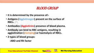
• It is determined by the presence of:-
• Antigens (Agglutinogen) present on the surface of
RBCs.
• Antibodies (Agglutinins) presence of blood plasma.
• Antibody can bind to RBC antigens, resulting in
agglutination (clumping) or haemolysis of RBCs.
• 2 types of blood groups:
-ABO and Rh factor
Free Educational YouTube Channel Md Nursing Education
ABOBLOODGROUPINGSYSTEM
• Landsteiner (1900)
• According to ABO blood grouping system :-
4 types of blood groups:
1. Blood group –A
2. Blood group –B
3. Blood group –AB
4. Blood group -O
Free Educational YouTube Channel Md Nursing Education
ABOBLOODGROUP
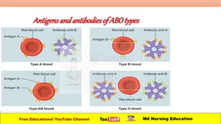
Blood group -A
• A antigen will present on
the surface of RBCs.
• B antibodies will present
in the plasm.
Free Educational YouTube Channel Md Nursing Education
ABO BLOOD GROUP
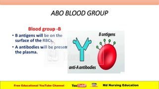
Blood group -B
• B antigens will be on the
surface of the RBCs.
• A antibodies will be present in
the plasma.
Free Educational YouTube Channel Md Nursing Education
ABOBLOODGROUP
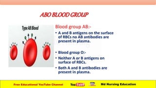
Blood group AB:-
• A and B antigens on the surface
of RBCs no AB antibodies are
present in plasma.
• Blood group O:-
• Neither A or B antigens on
surface of RBCs.
• Both A and B antibodies are
present in plasma.
Free Educational YouTube Channel Md Nursing Education
ABOBLOODGROUP
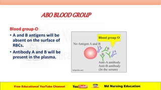
Blood group-O
• A and B antigens will be
absent on the surface of
RBCs.
• Antibody A and B will be
present in the plasma.
Free Educational YouTube Channel Md Nursing Education
Antigensand antibodies of ABOtypes
Free Educational YouTube Channel Md Nursing Education
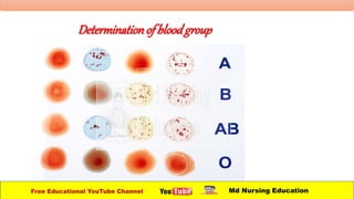
Determinationof bloodgroup
Free Educational YouTube Channel Md Nursing Education
• Blood group 'O’ is called “Universal donar” because it
has no antigens on RBCs.
• Blood group AB is called “universal receivers” because
it has no anti-bodies in plasma.
Free Educational YouTube Channel Md Nursing Education
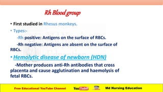
RhBloodgroup
• First studied in Rhesus monkeys.
• Types:-
-Rh positive: Antigens on the surface of RBCs.
-Rh negative: Antigens are absent on the surface of
RBCs.
•Hemolytic disease of newborn (HDN)
-Mother produces anti-Rh antibodies that cross
placenta and cause agglutination and haemolysis of
fetal RBCs.
Free Educational YouTube Channel Md Nursing Education
RhBloodGroup
• person with Rh- Blood develops Rh antibodies in the
blood if receives blood from Rh+ person.
• There is development of Rh-antibodies, that react with
donor’s Rh Antigens and agglutinate the blood.
• A person with Rh+ blood can receive blood from a
person with Rh blood about any problems.
Free Educational YouTube Channel Md Nursing Education
Haemostasis- Stoppage of bleeding
• When the blood vessels get damaged, platelets plays a vital role in
Haemostasis.
• 3 mechanisms are involved in Haemostasis:
1. Vascular spasm
2. Blood clotting
3. Platelet plug formation
Free Educational YouTube Channel Md Nursing Education
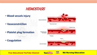
HEMOSTASIS
• Blood vessels injury
• Vasoconstriction
• Platelet plug formation
• Coagulation
Free Educational YouTube Channel Md Nursing Education
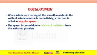
VASCULARSPASM
• When arteries are damaged, the smooth muscles in the
walls of arteries contracts immediately, a reaction is
called as vascular spasm.
• The spasm is caused due to release of mediators from
the activated platelets.
Free Educational YouTube Channel Md Nursing Education
BLOODCLOTTING
• Blood clots consists of network of insoluble protein
fibres called fibrin in which the formed elements (RBCs,
WBCs and platelets) of blood are trapped.
• The process of clot forming called as clotting.
Free Educational YouTube Channel Md Nursing Education
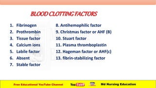
BLOODCLOTTINGFACTORS
1. Fibrinogen
2. Prothrombin
3. Tissue factor
4. Calcium ions
5. Labile factor
6. Absent
7. Stable factor
8. Antihemophilic factor
9. Christmas factor or AHF (B)
10. Stuart factor
11. Plasma thromboplastin
12. Hageman factor or AHF(c)
13. fibrin-stabilizing factor
Free Educational YouTube Channel Md Nursing Education
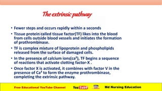
Theextrinsicpathway
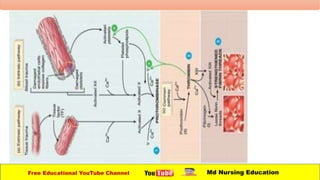
• Fewer steps and occurs rapidly within a seconds
• Tissue protein called tissue factor(TF) likes into the blood
from cells outside blood vessels and initiates the formation
of prothrombinase.
• TF is complex mixture of lipoprotein and phospholipids
released from the surface of damaged cells.
• In the presence of calcium ions(ca²), TF begins a sequence
of reactions that activate clotting factor-X .
• Once factor X is activated, it combines with factor V in the
presence of Ca² to form the enzyme prothrombinase,
completing the extrinsic pathway.
Free Educational YouTube Channel Md Nursing Education
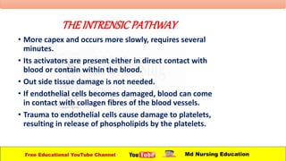
THEINTRENSICPATHWAY
• More capex and occurs more slowly, requires several
minutes.
• Its activators are present either in direct contact with
blood or contain within the blood.
• Out side tissue damage is not needed.
• If endothelial cells becomes damaged, blood can come
in contact with collagen fibres of the blood vessels.
• Trauma to endothelial cells cause damage to platelets,
resulting in release of phospholipids by the platelets.
Free Educational YouTube Channel Md Nursing Education
Theintrinsicpathways
• Contact with collagen fibres activates clotting factor
XII, which begins a sequence of reactions that activates
clotting factor X.
• Platelet phospholipids and Ca² can also participate in
the activation of factors X.
• Once factor X is activated, it combines with factor V to
form the active enzyme prothrombinase, completing
the intrinsic pathway.
Free Educational YouTube Channel Md Nursing Education
THECOMMONPATHWAY
• The formation of prothrombinase starts the beginning
of the common pathway
• In the second stage of blood clotting, prothrombinase
and Ca² catalyze the conversion of prothrombin to
thrombin.
• In the third stage thrombin in presence of Ca², converts
fibrinogen also activates factor Xlll (fibrin stabilizing
factor) which strengthens and stabilizes the fibrin
threads into a stuart clot.
Free Educational YouTube Channel Md Nursing Education
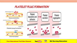
PLATELETPLUGFORMATION
Free Educational YouTube Channel Md Nursing Education
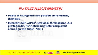
PLATELETPLUGFORMATION
• Inspite of having small size, platelets store lot many
chemicals.
• It contains ADP, ATP,Ca², serotonin, thromboxane A, a
prostaglandin, fibrin-stabilizing factor and platelet-
derived growth factor (PDGF).
Free Educational YouTube Channel Md Nursing Education
Platelets plugformation
• Initially, platelets stick to part of a damaged blood
vessel, such as collagen fibres of damaged endothelial
cells.
• This process is called as platelet adhesion.
• Due to adhesion, the platelets become activated, and
their characteristics change dramatically.
• They extend many projections and they being to
liberate the contents of their vesicles.
• This phase is called platelet release reaction.
Free Educational YouTube Channel Md Nursing Education
Plateletsplugformation
• Liberated ADP and play, a major role of activating nearby platelets.
• Serotonin and thromboxane A² functions as vasoconstriction,
causing congratulations of vascular smooth muscle, which decrease
blood flow through the injured vessels.
Free Educational YouTube Channel Md Nursing Education
Platelets plugformation
• The release of ADP makes others platelets sticky , and
adhere to the originally activated platelets.
• This gathering of platelets is called as platelet
aggregation.
• The accumulation and attachment of large numbers of
platelets to the site of injury to form a solid mass called
as platelet plug.
Free Educational YouTube Channel Md Nursing Education
INFLAMMATION
• Inflammation of the reaction of vascularized
living tissue to local injury.
• This reaction results in accumulation of fluid and
leukocytes in the extracellular tissue.
• It is a defence mechanism to eliminate or limit
the spread of injury or injurious agent.
Free Educational YouTube Channel Md Nursing Education
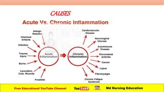
CAUSES
Free Educational YouTube Channel Md Nursing Education
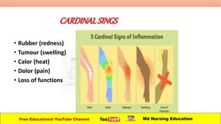
CARDINALSINGS
• Rubber (redness)
• Tumour (swelling)
• Calor (heat)
• Dolor (pain)
• Loss of functions
Free Educational YouTube Channel Md Nursing Education
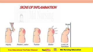
SIGNSOFINFLAMMATION
Free Educational YouTube Channel Md Nursing Education
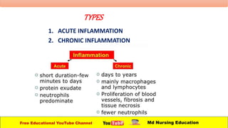
TYPES
1. ACUTE INFLAMMATION
2. CHRONIC INFLAMMATION
Free Educational YouTube Channel Md Nursing Education
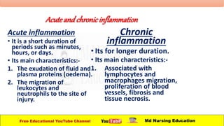
Acute and chronicinflammation
Acute inflammation
• It is a short duration of
periods such as minutes,
hours, or days.
• Its main characteristics:-
1. The exudation of fluid and
plasma proteins (oedema).
2. The migration of
leukocytes and
neutrophils to the site of
injury.
Chronic
inflammation
• Its for longer duration.
• Its main characteristics:-
1. Associated with
lymphocytes and
macrophages migration,
proliferation of blood
vessels, fibrosis and
tissue necrosis.
Free Educational YouTube Channel Md Nursing Education
Tissueandcellsininflammation
• The circulating cells:-Neutrophils, monocytes,
eosinophils, lymphocytes, basophils and platelets.
• The connective tissue cells:-Mast cells, fibroblasts,
macrophages and lymphocytes.
• The extracellular matrix:-fibrous protein(collagen,
elastin) and glycoproteins(fibronectin, laminin,
collagen) and proteoglycans.
Free Educational YouTube Channel Md Nursing Education
Inflammatorymediators
1. Plasma:-
• Complimentary system
• Kinin system
• Clotting system
2. Cells :-
• Histamine
• Serotonin
• Lysosomal enzymes
• Prostaglandins
• Leukotrienes
• Cytokines(IL1, IL6, IL8)
• Platelet activating factor
Free Educational YouTube Channel Md Nursing Education
Vascularchanges
1. Change in vascular blood flow:
• Transient vasoconstriction of the arterioles followed by
vasoconstriction.
• Vasodilation involves arterioles first then
microvascular bed (leading to redness and heat).
• It leads to slowing of blood circulation (blood stasis).
Free Educational YouTube Channel Md Nursing Education
Vascularchanges
2. Increase in vascular permeability :
• Vascular permeability:-capacity of the blood vessels
wall to allow the entry of small molecules or
lymphocytes in and out of the vessels.
• Increase in hydrostatic pressure leading to leakage of
fluid to the extravascular space.
• Increase in osmotic pressure leading to leakage of
protein-rich fluid.
• The end result is Oedema.
Free Educational YouTube Channel Md Nursing Education
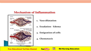
Cellularevents
• Decided into five steps:-
• Margination
• Adhesion to endothelium
• Emigration
• Chemotaxis
• Phagocytosis
Free Educational YouTube Channel Md Nursing Education
Cellularevents
1. Migration:-
• sticking of leucocytes to the endothelial lining of blood
vessels (pavementation).
2. Adhesion:-
• Mediators of inflammation activates the adhesion
molecules on the surface of leukocytes and endothelial
cells and facilities their adhesion.
• Adhesion molecules are bacterial toxins, complement
fragments, chemotactic peptides and cytokines.
Free Educational YouTube Channel Md Nursing Education
Cellularevents
3. Emigration:-
• Emigration refers to the process by which leucocytes
escape from blood vessels to the perivascular tissue.
4. Chemotaxis:-
• It is defined as unidirectional migration of leukocyte
towards an attractant (chemotactic factors).
Free Educational YouTube Channel Md Nursing Education
Cellularevents
5. Phagocytosis:-
• It is the process of engulfment of pathogens and damaged cells.
• 3 distinct steps:
• Recognition and attachment
• Engulfment
• Killing or degradation
Free Educational YouTube Channel Md Nursing Education
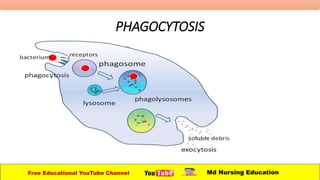
Phagocytosis
1. Recognition and attachment:-
• WBCs have specific receptors on the surface.
2. Engulfment:-
• The particle is engulfed by pseudopodia and enclosed
in membrane bound vesicle called as phagosome.
1. 3. Killing and degradation:-
• Phagosome fuses to lysosome to form phagolysosome .
Killing is facilitated by:
• Oxygen free radicals (oxidative burst).
• Lysosomal enzymes (myeloperoxidase).
Free Educational YouTube Channel Md Nursing Education
PHAGOCYTOSIS
Free Educational YouTube Channel Md Nursing Education