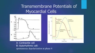
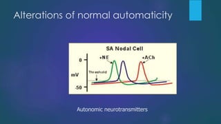
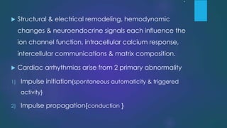
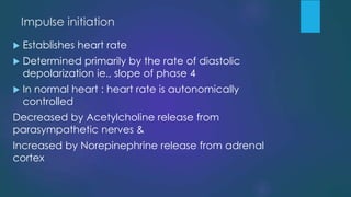
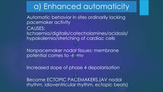
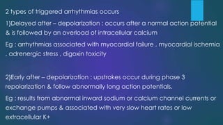
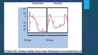
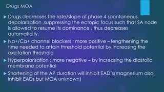
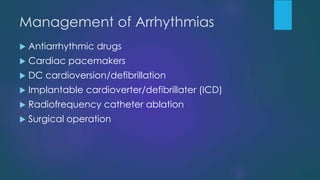
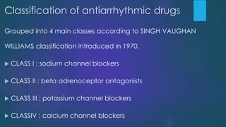
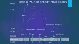
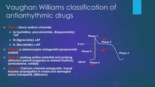
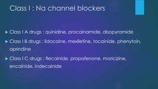
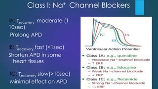
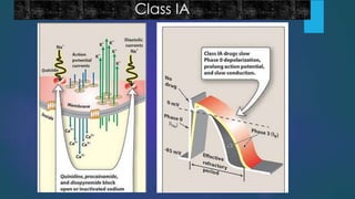
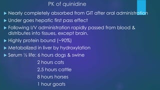
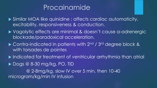
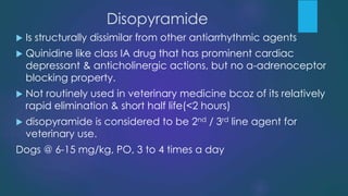
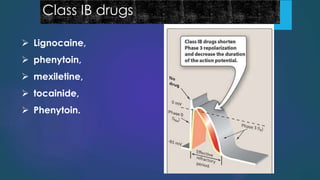
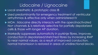
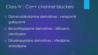
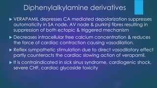
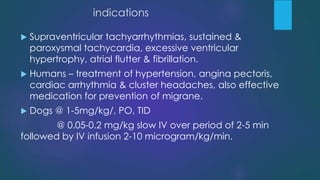
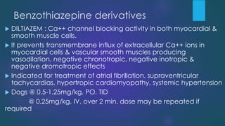
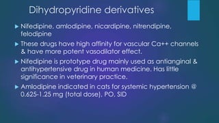
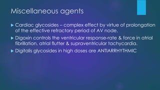
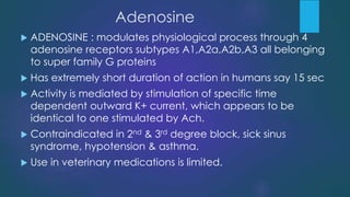
This document summarizes arsenic and lead poisoning in animals. It discusses sources of exposure, factors affecting toxicity, absorption and distribution in the body, mechanisms of toxicity, clinical signs, post-mortem findings, diagnosis, and treatment for both arsenic and lead poisoning. For arsenic, common sources of exposure include improper use of drugs, contaminated water or herbage, and overdose of feed additives. Clinical signs can be acute, subacute, or chronic and include gastrointestinal issues, neurological effects, and poor condition. Treatment involves chelating agents like Dimercaprol. For lead, sources include contaminated grass or foods, and exposure increases toxicity. Clinical signs vary by species but include neurological, gastrointestinal, and hematological
This document discusses blood transfusion in animals. It covers the basics of blood transfusion including indications, components transfused, blood typing and donor selection. It then discusses specific details regarding canine, feline, equine and bovine blood groups. It also covers cross-matching, collection sites, dose calculation, transfusion procedures and potential complications. The key aspects are blood typing and donor selection to avoid transfusion reactions, and monitoring for side effects during and after transfusion.
This document provides an overview of colic in horses. It begins by defining colic as acute abdominal pain in horses. Colic can be classified as spasmodic, tympanitic, obstructive, or impactive. Spasmodic colic involves hypermotility of the intestines. Tympanitic colic is caused by gas accumulation in the intestines. Obstructive colic blocks intestinal passages, while impactive colic specifically involves food or other material blocking the stomach or intestines. The document discusses causes, signs, diagnosis, and treatment for each type of colic. Common signs of colic include pawing, looking at the flank, lip curling, rolling, and abdominal distension
This document provides information about epilepsy in dogs. It defines epilepsy as a brain disorder causing repeated seizures. There are two main types - primary/idiopathic epilepsy which may have unknown causes or genetic factors, and secondary/symptomatic epilepsy which has identifiable causes like infections, tumors, trauma or metabolic diseases. Signs include seizures of different types as well as postictal phases. Diagnosis involves a patient history, physical exam, clinical pathology tests and diagnostic imaging. Treatment primarily involves use of anticonvulsant medications. The causes of epilepsy can vary depending on the dog's age or breed.
Azoturia, also known as Monday morning disease or tying-up syndrome, is a metabolic muscular disorder in horses characterized by stiffness, lameness, and muscle swelling. It occurs after a period of at least 2 days rest when horses return to exercise on a full ration. The major cause is carbohydrate overloading from excessive glycogen buildup in muscles during rest, leading to lactic acid accumulation during subsequent exercise and muscle damage. Clinical signs range from poor performance to an inability to rise. Diagnosis involves detecting myoglobin in the urine and elevated muscle enzyme levels in blood. Treatment focuses on rest, pain relief, intravenous fluids, and thiamine supplementation.
Kennel cough, also known as canine infectious tracheobronchitis, is a highly contagious disease of the canine respiratory tract that causes sudden onset of a paroxysmal cough lasting several days. The two most common causes are canine parainfluenza virus and Bordetella bronchiseptica. Clinical signs include a dry, hacking cough that is more frequent during exercise or changes in temperature/humidity. Diagnosis is based on exposure history and cough, with radiography and cytology used in severe cases to check for pneumonia. Treatment involves antibiotics, cough suppressants, bronchodilators and supportive care. Prevention focuses on vaccination and sanitary kennel practices like isolation, disinfection and
The document discusses canine dilated cardiomyopathy (DCM), including its definition, classification, clinical progression, diagnosis and treatment. DCM is characterized by ventricular dilation and reduced contractility. It most commonly affects large breed dogs and has an asymptomatic occult phase followed by an overt clinical phase with signs of congestive heart failure. Diagnosis involves ECG, Holter monitoring, echocardiography and bloodwork. Treatment depends on the stage of disease and aims to manage congestive heart failure and arrhythmias using diuretics, inotropes, ACE inhibitors and antiarrhythmics. Nutritional deficiencies like taurine can also contribute to DCM in some breeds.
The document discusses seizures and epilepsy, defining them as brief disturbances in brain electrical activity and a disorder characterized by recurring seizures respectively. It covers types of seizures including generalized and focal seizures, their causes, pathophysiology, diagnosis through history, tests and imaging, as well as treatment including emergency protocols, anticonvulsant drugs, and management of status epilepticus.
A presentation by Dr. Renee Streeter, DVM, DACVN, and Dr. Bradley Quest, DVM giving a detailed overview of dilated cardiomyopathy in dogs and what the recent reports from the FDA mean for the pet industry at large.
Impaction in dairy animals refers to the failure of digestion and accumulation of feed in the stomach compartments, leading to constipation or absence of dung. Symptoms of impaction include off food, no rumen sounds, lameness, respiratory distress, low milk production, and sudden death.
This document discusses fluid and electrolyte therapy in veterinary medicine. It describes the normal distribution of body water and outlines causes of abnormal fluid balance like decreased intake, increased output, and third spacing of fluids. The key aspects of fluid therapy are estimating fluid deficits, calculating fluid requirements, selecting appropriate replacement or maintenance fluids, and administering fluids via various routes. Common intravenous fluids used include lactated Ringer's, normal saline, plasmalyte, and colloids like hetastarch. Guidelines are provided for administering fluids and supplementing electrolytes like potassium and bicarbonate as needed. Signs of overhydration are also reviewed.
Local anesthesia and nerve blocks in large animals.GangaYadav4
Local anesthesia involves the reversible loss of sensation in a limited area of the body using chemical agents without loss of consciousness. It is useful for reducing pain and stress during surgery. The document discusses various local anesthetics used in veterinary practice like lidocaine, bupivacaine and mepivacaine. It also covers the mechanisms of action, classifications, advantages, disadvantages and methods of administering local anesthesia like infiltration, regional and intravenous regional anesthesia. Toxicities can occur if the anesthetic is absorbed systemically. Proper technique and dose are important for safe use of local anesthesia.
This document describes left displaced abomasum (LDA) in cattle. LDA typically occurs in dairy cows within a month of calving, often due to nutritional management issues. The abomasum is displaced to the left side of the abdominal cavity due to atony from volatile fatty acids in the diet. Clinical signs include reduced appetite and milk production. Treatment involves surgical correction, usually using right flank omentopexy to fix the abomasum, along with supportive care like fluids and anti-inflammatories. Proper nutritional management can help prevent LDA.
This document provides information on endotracheal intubation in pets. It discusses the uses of endotracheal tubes, including maintaining an open airway and facilitating oxygen administration. It describes the materials tubes are made from and appropriate sizes for dogs and cats. Steps for the intubation procedure are outlined, including preparing the tube, positioning the patient, visualizing the larynx, and properly placing and securing the tube. The document also covers checking proper tube placement and potential complications like laryngospasms.
1. Renal failure occurs when 75% of kidney function is lost and can be classified as pre-renal, primary renal, or post-renal depending on the underlying cause.
2. Acute renal failure is a sudden loss of kidney function that is potentially reversible, while chronic renal failure is a progressive and irreversible condition resulting from gradual nephron death over time.
3. Clinical signs of acute renal failure include decreased urine output and increased BUN and creatinine levels that can progress through oliguric and polyuric phases before possible functional recovery. Treatment focuses on identifying and removing the cause while maintaining fluid, electrolyte, and
Enterotoxemia is caused by Clostridium perfringens type D bacteria. It occurs commonly in young lambs and kids and is characterized by diarrhea, depression, and nervous signs. The bacteria produces alpha and epsilon toxins that damage the intestinal epithelium, causing necrosis and toxemia. Clinical signs include fever, abdominal pain, diarrhea, and neurological signs such as tremors and convulsions. Treatment involves antibiotics, antitoxin serum, supportive therapy, and prevention through gradual diet changes and vaccination.
This document discusses fluid therapy in canines. It describes the reasons for fluid administration including correcting dehydration, acid-base abnormalities, and electrolyte imbalances. Ringer's lactate solution is preferred for correcting dehydration from diarrhea as the lactate is converted to bicarbonate, while isotonic saline is better for vomiting-induced dehydration to prevent further potassium loss. The document also covers fluid types, calculating fluid deficits, monitoring parameters, and guidelines for fluid administration.
This document provides information on various diseases that affect equines in India. It begins with background on the equine population in India and then lists and describes several important viral diseases (Hendra, equine influenza, equine herpes virus, equine infectious anemia, African horse sickness, equine viral arteritis, West Nile fever, equine encephalitis) and bacterial diseases (glanders, strangles, tetanus, Rhodococcus equi, leptospirosis, botryomycosis). For each disease, it discusses the causative agent, transmission, pathogenesis, clinical signs, lesions, and current status or outbreaks in India. Considerable detail is provided for Hendra virus, equ
This document discusses tail amputation procedures in various animal species. It describes the anatomy of the tail and indications for amputation such as trauma, infection, or cosmetic purposes. The procedure typically involves clipping and preparing the tail, retracting the skin, identifying the desired transection site, ligating vessels, disarticulating the tail between vertebrae, and suturing skin edges. Considerations for puppies, adults, and different animal species are provided. Complications can include tetanus or vertebral necrosis.
This document discusses various medicinal plants, herbs, and minerals that are commonly used in herbal medicine systems by different human cultures. It notes that the World Health Organization found around 20,000 plant species are used for medicinal purposes globally, but only about 250 have been thoroughly studied for their active chemical compounds. The document then provides a list of 52 common drugs that are standardized for bulk purchase by the Animal Husbandry Department, including details on their actions, uses, dosages and incompatibilities.
This document discusses fluid therapy in animals. It begins by describing the distribution of water in the body and the composition of intracellular and extracellular fluids. It then discusses three types of fluid disturbances: changes in volume, content, and distribution. The document outlines different types of fluid therapy including replacement, adjunctive, and supportive therapies. It provides details on routes of fluid administration and indications for intravenous fluids. Throughout, it discusses evaluating and monitoring fluid therapy, diagnosing and treating dehydration, and complications of intravenous fluids.
This document provides guidance on performing a clinical examination of cattle. It outlines examining the animal's signalment and history, general appearance, vital signs, and performing a systems-based physical exam. The systems examined include cardiovascular, respiratory, gastrointestinal, urogenital, lymphatic, musculoskeletal, nervous, skin, and head and neck. Basic examination techniques are described for evaluating posture, gait, ears, eyes, body condition, and specific regions of the animal. The goal of the clinical exam is to develop a differential diagnosis and plan appropriate tests, treatment, and information for the owner.
This document discusses antimicrobial contraindications in dogs and cats. It notes that antimicrobials can cause serious side effects in some species. Various antimicrobial classes are associated with issues like nephrotoxicity, hepatotoxicity, vomiting, and joint problems. Certain antimicrobials should be avoided in conditions like renal failure, pregnancy, and for neonates. Safer alternatives like penicillins and cephalosporins are recommended when possible. The document emphasizes that the choice of antimicrobial depends on the individual animal's situation.
APPROACH TO A NEUROLOGICAL EMERGENCY CASE STARTS WITH THE BASIC TRIAGE APPROACH AS IN ANY OTHER EMERGENCY CASE. A NEUROLOGICAL ASSESSMENT IS ONLY DONE AFTER THE STABILIZATION OF THE PATIENT. THERE CAN BE MANY DIFFERENT APPROACHES BUT ALL BASICALLY AIM AT FIRST CONFIRMING IF AT ALL IT IS A NEURO CASE AND IF YES, WHERE IS THE LESION..IS IT IN THE CRANIUM OR BRAINSTEM OR THE SPINAL CORD? LESION LOCALISATION WILL NOT ONLY HELP TO UNDERSTAND BETTER THE TYPE OF THERAPY TO BE CHOSEN BUT WILL ALSO HELP TO TELL ABOUT THE PROGNOSIS OF THE CASE. MOST COMMONLY WE GET STATUS EPILEPTICUS, TRAUMATIC BRAIN INJURY, POISONING, SPINAL CORD INJURIES AND ACUTE VESTIBULAR DISEASES AS THOUGHT TO LINKED WITH NEUROLOGICAL EMERGENCY SITUATIONS. AN EMERGENCY MUST BE FAMILIAR WITH WITH THE RELEVANT HISTORY OF THE PATIENT. HE SHOULD ALSO BE KEEP A TEAM READY WHO CAN HELP HIM PUT THE IV ACCESS AND SEDATIONS WHILE HE CAN COLLECT THE BLOOD FOR BASIC ROUTINE BLOOD ANALYSIS. COUNTERING THE ONGOING STAGE OF SHOCK TO BRING IT TO NORMAL, CHECKING THE SYSTEMIC BLOOD PRESSURE, RECTAL TEMPERATURE AND OXYGEN CONCENTRATION ARE FEW OF THE MOST IMPORTANT FACTORS A CLINICIAN HAS TO DO WHILE ADMINISTERING THE MEDICS. SCORING SYSTEMS LIKE MODIFIED GLASSGOW COMA SCALE AS SUGGESTED BY DR PLATT ARE REALLY HELPFUL TO GIVE A PROGNOSTIC IDEA IN CASES LIKE CRANIO-CERBRAL INJURIES. RECENT TREATMENT UPDATES ARE REALLY HELPFUL TO KEEP HAVING BETTER OPTIONS IN CASE THE ROUTINE PROTOCOL FOR STABILIZING A SEIZURE PATIENT IS NOT WORKING.
The document provides information about the liver anatomy and functions in various animal species. It discusses the liver's role in metabolizing hemoglobin and bilirubin, maintaining glucose homeostasis, processing amino acids and lipids, detoxifying ammonia, storing vitamins and producing clotting factors. Common liver diseases mentioned include infectious hepatitis, cirrhosis, and red water disease caused by Clostridium haemolyticum bacteria. A number of liver function tests and their reference ranges in different animals are also outlined.
Osteodystrophy and osteomyelitis in domestic animalsAjith Y
This document discusses various bone diseases including osteodystrophy, rickets, osteomalacia, and osteoporosis. It describes their causes such as nutritional deficiencies, physical trauma, tumors, and endocrine disorders. Clinical signs, diagnostic testing, and treatment approaches are covered. Specific conditions like renal osteodystrophy, laminitis, osteomyelitis, and bone tumors are also summarized.
This document discusses cystitis, or inflammation of the urinary bladder. It notes that cystitis can be caused by trauma, ascending or descending infections, iatrogenic factors, nutritional issues, urinary stasis, hyperadrenocorticism, diabetes mellitus, and rare neoplasms. Common symptoms in dogs and cats include frequent and painful urination, cloudy urine, abdominal pain, and dullness. Diagnosis involves urinalysis, culture and sensitivity testing, ultrasound or cystoscopy. Treatment focuses on removing the cause, managing pain and inflammation, flushing out organisms, correcting urine pH, and controlling infections with antibiotics based on sensitivity testing.
This document discusses adrenergic agonists and antagonists. It begins by classifying adrenergic agonists as direct acting, mixed acting, or indirect acting. It then discusses specific alpha-1, alpha-2, beta-1, and beta-2 adrenergic receptor agonists like phenylephrine, clonidine, dobutamine, and terbutaline. It details their mechanisms of action and clinical indications. The document concludes by discussing classes of adrenergic antagonists including alpha receptor antagonists, beta receptor antagonists, and specific drugs like propranolol, prazocin, and yohimbine.
This document discusses the contributions of various scientists to the field of veterinary pharmacology over history. It describes important early contributors from ancient civilizations like Ayurveda, Shennong Pen Ts'ao Ching, and texts like the Kahun and Ebers papyruses. It then discusses the contributions of figures like Hippocrates, Galen, Aristotle, Theophrastus, Ibn Hajar, and Razi from ancient Greece and the Islamic Golden Age. It continues with contributors from the 17th-18th centuries who discovered or isolated important drugs. These include Withering and digitalis, Jenner and vaccination, Harvey and blood circulation. The document concludes discussing 19th century physi
This document provides an overview of various experimental animal models that are used to induce different disease conditions and evaluate potential treatments. It discusses models for inflammatory diseases, pyrexia, arrhythmias, hypertension, diabetes, hyperlipidemia, and tests for assessing central nervous system activity, muscle relaxation, sedation, anxiety, seizures, convulsions, and analgesia. Examples of specific animal models and procedures are provided for each condition. The models described allow for studying disease pathogenesis and testing new drug candidates before human trials.
This document discusses hemostatic drugs used to control bleeding. It describes topical agents like coagulants, occlusive agents, and vasoconstrictors that work locally at the site of bleeding. It also discusses systemic hemostatic drugs like vitamin K, blood and blood components, and fibrinolytic inhibitors. Specific drugs are explained in detail, along with their mechanisms of action and protocols for use in different animal species. Commercial hemostatic products and hemostatic testing methods are also mentioned.
This document provides an overview of clinical and pathological effects of toxic plants. It begins with an introduction and classifications of toxic plants. It then discusses the clinical and pathophysiological effects of toxic plants, including specific plants that can cause various types of poisonings. The document covers various plant metabolites and toxins, including alkaloids, terpenes, glycosides, and others. It concludes with treatments and management of plant poisonings.
This document discusses the classification, identification, and chemical constituents of poisonous plants. It outlines several categories of toxic plant compounds including alkaloids, terpenes, glycosides, proteinaceous compounds, organic acids, and resins. Specific poisonous plants and their toxic principles are provided as examples for each compound category. The document serves as an introduction to plant toxicology and identification of poisonous plants.
This document discusses catecholamines and non-catecholamines used as autonomic drugs. It describes the classifications of autonomic drugs and their mechanisms of action. Specific catecholamines discussed include norepinephrine, epinephrine, isoproterenol, and dopamine. Non-catecholamines discussed include ephedrine, pseudoephedrine, amphetamine, methylphenidate, phenylpropanolamine, and oxymetazoline. Their pharmacological effects, clinical uses, and dosages are summarized for various conditions and species. The document provides an overview of important adrenergic drugs and their mechanisms and applications in veterinary medicine.
An Over view on Bioassay, structure & principles, types & methods of bioassay. Also mention of other assay's like biotechnology, microbio assay, immunoassay etc.
This document discusses the phytochemical screening and analysis of medicinal plants. It describes the qualitative and quantitative analysis methods used to detect primary and secondary metabolites such as alkaloids, carbohydrates, saponins, phytosterols, phenolic compounds, tannins, flavonoids, proteins, amino acids and terpenoids. Standard procedures are provided for the quantitative determination of total phenols, alkaloids, flavonoids, tannins, and saponins. The extraction, drying, packing and storage of crude drugs is also summarized.
The document discusses various cardiac rhythm disorders and mechanisms including:
1. Abnormal automaticity, triggered activity, and reentry can cause arrhythmias. Reentry requires both a substrate and a trigger.
2. Bundle branch blocks are conduction disorders involving the left or right bundle branch and are diagnosed based on specific ECG criteria including QRS width and morphology.
3. Bradyarrhythmias are classified based on whether they involve impaired impulse formation in the sinus node or conduction blocks in the AV node. Examples of each type are described briefly.
The document discusses the cardiovascular system, including its major components and functions. It describes the three main divisions of the cardiovascular system: the distribution system, perfusion system, and collecting system. It also discusses the origin and types of blood cells, the mechanisms of circulation and hemostasis, the coagulation cascade, and common screening tests used to detect abnormalities.
This document discusses veterinary anesthesia. It begins by defining anesthesia and anesthesiology. It notes that anesthesia aims to minimize or eliminate pain, relax muscles, and facilitate patient restraint during procedures. Various types of anesthesia are discussed, including local, regional, and general anesthesia. Common drugs used in veterinary anesthesia are also outlined, including sedatives, analgesics, dissociatives, and neuromuscular blocking agents. The document also covers anesthesia administration techniques and important considerations for patient monitoring and recovery.
This document discusses the contributions of various scientists and scholars to the field of veterinary pharmacology throughout history. It begins with Ayurveda in ancient India and mentions important ancient texts like the Kahun Papyrus, Ebers Papyrus, and Shen Nung Pen Ts'ao Ching that documented early uses of herbal medicines and treatments. It then outlines the contributions of figures like Hippocrates, Galen, Aristotle, Theophrastus, Ibn Hajar, and Razi from ancient Greece and the Islamic world. The document notes advances made during the 17th-18th centuries with the discovery of various drugs and mentions scientists like Withering, Jenner, Harvey, and W
The document discusses principles of acid-base balance in veterinary practice. It covers topics like water balance, electrolytes, acid-base balance, renal functions, fluid compartments, fluid therapy, dehydration assessment and treatment, electrolyte imbalances, and commercially available fluids. Key points include the importance of water and electrolytes for life, roles of kidneys and blood in acid-base balance maintenance, classification and assessment of dehydration severity, and fluid therapy considerations like cause, degree of dehydration, and patient condition.
The document discusses various classes of anti-arrhythmic drugs, their mechanisms of action, therapeutic uses, and side effects. It covers class IA drugs like quinidine, procainamide, and disopyramide; class IB drugs like lidocaine, mexiletine, and tocainide; class IC drugs like flecainide, propafenone, and moricizine; class II beta blockers; class III potassium channel blockers like amiodarone, bretylium, and sotalol; and class IV calcium channel blockers. Common side effects include cardiac toxicity, arrhythmia exacerbation, hypotension, and gastrointestinal issues.
Class II antiarrhythmic drugs are beta blockers that reduce sympathetic tone in the heart by blocking beta-1 and beta-2 receptors. They are useful for treating supraventricular arrhythmias by slowing heart rate and conduction through the AV node. Common Class II drugs include propranolol, metoprolol, and atenolol which are effective at preventing recurrence of atrial fibrillation and reducing ventricular rate during atrial fibrillation.
Herbal Foods and its Medicinal Values (CARBOHYDRATES, PROTEINS, FATS, MINER...Ajjay Kumar Gupta
Food has been a basic part of our existence. Through the centuries we have acquired a wealth of information about the use of food as a part of our community, social, national and religious life. It has been used as an expression of love, friendship and social acceptance without knowing the medicinal values of such food. India is one of the leading herbal food producer and exporter in the world. Traditional use of herbal medicines is recognized as a way to learn about potential future medicines. Several meticulous researches were conducted and experimented with herbal food.
See more:-
https://goo.gl/B5sgQQ
https://goo.gl/HdATS6
Contact us
Niir Project Consultancy Services
106-E, Kamla Nagar, Opp. Spark Mall,
New Delhi-110007, India.
Email: npcs.ei@gmail.com , info@entrepreneurindia.co
Tel: +91-11-23843955, 23845654, 23845886, 8800733955
Mobile: +91-9811043595
Fax: +91-11-23845886
Website : www.entrepreneurindia.co , www.niir.org
Take a look at NIIR PROJECT CONSULTANCY SERVICES on #Street View
https://goo.gl/VstWkd
This lecture covers the electrophysiology of the heart and classification of arrhythmias and antiarrhythmic drugs. Key points:
1. The lecture outlines topics on electrophysiology, arrhythmia mechanisms/types, classification of antiarrhythmic drugs, and treatment of some arrhythmias.
2. Arrhythmias are caused by abnormalities in impulse formation and conduction in the myocardium. They can be classified based on the anatomical site of abnormality: atria, AV node, or ventricles.
3. Antiarrhythmic drugs are classified into four classes based on their effects on the cardiac action potential. Class I drugs block sodium channels, class II are beta blockers,
This document summarizes the normal mechanisms of cardiac cellular electrophysiology and the three main mechanisms responsible for cardiac arrhythmias: automaticity, triggered activity, and reentry. It describes how cardiac myocytes generate action potentials through the coordinated flow of ions like sodium, calcium, and potassium. Automaticity refers to the ability of pacemaker cells to spontaneously depolarize. Triggered activity occurs when early or delayed afterdepolarizations trigger premature action potentials. Reentry involves abnormal conduction that allows impulse propagation to reactivate previously excited tissue. Understanding these mechanisms is important for properly diagnosing and treating different types of arrhythmias.
Antiarrhythmic drugs work by altering the conduction of electrical signals in the heart and changing the refractory periods of cardiac cells. They are classified into four classes based on their effects. Class IA drugs like quinidine and procainamide work by slowing the rise of the action potential upstroke, decreasing conduction velocity, and prolonging the refractory period. They have moderate potassium channel blocking effects. Class IA drugs are used for supraventricular arrhythmias and ventricular tachycardia but can cause toxicity like heart block or dangerous arrhythmias.
Antiarrhythmic drugs are used to suppress abnormal heart rhythms known as arrhythmias or dysrhythmias. Arrhythmias can cause the heart rate to be too fast, too slow, or irregular. They are caused by problems with impulse generation or conduction in the heart's electrical system. Common types of arrhythmias include sinus tachycardia, atrial tachycardia, and various types of heart block such as first-degree, second-degree, and third-degree heart block. Antiarrhythmic drugs work to normalize the heart's rhythm and rate.
This document discusses heart arrhythmias and how anti-arrhythmic drugs work to treat them. It provides details on:
1) The electrophysiology of normal heart contraction and how disturbances can cause arrhythmias.
2) Classification systems for anti-arrhythmic drugs based on their mechanisms of action, such as blocking specific ion channels.
3) How different classes of drugs can alter properties like conduction velocity, refractoriness, and automaticity to restore normal rhythm or prevent dangerous arrhythmias.
Since I couldn't find a good enough book on arrhythmia which included everything, I decided to make one. In the hope that it helps someone, since collecting notes is time-consuming (I've been there), I'm posting this in here.
I collected data from various books of Electrocardiography and arrhythmia, various sites, a few research studies and some people's public notes.
I think I included all types of arrhythmia and heart blocks, let me know what do you think of it or in case I left something out.
This document discusses antiarrhythmic drug therapy and summarizes the following key points:
- Antiarrhythmic drugs are classified into four classes based on their mechanism of action and effects on the cardiac action potential. Classes I-III work by blocking sodium, calcium or potassium channels.
- Class I drugs like quinidine and procainamide work by blocking fast sodium channels, reducing the rate of depolarization. Class II drugs like propranolol are beta blockers that reduce heart rate and conduction velocity.
- Common arrhythmias treated include atrial fibrillation, ventricular tachycardia, and supraventricular tachycardias. Drug choice is based on the arrhythmia type
The document discusses the heart and cardiac arrhythmias. It begins by describing the structure and function of the heart, including the specialized conducting system that coordinates contraction and pumping of blood. It then discusses factors that can disrupt normal cardiac rhythm and cause arrhythmias through various mechanisms like changes in automaticity, triggered activity, and abnormal conduction. Various types of arrhythmias are described like tachycardias and bradycardias. Treatment of arrhythmias aims to restore normal rhythm and cardiac output when compromised. Antiarrhythmic drugs are classified based on their mechanisms of action like sodium channel blockade, calcium channel blockade, and beta blockade.
Cardiac arrhythmias are irregularities in the heart's rhythm that can be caused by issues with impulse formation, conduction, or both. Common arrhythmias include atrial fibrillation, atrial flutter, supraventricular tachycardia, ventricular tachycardia, ectopic beats, and various types of atrioventricular block. Antiarrhythmic drugs are used for management but can paradoxically cause arrhythmias, so careful monitoring is needed. Non-pharmacological treatments include cardioversion, ablation, and implantable defibrillators.
Preclinical Screening of Antiarrhythmatic Agents- Mpharm-edited.pptxAshish Kumar Jha
This document provides an overview of preclinical screening methods for antiarrhythmic agents. It discusses various in vivo and in vitro models used to study cardiac arrhythmias and evaluate potential antiarrhythmic drugs. In vivo models include chemically-induced arrhythmias using drugs like aconitine or digoxin in rats or guinea pigs. Electrically-induced models use techniques like ventricular fibrillation threshold testing in dogs. Mechanically-induced models involve coronary artery ligation and reperfusion in rats. In vitro preparations include isolated tissues like guinea pig papillary muscle to study action potentials. The goal is to find safe and effective therapies for treating abnormal heart rhythms.
This document discusses normal cardiac electrophysiology and the mechanisms of arrhythmias. It begins by describing the normal sinus rhythm and the role of ion channels and gap junctions. It then explains the cardiac action potential and factors that determine the resting membrane potential. Different types of abnormal impulse formation are described, including abnormal automaticity, triggered activity, and reentry. The document outlines evaluation and management of arrhythmias and discusses specific arrhythmias like atrial fibrillation, SVT, and VT. In summary, it provides an overview of cardiac electrophysiology, mechanisms of arrhythmias, evaluation, and treatment approaches.
Individualized Webcam facilitated and e-Classroom USMLE Step 1 Tutorials with Dr. Cray. 1 BMS Unit is 4 hr. General Principles and some Organ System require multiple units to complete in preparation for the USMLE Step 1 A HIGH YIELD FOCUS IN Biochemistry / Cell Biology, Microbiology / Immunology and the 4 P’s-Phiso, Pathophys, Path and Pharm. Webcam Facilitated USMLE Step 2 Clinical Knowledge and Clinical Skills diadactic tutorials /1 Unit is 4 hours, individualized one-on-one and group sessions, Including all Internal Medicine sub-sub-specitialities. For questions or more information.. drcray@imhotepvirtualmedsch.com
This document discusses arrhythmias and their treatment. It defines arrhythmias as abnormalities in heart rhythm that result in insufficient cardiac output. The document describes the normal physiology of cardiac rhythm controlled by the sinoatrial node. It outlines different types of arrhythmias caused by issues with pacemaker impulse formation or conduction. The mechanisms of various arrhythmias are explained including reentry circuits and abnormal pacemaking. Finally, the document discusses pharmacological treatments for arrhythmias including classes I-IV antiarrhythmic drugs that act on ion channels and membranes to normalize heart rhythm.
Arrhythmias are abnormal heart rhythms that can be caused by issues with the heart's electrical conduction system or structural problems with heart muscles or valves. Common causes include ischemia, electrolyte imbalances, cardiomyopathy, and genetic conditions. Arrhythmias are classified based on heart rate, regularity, site of origin in the heart, and ECG characteristics. They are often due to reentry circuits, abnormal automaticity, or heart block. Treatment involves medications to suppress arrhythmias or restore normal rhythm, ablation procedures, or pacemakers.
This document discusses various cardiac arrhythmias including their mechanisms and treatment. It begins by describing the three main mechanisms of cardiac arrhythmia: alterations in impulse initiation (automaticity), afterdepolarizations and triggered automaticity, and abnormal impulse conduction (reentry). It then discusses various specific arrhythmias in more detail, including types of heart block, tachycardias like atrial fibrillation, flutter and sinus tachycardia, as well as treatment options like antiarrhythmic drugs, catheter ablation, and pacemakers. In summary, the document provides an overview of the conduction system of the heart and covers the pathophysiology, classification, evaluation and management of different cardiac arrhythmias.
Antiarrhythmic drugs are used to prevent or treat irregularities in cardiac rhythm caused by disturbances in the heart's electrical impulses. The drugs work by reducing abnormal pacemaker activity or modifying conduction to disable reentrant circuits. Quinidine is a Class IA drug that blocks sodium channels, prolonging the action potential and increasing the refractory period. It can treat both atrial and ventricular arrhythmias but causes side effects like cinchonism. Procainamide is also a Class IA drug that works similarly to quinidine to treat ventricular and some supraventricular arrhythmias but has more ganglionic blocking effects and can cause lupus-like symptoms.
This document provides an overview of cardiac arrhythmias including their classification, mechanisms, clinical manifestations, diagnostic approaches and management strategies. It discusses various specific arrhythmias in detail such as atrial fibrillation, atrial flutter, supraventricular tachycardia, ventricular arrhythmias, sick sinus syndrome and heart block. Treatment options covered include pharmacological therapies using different classes of antiarrhythmic drugs, procedures like cardiac ablation and use of devices like pacemakers.
This document discusses the pharmacology of antiarrhythmic drugs. It begins by defining cardiac arrhythmias and their underlying mechanisms, including abnormal automaticity, impaired conduction, afterdepolarizations, and reentry. It then classifies antiarrhythmic drugs according to their primary electrophysiological actions on sodium, potassium, or calcium channels. Several example drugs are discussed in depth, including their mechanisms of action, effects, uses, and adverse effects. The document provides an overview of important cardiac arrhythmias and categorizes antiarrhythmic drugs into four classes based on their predominant mechanisms and sites of action.
This document provides information about arrhythmias including:
1) It defines arrhythmias as heart rhythm disorders divided into tachyarrhythmias due to increased electrical activity and bradyarrhythmias due to decreased electrical activity.
2) It describes the physiology of the heart's electrical activity including the phases of the transmembrane action potential.
3) It outlines methods for studying cardiac arrhythmias including anamnesis, physical examination, and complementary explorations like ECG, Holter monitoring, and electrophysiological studies.
Antiarrhythmic drugs work by altering the electrophysiology of the heart. They are classified into four main classes based on their mechanisms of action: Class I drugs block sodium channels, Class II block beta-adrenergic receptors, Class III prolong the heart's repolarization, and Class IV block calcium channels. While these drugs can treat arrhythmias, they may also paradoxically cause arrhythmias due to their effects on the heart's electrical activity. Pacemakers provide an alternative treatment for arrhythmias by using implanted leads and a pulse generator to electrically stimulate the heart and maintain a normal rhythm.
Similar to ANTIARRHYTHMIC AGENTS IN VETERINARY PRACTICE. (20)
How to Fix the Import Error in the Odoo 17Celine George
An import error occurs when a program fails to import a module or library, disrupting its execution. In languages like Python, this issue arises when the specified module cannot be found or accessed, hindering the program's functionality. Resolving import errors is crucial for maintaining smooth software operation and uninterrupted development processes.
A review of the growth of the Israel Genealogy Research Association Database Collection for the last 12 months. Our collection is now passed the 3 million mark and still growing. See which archives have contributed the most. See the different types of records we have, and which years have had records added. You can also see what we have for the future.
Main Java[All of the Base Concepts}.docxadhitya5119
This is part 1 of my Java Learning Journey. This Contains Custom methods, classes, constructors, packages, multithreading , try- catch block, finally block and more.
Physiology and chemistry of skin and pigmentation, hairs, scalp, lips and nail, Cleansing cream, Lotions, Face powders, Face packs, Lipsticks, Bath products, soaps and baby product,
Preparation and standardization of the following : Tonic, Bleaches, Dentifrices and Mouth washes & Tooth Pastes, Cosmetics for Nails.
This presentation was provided by Steph Pollock of The American Psychological Association’s Journals Program, and Damita Snow, of The American Society of Civil Engineers (ASCE), for the initial session of NISO's 2024 Training Series "DEIA in the Scholarly Landscape." Session One: 'Setting Expectations: a DEIA Primer,' was held June 6, 2024.
How to Add Chatter in the odoo 17 ERP ModuleCeline George
In Odoo, the chatter is like a chat tool that helps you work together on records. You can leave notes and track things, making it easier to talk with your team and partners. Inside chatter, all communication history, activity, and changes will be displayed.
it describes the bony anatomy including the femoral head , acetabulum, labrum . also discusses the capsule , ligaments . muscle that act on the hip joint and the range of motion are outlined. factors affecting hip joint stability and weight transmission through the joint are summarized.
This presentation includes basic of PCOS their pathology and treatment and also Ayurveda correlation of PCOS and Ayurvedic line of treatment mentioned in classics.
Thinking of getting a dog? Be aware that breeds like Pit Bulls, Rottweilers, and German Shepherds can be loyal and dangerous. Proper training and socialization are crucial to preventing aggressive behaviors. Ensure safety by understanding their needs and always supervising interactions. Stay safe, and enjoy your furry friends!
Liberal Approach to the Study of Indian Politics.pdf
ANTIARRHYTHMIC AGENTS IN VETERINARY PRACTICE.
1. ANTIARRHYTHMIC AGENTS IN VETERINARY PRACTICE
PRESENTED BY
SINDHU K
MVSC SCHOLAR,
DEPT OF VPT,
COVAS, POOKODE.
2. ARRYTHMIA
Is an abnormality in the rate , regularity , or site of
origin of cardiac impulse or a disruption in impulse
conduction such that the normal sequence of atrial
& ventricular activation is changed.
5. Cardiac Action Potential
Divided into five phases (0,1,2,3,4)
Phase 4 - resting phase (resting membrane potential)
Phase cardiac cells remain in until stimulated
Associated with diastole portion of heart cycle
Addition of current into cardiac muscle (stimulation) causes
Phase 0 – opening of fast Na channels and rapid depolarization
Drives Na+ into cell (inward current), changing membrane potential
Transient outward current due to movement of Cl- and K+
Phase 1 – initial rapid repolarization
Closure of the fast Na+ channels
Phase 0 and 1 together correspond to the R and S waves of the ECG
6. Cardiac Action Potential (con’t)
Phase 2 - plateau phase
sustained by the balance between the inward movement of Ca+ and outward
movement of K +
Has a long duration compared to other nerve and muscle tissue
Normally blocks any premature stimulator signals (other muscle tissue can accept
additional stimulation and increase contractility in a summation effect)
Corresponds to ST segment of the ECG.
Phase 3 – repolarization
K+ channels remain open,
Allows K+ to build up outside the cell, causing the cell to repolarize
K + channels finally close when membrane potential reaches certain level
Corresponds to T wave on the ECG
7. +30 mV
0 mV
-80 mV
-90 mV
OUTSIDE
MEMBRANE
INSIDE
Na+
0
4
3
2
1
K+
Ca++K+
At
p
K+
Na+
K+
Ca++
Na+
K+
Na+
Resting
open
Inactivated
Phase zero
depolarization
Early
repolarization
Plateau
phase
Rapid
Repolarization
phase
Phase 4
depolarization
8. etiology
Ischemia/hypoxemia
Imbalance of the parasympathetic & sympathetic
branches of the ANS
Serum electrolyte imbalance[K+ & Ca ++]
Activation of RAAS
Pharmacologic therapy
Inherited causes (rare)
Arrhythmia associated with acquired heart diseases viz
CHF, viral myocarditis etc
Infarction of the heart muscle
9. A: Contractile cell
B: Autorhythmic cell:
spontaneous depolarization at phase 4
Transmembrane Potentials of
Myocardial Cells
12. Impulse initiation
Establishes heart rate
Determined primarily by the rate of diastolic
depolarization ie., slope of phase 4
In normal heart : heart rate is autonomically
controlled
Decreased by Acetylcholine release from
parasympathetic nerves &
Increased by Norepinephrine release from adrenal
cortex
13. b) Trigerred automaticity
+30 mV
0 mV
-80 mV
-90 mV
Delayed After
Depolarisation
(DAD)
Intracellular cal. Overload (Ischemia
reperfusion, adr.stress, digitalis intoxication or
heart failure)
14. Disturbances in automaticity
Automatic cells of the SA node are dominant pacemaker ,
reaching threshold first with the resultant propagating impulse
exciting all other potential pacemaker cells before they
spontaneously attain threshold values
If automaticity of the SA node is depressed/the spontaneous
firing rate in some other tissue (latent pacemaker)is accelerated ,
region of the heart other than SA node serves as pace maker &
initiates ectopic impulses
15. 1.The slope of phase 4 can be affected by a no of abnormal
conditions.
Enhanced automaticity occurs when the rate of spontaneous
diastolic depolarization increases sufficiently to allow
emergence of pathologically slowed or increased rates { sinus
tachycardia }
2.Ectopic foci(pace makers that normally are latent)
May emerge & may cause tachycardia if the frequency
exceeds that of sinoatrial node.
17. a) Enhanced automaticity
Automatic behavior in sites ordinarily lacking
pacemaker activity
CAUSES:
Ischaemia/digitalis/catecholamines/acidosis/
hypokalemia/stretching of cardiac cells
Nonpacemaker nodal tissues: membrane
potential comes to -60mv
Increased slope of phase 4 depolarisation
Become ECTOPIC PACEMAKERS.(AV nodal
rhythm, idioventricular rhythm, ectopic beats)
18. 2 types of triggered arrhythmias occurs
1}Delayed after – depolarization : occurs after a normal action potential
& is followed by an overload of intracellular calcium
Eg : arrhythmias associated with myocardial failure , myocardial ischemia
, adrenergic stress , digoxin toxicity
2}Early after – depolarization : upstrokes occur during phase 3
repolarization & follow abnormally long action potentials.
Eg : results from abnormal inward sodium or calcium channel currents or
exchange pumps & associated with very slow heart rates or low
extracellular K+
20. Drugs MOA
Drugs decreases the rate/slope of phase 4 spontaneous
depolarization ,suppressing the ectopic focus such that SA node
is allowed to resume its dominance , thus decreases
automaticity.
Na+/Ca+ channel blockers ; more positive – lengthening the
time needed to attain threshold potential by increasing the
excitation threshold
Hyperpolarisation ; more negative – by increasing the diastolic
membrane potential
Shortening of the AP duration will inhibit EAD`s(magnesium also
inhibit EADs but MOA unknown)
22. Disturbances in impulse conduction
Associated with a phenomenon of REENTRY or CIRCUS movement.
REENTRANT arrhythmias : anatomic
functional
Anatomic arrhythmias involves 2/more pathways that travel to the
same region of the heart but differ in electrophysiology.
Functional reentrant arrhythmias are exemplified by pathologies viz
ISCHEMIA that markedly slows conduction.
23. The concept of REENTRY(Schmidt & Erlanger 1929)
Based on very slow conduction velocity
An area of heart demonstrating unidirectional block of impulse
conduction
Abnormally brief refractory period
This theory holds that a cardiac impulse can travel circuitously around
an anatomic loop of fibers in which slowed conduction velocity & brief
refractoriness permit the impulse to arrive at cells that are no longer
refractory , there by permitting perpetual reexcitation
26. Drugs MOA
Reentry can be controlled by drug that either creates bidirectional
block or bidirectional conduction through the region of cells causing
the unidirectional block
Blocking specific ion channels {suppress initiation & automaticity}/ by
targeting autonomic functions thus altering initiation or conduction or
AP duration {thus refractory period}
Drugs that facilitates adenosine / acetylcholine – thus increasing the
maximum diastolic or resting potential
Drugs used to antagonize adrenergic receptors – thus decreasing the
slope of phase 4
31. Classification of antiarrhythmic drugs
Grouped into 4 main classes according to SINGH VAUGHAN
WILLIAMS classification introduced in 1970.
CLASS I : sodium channel blockers
CLASS II : beta adrenoceptor antagonists
CLASS III : potassium channel blockers
CLASSIV : calcium channel blockers
32. +30 mV
0 mV
-80 mV
-90 mV
OUTSIDE
MEMBRANE
INSIDE
Na+
0
4
3
2
1
K+
Ca++K+
At
p
K+
Na+
K+
Ca++
Na+
Na+Ca++K+
RATE
SLOPE
Effective Refractory Period
RMP
THRESHOLD POTENTIAL
Possible MOA of antiarrythmic agents
33. Vaughan Williams classification of
antiarrhythmic drugs
Class I: block sodium channels
Ia (quinidine, procainamide, disopyramide)
AP
Ib (lignocaine) AP
Ic (flecainide) AP
Class II: β-adrenoceptor antagonists (propranolol,
sotalol)
Class III: prolong action potential and prolong
refractory period (suppress re-entrant rhythms)
(amiodarone, sotalol)
Class IV: Calcium channel antagonists. Impair
impulse propagation in nodal and damaged
areas (verapamil, diltiazem)
.
Phase 4
Phase 0
Phase 1
Phase 2
Phase 3
0 mV
-80mV
II
I
III
IV
`
34. Class I : Na channel blockers
Class I A drugs : quinidine, procainamide, disopyramide
Class I B drugs : lidocaine, mexiletine, tocainide, phenytoin,
aprindine
Class I C drugs : flecainide, propafenone, moricizine,
encainide, indecainide
35. Class I: Na+ Channel Blockers
IA: Ʈrecovery moderate (1-
10sec)
Prolong APD
IB: Ʈrecovery fast (<1sec)
Shorten APD in some
heart tissues
IC: Ʈrecovery slow(>10sec)
Minimal effect on APD
37. QUINIDINE
Prototype class IA
Quinidine is an alkaloid obtained from cinchona bark & is
dextro-isomer of antimalarial drug quinine
MOA
1) Blocks myocardial Na+ channels in frequency of use
dependent manner
2) Intermediate association with open/inactivated Na channels &
intermediate rate of dissociation from resting channels.
3) Prolongs AP due to K+ channel block
4) @ high conc, quinidine also inhibits L type Ca++ channels
38. PK of quinidine
Nearly completely absorbed from GIT after oral administration
Under goes hepatic first pass effect
Following I/V administration rapidly passed from blood &
distributes into tissues, except brain.
Highly protein bound (~90%)
Metabolized in liver by hydroxylation
Serum ½ life: 6 hours dogs & swine
2 hours cats
2.5 hours cattle
8 hours horses
1 hour goats
39. Adverse effects
Dogs & cats : anorexia, nausea, vomiting, diarrhea.
Horses : swelling of nasal mucosa, urticarial wheals & laminitis
Sinus tachycardia, increased ventricular rates in patients atrial
fibrillation, hypotension, syncope, pro arrhythmic effects
OVER DOSAGE : depressed automaticity & conduction/
tachyarrhytmias
QUINIDINE TOXICITY : 25% increase in duration of QRS complex,
atrioventricular block, acceleration of ventricular tachyarrythmia
Dogs : therapeutic range @ 2.5 – 5 micro g/ml
toxic range @ > 10 micro g/ml
40. TREATMENT OF QUINIDINE TOXICITY
1)I/V administration of sodium lactate 1/6 M or sodium
bicarbonate may reduce cardiotoxic effects by increasing
quinidine protein binding
2)Supportive & symptomatic measures
Forced diuresis using fluids & diuretics along with reduction of
urinary pH may enhance the renal excretion of quinidine
41. Contraindications & interactions
CI in complete AV block, intraventricular conduction defect,
aberrant ectopic impulses, myasthenia gravis, hepatic
impairment & drug sensitivity.
DRUG INTERACTIONS
Increases digoxin, amiodarone, verapamil serum
concentration.
Enhances negative inotropic & hypotensive effects of beta
adrenoceptor antagonists & calcium agonists
Alkalinisation of urine decreases excretion
Acidification of urine increases quinidine excretion
42. CLINICAL INDICATIONS
EQUINES : treatment for supraventricular arrhythmias
Dose @ 20mg/kg, PO, every 2 hours (maximal dose 60g
daily) by stomach tube until arrhythmia is abolished.
SMALL ANIMALS : supraventricular arrhythmias associated
with anomalous conduction in WOLFF-PARKINSON WHITE
SYNDROME.
Acute atrial fibrillation
Dogs @ 6-20 mg/kg, PO, 3 to 4 times daily
Cats @ 4-8 mg/kg, IM, TID.
43. Procainamide
Similar MOA like quinidine ; affects cardiac automaticity,
excitability, responsiveness & conduction.
Vagolytic effects are minimal & doesn’t cause a-adrenergic
blockade/paradoxical acceleration.
Contra-indicated in patients with 2nd / 3rd degree block &
with torsades de pointes
Indicated for treatment of ventricular arrhythmia than atrial
Dogs @ 8-30 mg/kg, PO, TID
@ 2-8mg/kg, slow IV over 5 min, then 10-40
microgram/kg/min IV infusion
44. Disopyramide
Is structurally dissimilar from other antiarrhythmic agents
Quinidine like class IA drug that has prominent cardiac
depressant & anticholinergic actions, but no a-adrenoceptor
blocking property.
Not routinely used in veterinary medicine bcoz of its relatively
rapid elimination & short half life(<2 hours)
disopyramide is considered to be 2nd / 3rd line agent for
veterinary use.
Dogs @ 6-15 mg/kg, PO, 3 to 4 times a day
46. Lidocaine / Lignocaine
Local anesthetic & prototypic class IB
Used predominantly for emergency treatment of ventricular
arrhythmias & effective only when administered IV
MOA : lidocaine directly interacts with the open/inactivated
Na+ channels & is relatively selective for partially depolarized
cells & those with longer AP duration.
Markedly suppresses automaticity in purkinje fibres, improves
conduction in depolarized/stretched fibres by increasing RMP
to near normal values, as a result of improved conduction,
normal transmission is restored in areas of unidirectional blocks.
47. Indications
Following a therapeutic IV bolus, the onset of action is seen
with in 2 min & lasts for 10-20 min.
Lidocaine is one of the best antiarrhythmic drugs & first choice
for life saving tachyarrhythmias & for most ventricular
arrhythmias, principally ventricular tachycardia & ventricular
premature complexes
Dogs @ 1-2mg/kg, IV bolus, followed by 30-50 microg/kg/min,
IV infusion
Large animals @ 0.25-0.5 mg/kg, IV.
48. Mexiletine hcl
Is structural analogue of lidocaine & reported to produce
enhanced antiarrhythmic effects when combined with either
quinidine or procainamide therapy
Indicated especially frequent ventricular premature beats,
ventricular tachycardia & those induced by digitalis toxicity
Mexiletine is contraindicated for cats
Dogs @ 4-8 mg/kg, PO, TID
@ 3-5 mg/kg, IV, followed by 5-10 microg/kg/min, iv infusion
49. 1) TOCAINIDE : amide type local anesthetic
Indicated in dogs for long term control of ventricular
arrhythmias @ 15-20 mg/kg, PO, TID.
2) PHENYTOIN : an antiepileptic drug with class IB
antiarrhythmic effects.
Indicated for ventricular arrhythmia in dogs @
10mg/kg, IV, TID, usually in increments of 2-4mg/kg.
3) APRINDINE : effective in controlling pre-mature
ventricular beats & ventricular tachycardia
Dogs @ 0.1mg/kg IV infusion for 5 min, repeated at 10
min intervals till arrhythmia is controlled, followed by
3mg/kg PO TID
50. Class I C drugs
Encainide, Flecainide, Propafenone
Have minimal effect
on repolarization
Are most potent
sodium channel
blockers
• Risk of cardiac arrest
, sudden death so not
used commonly
• May be used in
severe ventricular
arrhythmias
51. Ia Ib Ic
Moderate Na
channel blockade
Mild Na channel
blockade
Marked Na channel
blockade
Slow rate of rise of
Phase 0
Limited effect on
Phase 0
Markedly reduces
rate of rise of phase 0
Prolong refractoriness
by blocking several
types of K channels
Little effect on
refractoriness as there
is minimal effect on K
channels
Prolong refractoriness
by blocking delayed
rectifier K channels
Lengthen APD &
repolarization
Shorten APD &
repolarization
No effect on APD &
repolarization
Prolong PR, QRS QT unaltered or
slightly shortened
Markedly prolong PR
& QRS
52. Class II : beta – adrenoceptor
antagonists
Depress phase 4 depolarization of pacemaker cells,
Slow sinus as well as AV nodal conduction :
↓ HR, ↑ PR
↑ ERP, prolong AP Duration by ↓ AV conduction
Reduce myocardial oxygen demand
Well tolerated, Safer
Propranolol, atenolol, esmolol, sotalol, timolol, carazol,
bisoprolol.
53. MOA
Increasing the magnitude of Ca++ current & slowing its
inactivation
Increases the magnitude of repolarizing K+ & Ca++ current
Increases the pace maker current & under
pathophysiological conditions increases both DAD & EAD
mediated currents
Positive inotropic effect
In heart activity is mainly through inhibition of beta1
adrenergic receptors, inhibits effect of sympathetic NS by
reducing heart rate, decreases intracellular Ca++overload &
inhibiting after depolarization mediated automaticity.
54. β Adrenergic
Stimulation
β Blockers
↑ magnitude of Ca2+
current & slows its
inactivation
↓ Intracellular Ca2+
overload
↑ Pacemaker current→↑
heart rate
↓Pacemaker current→↓
heart rate
↑ DAD & EAD mediated
arrhythmias
Inhibits after-
depolarization mediated
automaticity
Epinephrine induces
hypokalemia (β2 action)
Propranolol blocks this
action
56. Class III : K+ channel blockers
Amiodarone, dronedarone, bretylium,
bunaftine, ibutilide, nifekalant
Amiodarone is Iodone containing agent
structurally related to thyroid hormone
thyroxine
indicated in horses to treat atrial fibrillation @
5mg/kg/hr, IV, for 1 hour followed by 0.8
mg/kg/hr for 23 hour
57. Bretylium
Complex electrophysiological effects : partly result of
blockade of norepinephrine release release fron adrenergic
nerve terminals in heart but major direct action is
prolongation of action potential duration & effective
refractory period due to K+ channel blockade.
Indicated in life threatening ventricular arrhythmias in dogs
@ 5-20 mg/kg, IV
In pigs used for protection against tachyarrhythmias
induced by general anesthesia @ 0.02mg/kg/min, IV
infusion.
58. Calcium channel blockers (Class IV)
• Inhibit the inward
movement of
calcium ↓
contractility,
automaticity , and
AV conduction.
• Verapamil &
diltiazem
60. Diphenylalkylamine derivatives
VERAPAMIL depresses CA mediated depolarization suppresses
automaticity in SA node, AV node & purkinji fibres resulting in
suppression of both ectopic & triggered mechanism
Decreases intracellular free calcium concentration & reduces
the force of cardiac contraction causing vasodilation.
Reflex sympathetic stimulation due to direct vasodilatory effect
partly counteracts the cardiac slowing action of verapamil.
It is contraindicated in sick sinus syndrome, cardiogenic shock,
severe CHF, cardiac glycoside toxicity
61. indications
Supraventricular tachyarrhythmias, sustained &
paroxysmal tachycardia, excessive ventricular
hypertrophy, atrial flutter & fibrillation.
Humans – treatment of hypertension, angina pectoris,
cardiac arrhythmia & cluster headaches, also effective
medication for prevention of migrane.
Dogs @ 1-5mg/kg/, PO, TID
@ 0.05-0.2 mg/kg slow IV over period of 2-5 min
followed by IV infusion 2-10 microgram/kg/min.
62. Benzothiazepine derivatives
DILTIAZEM : Ca++ channel blocking activity in both myocardial &
smooth muscle cells.
It prevents transmembrane influx of extracellular Ca++ ions in
myocardial cells & vascular smooth muscles producing
vasodilation, negative chronotropic, negative inotropic &
negative dromotropic effects
Indicated for treatment of atrial fibrillation, supraventricular
tachycardias, hypertropic cardiomyopathy, systemic hypertension
Dogs @ 0.5-1.25mg/kg, PO, TID
@ 0.25mg/kg, IV, over 2 min. dose may be repeated if
required
63. Dihydropyridine derivatives
Nifedipine, amlodipine, nicardipine, nitrendipine,
felodipine
These drugs have high affinity for vascular Ca++ channels
& have more potent vasodilator effect.
Nifedipine is prototype drug mainly used as antianginal &
antihypertensive drug in human medicine. Has little
significance in veterinary practice.
Amlodipine indicated in cats for systemic hypertension @
0.625-1.25 mg (total dose), PO, SID
64. Miscellaneous agents
Cardiac glycosides – complex effect by virtue of prolongation
of the effective refractory period of AV node.
Digoxin controls the ventricular response-rate & force in atrial
fibrillation, atrial flutter & supraventricular tachycardia.
Digitalis glycosides in high doses are ANTIARRHYTHMIC
65. Adenosine
ADENOSINE : modulates physiological process through 4
adenosine receptors subtypes A1,A2a,A2b,A3 all belonging
to super family G proteins
Has extremely short duration of action in humans say 15 sec
Activity is mediated by stimulation of specific time
dependent outward K+ current, which appears to be
identical to one stimulated by Ach.
Contraindicated in 2nd & 3rd degree block, sick sinus
syndrome, hypotension & asthma.
Use in veterinary medications is limited.
66. references
HS SANDHU Essentials of veterinary pharmacology and
toxicology, 2nd edition.
H RICHARD ADAMS Veterinary pharmacology and
therapeutics, 8th edition.
GOOGLE IMAGES
ONLINE SEARCH RELATED TOPICS
RMP IS -90 MV
Cardiac bounded by a lipoprotein membrane which has receptor channels crossing it
WHEN AN ATRIAL OR VENTRICULAR CELL RECIEVES An action potential it starts depolarising in response to it..and sodium starts entering it
Intracellular negativity starts diminishing
When such depolarisation reaches a threshold potential, the sodium channels open abruptly
Na enters cell in large quantities
CELL MEMBRANE ACTION POTENTIAL CHANGES FROM -90 TO ALMOST +30MV
Phase 0: rapid depolarisation…fast selective inflow of na ions
During latter part, ca ions also enter the cell via na channels
Frther in phase 1 and 2 ca ions enter thru slow ca channels
THE CONFORMATION OF THE SODIUM CHANNELS HENCE CHANGES TO INACTIVE STATE
The ca which enters the cell in dis manner causes release of ca from sarcoplasmic reticulumraising the conc of ca within the cells
This intracellular free ca interacts with actin myocin system and causes contraction of heart
Afetr this, phase 1: short rapid repolarisation due to beginning of outflow of potassium and entry of cloride ions into the cells, MEMBRANE CHARGE CHANGES FROM +30 TO ALMOST 0 MV IN VERY SHORT TIME
Phase 2 : prolonged plateau phase.. Balance bw ca enterin the cell and k leavin the cell..VOLTAGE SENSITIVE SLOW l type CA CHANNELS OPEN …SLOW INWARD CA CURRENT BALANCED BY SLOW OUTWARD K CURRENT..DEPOLARISATION = REPOLARISATION
Phase 3 : rapid repolarisation.. CA CHANNELS CLOSE…K CHANNELS OPEN..Contimued extrusion of k…RESUMES INITIAL NEGATIVITY
FROM PHASE 0 TO 3 THERE HAS BEEN A GAIN OF NA AND A LOSS OF K ..THIS IS NOW REVERTED AND BALANCED BY NA K ATPASE
Phase 4: resting phase..ELECTRICALLY STABLE… Ionic reconstitution of cell is reachieved by na k exchange pump
RMP MAINTAINED BY OUTWARD K LEAK CURRENTS AND NA CA EXCHANGERS
The cycle is then repeated
Inactivation gates of sodium channels in resting membranes close over the potential range of -75 to -55mv
Cardiac sodium channel protein shows 3 different conformations
Depolarisation to threshold voltage results in opening of the activation gates of sodium channel thus causing markerdly increased sodium permeability
Brief intense sodium current , conductance of fast sodium channel suddenly increases in response to depolarising stimulUs
Very large influx of na accounts for phase 0 depolarisation
Clusure of inactivation gates result
Remain inactivated till mid phase 3 to permit a new propagated response to external stimulus…refractory period..
Cardiac calcium channels are L type
Phase 1 and 2 : turning off nodium current, waxing and waning of calcium curent, slow development of repolarising potassium current, calcium enters ..potassium leaves..
Phase 3: complete inactivation of sodium and calcium currents and full opening of potassium
2 types of main potassium currents involved in phase 3 : ikr and iks
Certain potassium channels are open at rest also…”inward rectifier” channels
In addition there are 2 energy requiring exchange pumps in cardiac myocyte cell membrane…na k exchange pump…and and na-ca exchange pump
Normally na ions concentrated extracellularly and vice versa for k cions
Thus have a tendency odf diffusion along concentration gradient
This diffusion is opposed by na k pump
This pump operates contimuously and does not switch on and off during action potential of cardiac cells
Late Afterdepolarizations
Secondary deflection after attaining RMP
Increased intracellular Ca2+ overload
Adrenergic stress, digitalis intoxication, ischemia-reperfusion
AFTRE attaining Resting membrane potential, a secondary deflection occurs..
If this reaches threshold potential..it initiates a single premature AP
GENERALLY OCCURS FROM CALCIUM OVERLOAD..digitalis toxicity..ischaemia reperfusion
Ectopic pacemaker activity is encouraged by
Faster phase 4 depolarization due to ishemia
Less negative resting membrane potential
More negative threshold potential due to ishemia
Atrial fibrillation - Atria remains dilated and quiver like bag of worms
Torsades de pointes – polymorphic ventricular tachycardia
Surprisingly few mechanisms of antiarythmic action
In general these drugs have these action..they act by altering..
Rate of phase 0 depolarisation
Slope of phase 0 depolarisation..blocks reentrant impulses…quinidine, procainamide, disopyramide, lignocaine and verapamil posess this action
Increasing the effective refractory period..thus duration of action potential..and blocking reentrant impulses…quinidine, procainamide, propanolol and potassium posess this action
Making the resting membrane potential even more negative and decreasing the slope of phase 4..thus supressing automaticity…this action is shown by all antiarrythmic drugs….it supresses the enhanced automaticity of ectopic foci ..examples are lignocaine and phenytoin
Making the threshold potential less negative i.e. shifting it towards 0…again supresses enhanced automaticity of ectopic focii..quinidine..procainamide, propanolol and potassium posess this action
In general, altering the na and ca channels, alter the threshold potential and altering the potassium channels will alter the length of refractory period and thus duration of action potential
Class III drugs block outward K+ channels during phase III of action potential
These drugs prolong the duration of action potential without without affecting phase 0 of action potential or resting membrane potential they instead prolong ERP