
Download as PDF, PPTX


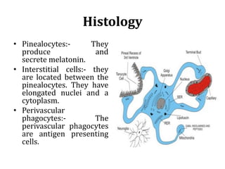


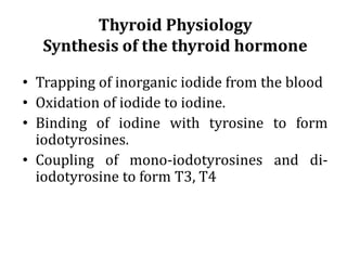


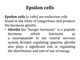
![Stimulators
• Decreased serum [Ca2+].
• Mild decreases in serum [Mg2+].
• An increase in serum phosphate
Inhibitors
• Increased serum [Ca2+].
• Severe decreases in serum [Mg2+]
• Calcitriol](https://image.slidesharecdn.com/endocrinesystem-200621083153/85/Endocrine-system-111-320.jpg)


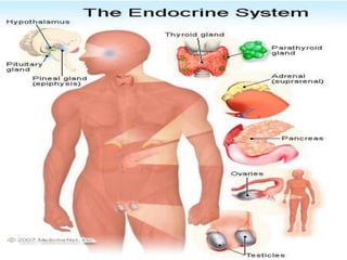
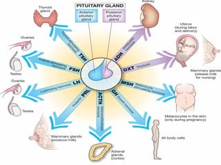
The document summarizes key aspects of the endocrine system. It describes how hormones are chemical messengers that influence metabolic activity by binding to receptors and initiating responses. The major endocrine glands - pituitary, thyroid, parathyroid, thymus, pancreas, adrenal glands, testes, and ovaries - are described along with their locations, secretions, and functions in processes like reproduction, growth, metabolism regulation, and stress response. The hypothalamus links the nervous system to the endocrine system by secreting hormones that stimulate or inhibit pituitary secretions.


