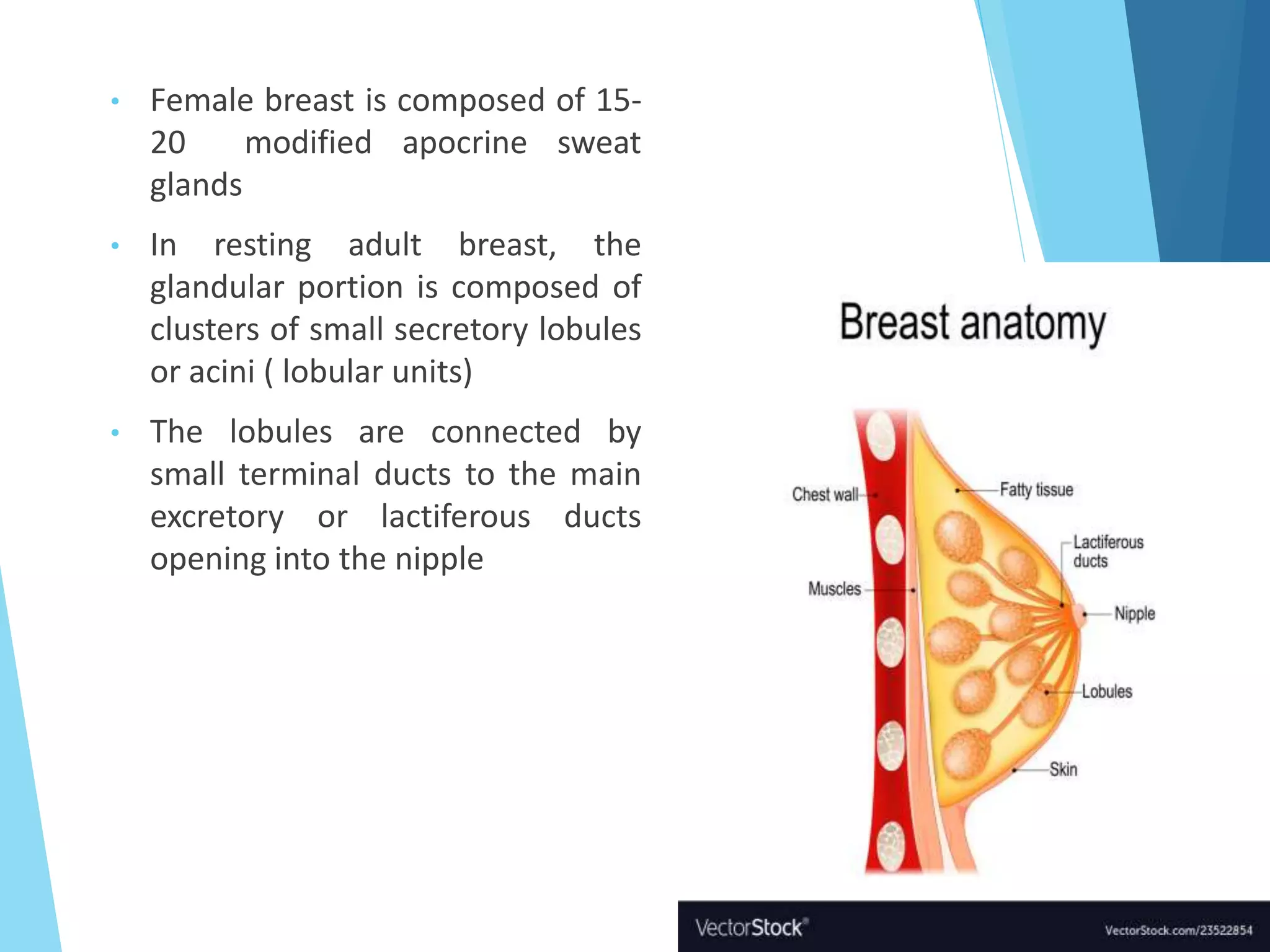
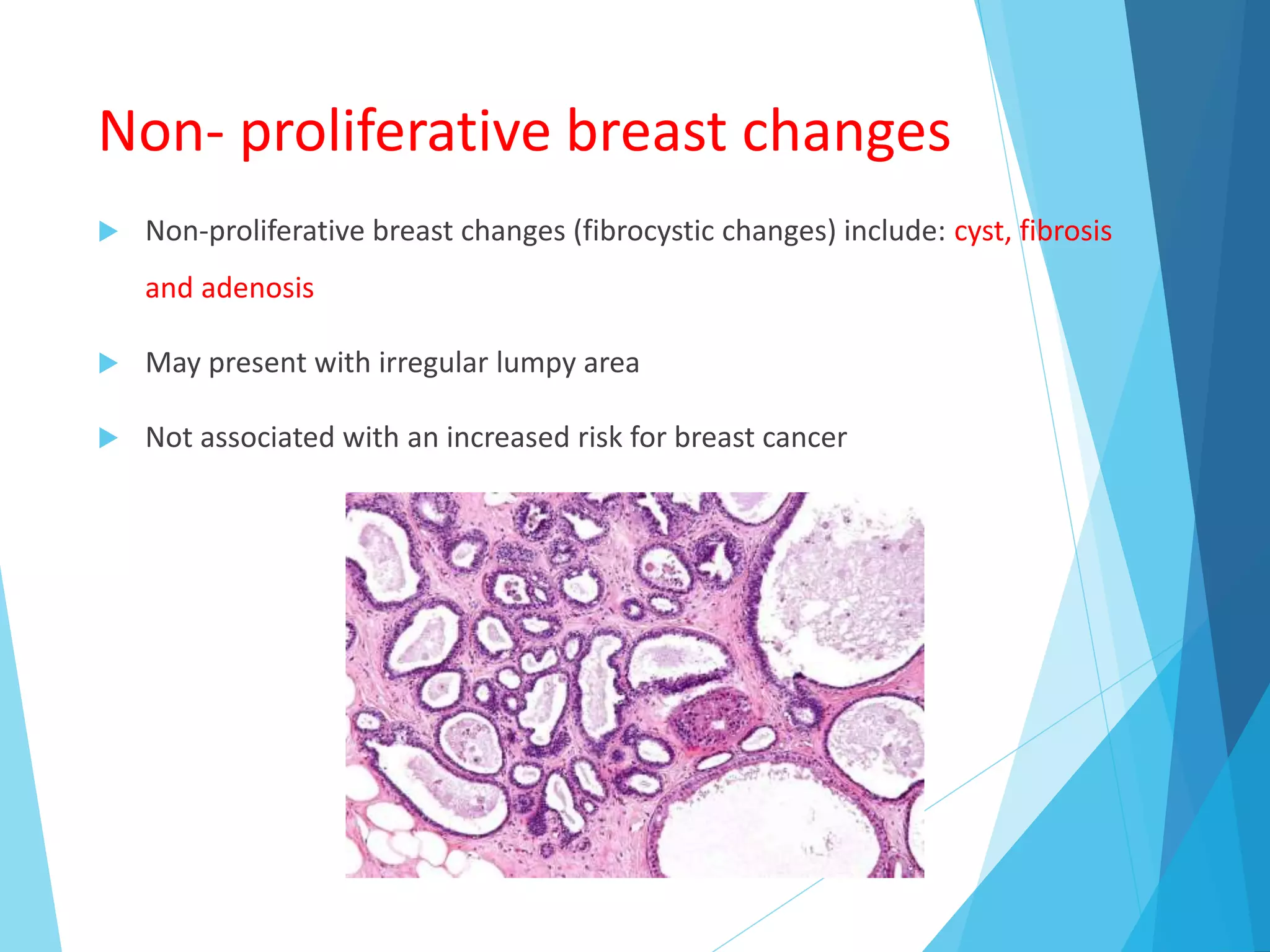
Diseases of Breast
Dr. Umme Kulsum Munmun
MD (Pathology)
Assistant Professor
Department of Pathology
Chandpur Medical College
• Female breast is composed of 15-
20 modified apocrine sweat
glands
• In resting adult breast, the
glandular portion is composed of
clusters of small secretory lobules
or acini ( lobular units)
• The lobules are connected by
small terminal ducts to the main
excretory or lactiferous ducts
opening into the nipple
 The surrounding stroma is of two types : intralobular and
interlobular
 Each elements are the source of both benign and malignant
lesions
 before puberty – breasts in both sexes – ducts
 after menopause – gradual and progressive involution
(lobular atrophy, increased fat, cystic dilatation of ducts)
Histology
• Two types of cells line the ducts and
lobules
 Luminal cuboidal cells-
produce milk
 Contractile
myoepithelial cells  lie on
the basement membrane 
assist in milk ejection
during lactation and
provide structural
support to the lobules
• The stroma is composed of loose
connective tissue and fat
• After menopause, the lactiferous
apparatus undergoes atrophy and
the stroma becomes more fibrous
Clinical Presentations of Breast
Disease
 The most common symptoms of disorders of breast are –
- pain
- palpable mass
- lumpiness (without a discrete mass)
- nipple discharge
Pain (Mastalgia or mastodynia)
 May be cyclic with menses or non-cyclic
 Diffuse cyclic pain may be due to premenstrual edema
 Non-cyclic pain – localized to one area, caused by
ruptured cysts, physical injury and infections
 Almost all painful masses are benign
 10% of breast cancers present with pain
Palpable mass
 Must be distinguished from the normal nodularity of
breast
 The most common palpable lesions are cysts,
fibroadenomas and invasive carcinomas
 Benign palpable masses are most common in
premenopausal women
 Likelihood of a malignancy increases with age
Nipple Discharge
 Less common
 Suggests carcinoma when it is spontaneous and unilateral
 Milky discharges associated with elevated prolactin levels
(pituitary adenoma, hypothyroidism or endocrine
anovulatory syndrome)
 Occurs in patients taking OCP, TCA, methyldopa or
phenothiazines
 Bloody or serous discharges are most commonly due to
large duct papillomas and cysts
Role of Mammography
 Detects small, non-palpable asymptomatic breast carcinomas
 Sensitivity and specificity of mammography increase with age as a
result of replacement of fibrous, radio-dense tissue of youth with the
fatty, radiolucent tissue of older women.
 The principal mammographic signs of breast carcinoma are densities
and calcifications
 Screening has increased the diagnosis of ductal carcinoma in situ
Disorders of Development
 Milk Line remnants- supernumerary nipples or breast may
form
 Accessory axillary breast tissue
 Congenital nipple inversion
Inflammatory Disorders
 Less than 1% of breast symptoms
 Include-
i - Acute mastitis : during the first month of breastfeeding due to
cracks or fissures in the nipples, the breast is erythematous, painful and
fever is often present. Initially one duct system is involved, may spread to the
entire breast if untreated
ii – Squamous metaplasia of lactiferous ducts: subareolar abscess/
periductal mastitis/ Zuska disease. Presents with painful erythematous,
subareolar mass. Associated with vitamin A difficiency, smoking/toxic
substances
 iii. Duct ectasia: fifth or sixth decades of life – palpable periareolar
mass often associated with thick, white nipple secretions and
occasionally with skin retraction.
 iv. Fat necrosis: presentation closely mimics cancer, painless palpable
mass, skin thickening or retraction, mammographic densities or
calcifications
 v. Lymphocytic mastopathy
 vi. Granulomatous mastitis: only occurs in parous women, may be
caused by a hypersensitivity reaction to antigens expressed during
lactation.
Benign Epithelial Proliferations
 Pathologic spectrum of seemingly related clinically benignbreast
abnormalitiy, palpably irregular and painful breasts
 Classified into three groups
- non-proliferative breast changes
- proliferative breast diseases without atypia
- proliferative breast diseases with atypia
Non- proliferative breast changes
 Non-proliferative breast changes (fibrocystic changes) include: cyst, fibrosis
and adenosis
 May present with irregular lumpy area
 Not associated with an increased risk for breast cancer
 Adenosis is defined as an increased number
of acini per lobule
 Preserved 2 cell layer (inner epithelial and
outer myoepithelial cells)
Proliferative breast disease
without atypia
 Lesions characterized by proliferation of epithelial cells without
atypia
 Associated with a small increase (1.5 to 2 times) in the risk of
subsequent carcinoma
 Commonly detected as mammographic densities, calcifications or
as incidental findings in biopsies performed for other reasons
 Also called ‘usual ductal hyperplasia’, ‘epithelial hyperplasia’,
‘epitheliosis’ etc
Usual Ductal Hyperplasia
The lumens are filled
with heterogenous ,
mixed population of
luminal and
myoepithelial cell types
Proliferative breast disease with
atypia
 Associated (4 to 5 times) with moderately increased risk of
carcinoma
 Includes two forms: atypical ductal hyperplasia and atypical lobular
hyperplasia
 ADH is present in 5% to 17% biopsy specimens from calcifications
Atypical Ductal Hyperplasia
In ALH, the lobules are normal sized and still contain
identifiable lumina. This image shows the spectrum of
findings from ALH to LCIS.
• ALH is found in fewer than 5% of biopsies
• Incidental finding , no radiologic correlates
• ALH consists of cells identical to those of LCIS but the cells do not fill or
distend more than 50% of the acini within a lobule
 Recent immunophenotypic and molecular studies indicated a
linear progression from normal epithelium through usual
hyperplasia, atypical hyperplasia and carcinoma in situ to invasive
cancer
Benign tumours of breast
Fibroadenoma
Tubular
adenoma
Lactating
Adenoma
Intraductal
papilloma
Benign tumours of breast
 Fibroadenoma (stromal)
• proliferation of epithelial and stromal elements
• most common breast tumor in adolescent and young adult
women (peak age = third decade)
• well-circumscribed, freely movable, nonpainful mass
• regress with age if left untreated
• ducts distorted elongated slit-like structures
 Tubular adenoma
• far less common than fibroadenomas
• young women, discrete, freely movable masses
• uniform sized ducts
 Lactating Adenoma
• enlarging masses during lactation or pregnancy
• prominent secretory change
 Intraductal papilloma
• in the mammary ducts, subareolar lactiferous ducts
• periductal inflammation, duct sclerosis
• serous or bloody nipple discharge
• fibrosis, infarction, squamous metaplasia
Phyllodes tumor (stromal)
• fleshy tumor, leaf-like pattern and cysts on cut
surface
• circumscribed, connective tissue and epithelial
elements (greater connective tissue cellularity
compared to fibroadenomas), 1-15 cm
• less than 1 % of breast tumors
• benign, borderline, malignant
• metastases are hematogenous
Stromal tumor
Fibroadenoma Phyllodes Tumour
Classification of Breast
Carcinoma
 In situ carcinoma:
- Ductal Carcinoma in situ (DCIS)
- Lobular Carcinoma in situ (LCIS)
 Invasive carcinoma:
- Invasive ductal carcinoma, not otherwise specified
- Invasive lobular carcinoma
- Tubular carcinoma
- Cribriform carcinoma
- Mucinous (Colloid) Carcinoma
- Medullary carcinoma
- Microinvasive carcinoma
- Papillary carcinoma
- Micropapillary carcinoma
- Apocrine carcinoma
- Metaplastic carcinoma
- Neuroendocrine carcinoma
- Inflammatory carcinoma
 Rare salivary gland type tumour:
- Acinic cell carcinoma
- Adenoid cystic carcinoma
Ductal Carcinoma In Situ
 Confined to the ductal-lobular system
 No evidence of invasion through the basement membrane into the
surrounding stroma Majority are non-palpable and detected
mammographically as microcalcifications (70 - 80%)
 Less frequently presents as palpable mass, nipple discharge, Paget
disease of the nipple
 8-10 times higher risk for subsequent development of invasive breast
carcinoma compared to general population
 Many morphologic variants exist:
- comedocarcinoma (High grade) and
- solid, cribriform, micropapillary, clinging etc
 Papillary carcinoma is a very distinct type, arise from large ducts
 Others originate in terminal duct lobular unit
Lobular Carcinoma in Situ (LCIS)
 No specific clinical features
 Most times incidental finding in biopsy for some other mass
producing lesion such as a fibroadonoma
 No specific clinical features
Invasive ductal carcinoma (NOS)
 Most common type of invasive breast carcinoma (75 - 80%)
 Lacks features of any other subtypes (i.e. is a diagnosis of exclusion)
Microscopic (histologic) description
 Sheets, nests, cords or individual cells
 Tubule formations are prominent in well differentiated tumors but absent
in poorly differentiated tumors
 Tumor cells are more pleomorphic than lobular carcinoma
 Calcification in 60% of cases, variable necrosis
 Often DCIS (up to 80%), perineural invasion (28%)
 Mitotic figures are often prominent
 No myoepithelial cell lining (as seen in DCIS or benign lesions)
 Angiolymphatic invasion: In 35%
Invasive Ductal Carcinoma (NOS)
Invasive Lobular Carcinoma
 Special subtype of invasive breast carcinoma characterized by
discohesive tumor cells arranged in single files or as individual
single cells (loss of E-cadherin)
Mucinous Carcinoma
 Rare tumor occurring in older women
 Also called-
Colloid carcinoma
Mucinoid carcinoma
Gelatinous carcinoma
Mucoid carcinoma
Mucinous adenocarcinoma
 Gross description
Well circumscribed mass of variable size (from < 1 cm to > 20 cm) with
gelatinous cut surface
Microscopic (histologic) description
 Clusters / nests of tumor cells with low or intermediate nuclear
grade floating in pools of extracellular mucin
Metaplastic Carcinoma
 Heterogeneous group of invasive breast carcinomas characterized
by differentiation of the neoplastic epithelium towards squamous
cells or mesenchymal looking elements, including spindle,
chondroid and osseous cells
Metaplastic carcinoma
Molecular Classification
 The diverse histologic appearances of breast carcinomas and
putative precursor lesions are the outward manifestations of the
complex genetic and epigenetic changes that drive
carcinogenesis.
 For prognostic and therapeutic purposes, the molecular
classification of breast cancer is more pronounced now-a-days
 There appear to be three major genetic pathways of
carcinogenesis
Diagnostic Approaches of CA Breast
 Approaches for diagnosis of CA breast
• Fine-needle aspiration cytology (FNAC)
• Core needle biopsy
• Lumpectomy
• Mastectomy
 ER, PR , Her2 immunohistochemistry can be performed on CNB sample if
representative tissue is provided
Tumours of Interlobular stroma
 Benign
- Lipoma
- Myofibroblastoma
- Fibromatosis
 Malignant
- Angiosarcoma
- Rhabdomyosarcoma
- Leiomayosarcoma
- Liposarcoma etc
Risk factors of breast carcinoma
 Germline mutations
 1st degree relatives with
breast cancer
 Race/ethnicity
 Age
 Age at menarche
 Age at 1st live birth
 Benign breast disease
 Estrogen exposure
 Breast density
 Radiation exposure
 Carcinoma of the
contralateral breast or
endometrium
 Diet
 Obesity
 Exercise
 Breastfeeding
 Environmental toxins
Prognostic and predictive factors
 Factors related to extent of
carcinoma:
- Invasive vs in situ
- Distant metastases
- Lymph node metastases
- Tunour size
- Locally advanced disease
- Inflammatory carcinoma
- Lymphovascular invasion
 Factors related to tumour
biology:
- Molecular subtype
- Special histologic
subtypes
- Histologic grade
- Proliferative rate
- Estrogen and
progesterone receptors
- HER 2
Familial breast cancer
 Approximately 12% of breast cancers
 Inheritance of gene mutations associated with breast cancer
( BRCA1, BRCA2, TP53, CHEK2)
 Multiple affected first degree relatives
 Early onset cancers
 Multiple cancers
 Family members with other specific cancers
Paget‘s disease of the nipple
• Result of intraepithelial spread of intraductal
carcinoma
• Large pale-staining cells within the epidermis
of the nipple
• Limited to the nipple or extend to the areola
• Pain or itching, scaling and redness, mistaken
for eczema
• Ulceration, crusting, and serous or bloody
discharge
Pathology of the male breast
 Gynecomastia
• most common clinical and pathologic abnormality of the
male breast
• increase in subareolar tissue
• in 30 to 40 percent of adult males, both breasts are
affected in many cases
• associated with hyperthyroidism, cirrhosis of the liver,
chronic renal failure, chronic pulmonary disease, and
hypogonadism, use of hormones - estrogens, androgens, and
other drugs (digitalis, cimetidine, spironolactone, marihuana, and
tricyclic antidepressants)
 Carcinoma of the male breast
• uncommon < 1 % of all breast cancers
Diseases of Breast.pptx

Diseases of Breast.pptx

  • 1.
    Diseases of Breast Dr.Umme Kulsum Munmun MD (Pathology) Assistant Professor Department of Pathology Chandpur Medical College
  • 2.
    • Female breastis composed of 15- 20 modified apocrine sweat glands • In resting adult breast, the glandular portion is composed of clusters of small secretory lobules or acini ( lobular units) • The lobules are connected by small terminal ducts to the main excretory or lactiferous ducts opening into the nipple
  • 3.
     The surroundingstroma is of two types : intralobular and interlobular  Each elements are the source of both benign and malignant lesions  before puberty – breasts in both sexes – ducts  after menopause – gradual and progressive involution (lobular atrophy, increased fat, cystic dilatation of ducts)
  • 4.
    Histology • Two typesof cells line the ducts and lobules  Luminal cuboidal cells- produce milk  Contractile myoepithelial cells  lie on the basement membrane  assist in milk ejection during lactation and provide structural support to the lobules • The stroma is composed of loose connective tissue and fat • After menopause, the lactiferous apparatus undergoes atrophy and the stroma becomes more fibrous
  • 6.
    Clinical Presentations ofBreast Disease  The most common symptoms of disorders of breast are – - pain - palpable mass - lumpiness (without a discrete mass) - nipple discharge
  • 7.
    Pain (Mastalgia ormastodynia)  May be cyclic with menses or non-cyclic  Diffuse cyclic pain may be due to premenstrual edema  Non-cyclic pain – localized to one area, caused by ruptured cysts, physical injury and infections  Almost all painful masses are benign  10% of breast cancers present with pain
  • 8.
    Palpable mass  Mustbe distinguished from the normal nodularity of breast  The most common palpable lesions are cysts, fibroadenomas and invasive carcinomas  Benign palpable masses are most common in premenopausal women  Likelihood of a malignancy increases with age
  • 9.
    Nipple Discharge  Lesscommon  Suggests carcinoma when it is spontaneous and unilateral  Milky discharges associated with elevated prolactin levels (pituitary adenoma, hypothyroidism or endocrine anovulatory syndrome)  Occurs in patients taking OCP, TCA, methyldopa or phenothiazines  Bloody or serous discharges are most commonly due to large duct papillomas and cysts
  • 10.
    Role of Mammography Detects small, non-palpable asymptomatic breast carcinomas  Sensitivity and specificity of mammography increase with age as a result of replacement of fibrous, radio-dense tissue of youth with the fatty, radiolucent tissue of older women.  The principal mammographic signs of breast carcinoma are densities and calcifications  Screening has increased the diagnosis of ductal carcinoma in situ
  • 13.
    Disorders of Development Milk Line remnants- supernumerary nipples or breast may form  Accessory axillary breast tissue  Congenital nipple inversion
  • 14.
    Inflammatory Disorders  Lessthan 1% of breast symptoms  Include- i - Acute mastitis : during the first month of breastfeeding due to cracks or fissures in the nipples, the breast is erythematous, painful and fever is often present. Initially one duct system is involved, may spread to the entire breast if untreated ii – Squamous metaplasia of lactiferous ducts: subareolar abscess/ periductal mastitis/ Zuska disease. Presents with painful erythematous, subareolar mass. Associated with vitamin A difficiency, smoking/toxic substances
  • 15.
     iii. Ductectasia: fifth or sixth decades of life – palpable periareolar mass often associated with thick, white nipple secretions and occasionally with skin retraction.  iv. Fat necrosis: presentation closely mimics cancer, painless palpable mass, skin thickening or retraction, mammographic densities or calcifications  v. Lymphocytic mastopathy  vi. Granulomatous mastitis: only occurs in parous women, may be caused by a hypersensitivity reaction to antigens expressed during lactation.
  • 16.
    Benign Epithelial Proliferations Pathologic spectrum of seemingly related clinically benignbreast abnormalitiy, palpably irregular and painful breasts  Classified into three groups - non-proliferative breast changes - proliferative breast diseases without atypia - proliferative breast diseases with atypia
  • 17.
    Non- proliferative breastchanges  Non-proliferative breast changes (fibrocystic changes) include: cyst, fibrosis and adenosis  May present with irregular lumpy area  Not associated with an increased risk for breast cancer
  • 18.
     Adenosis isdefined as an increased number of acini per lobule  Preserved 2 cell layer (inner epithelial and outer myoepithelial cells)
  • 19.
    Proliferative breast disease withoutatypia  Lesions characterized by proliferation of epithelial cells without atypia  Associated with a small increase (1.5 to 2 times) in the risk of subsequent carcinoma  Commonly detected as mammographic densities, calcifications or as incidental findings in biopsies performed for other reasons  Also called ‘usual ductal hyperplasia’, ‘epithelial hyperplasia’, ‘epitheliosis’ etc
  • 20.
    Usual Ductal Hyperplasia Thelumens are filled with heterogenous , mixed population of luminal and myoepithelial cell types
  • 21.
    Proliferative breast diseasewith atypia  Associated (4 to 5 times) with moderately increased risk of carcinoma  Includes two forms: atypical ductal hyperplasia and atypical lobular hyperplasia  ADH is present in 5% to 17% biopsy specimens from calcifications
  • 22.
  • 23.
    In ALH, thelobules are normal sized and still contain identifiable lumina. This image shows the spectrum of findings from ALH to LCIS. • ALH is found in fewer than 5% of biopsies • Incidental finding , no radiologic correlates • ALH consists of cells identical to those of LCIS but the cells do not fill or distend more than 50% of the acini within a lobule
  • 24.
     Recent immunophenotypicand molecular studies indicated a linear progression from normal epithelium through usual hyperplasia, atypical hyperplasia and carcinoma in situ to invasive cancer
  • 25.
    Benign tumours ofbreast Fibroadenoma Tubular adenoma Lactating Adenoma Intraductal papilloma
  • 26.
    Benign tumours ofbreast  Fibroadenoma (stromal) • proliferation of epithelial and stromal elements • most common breast tumor in adolescent and young adult women (peak age = third decade) • well-circumscribed, freely movable, nonpainful mass • regress with age if left untreated • ducts distorted elongated slit-like structures
  • 27.
     Tubular adenoma •far less common than fibroadenomas • young women, discrete, freely movable masses • uniform sized ducts  Lactating Adenoma • enlarging masses during lactation or pregnancy • prominent secretory change  Intraductal papilloma • in the mammary ducts, subareolar lactiferous ducts • periductal inflammation, duct sclerosis • serous or bloody nipple discharge • fibrosis, infarction, squamous metaplasia
  • 28.
    Phyllodes tumor (stromal) •fleshy tumor, leaf-like pattern and cysts on cut surface • circumscribed, connective tissue and epithelial elements (greater connective tissue cellularity compared to fibroadenomas), 1-15 cm • less than 1 % of breast tumors • benign, borderline, malignant • metastases are hematogenous
  • 29.
  • 30.
    Classification of Breast Carcinoma In situ carcinoma: - Ductal Carcinoma in situ (DCIS) - Lobular Carcinoma in situ (LCIS)  Invasive carcinoma: - Invasive ductal carcinoma, not otherwise specified - Invasive lobular carcinoma - Tubular carcinoma - Cribriform carcinoma - Mucinous (Colloid) Carcinoma
  • 31.
    - Medullary carcinoma -Microinvasive carcinoma - Papillary carcinoma - Micropapillary carcinoma - Apocrine carcinoma - Metaplastic carcinoma - Neuroendocrine carcinoma - Inflammatory carcinoma  Rare salivary gland type tumour: - Acinic cell carcinoma - Adenoid cystic carcinoma
  • 32.
    Ductal Carcinoma InSitu  Confined to the ductal-lobular system  No evidence of invasion through the basement membrane into the surrounding stroma Majority are non-palpable and detected mammographically as microcalcifications (70 - 80%)  Less frequently presents as palpable mass, nipple discharge, Paget disease of the nipple  8-10 times higher risk for subsequent development of invasive breast carcinoma compared to general population
  • 33.
     Many morphologicvariants exist: - comedocarcinoma (High grade) and - solid, cribriform, micropapillary, clinging etc  Papillary carcinoma is a very distinct type, arise from large ducts  Others originate in terminal duct lobular unit
  • 34.
    Lobular Carcinoma inSitu (LCIS)  No specific clinical features  Most times incidental finding in biopsy for some other mass producing lesion such as a fibroadonoma  No specific clinical features
  • 35.
    Invasive ductal carcinoma(NOS)  Most common type of invasive breast carcinoma (75 - 80%)  Lacks features of any other subtypes (i.e. is a diagnosis of exclusion)
  • 36.
    Microscopic (histologic) description Sheets, nests, cords or individual cells  Tubule formations are prominent in well differentiated tumors but absent in poorly differentiated tumors  Tumor cells are more pleomorphic than lobular carcinoma  Calcification in 60% of cases, variable necrosis  Often DCIS (up to 80%), perineural invasion (28%)  Mitotic figures are often prominent  No myoepithelial cell lining (as seen in DCIS or benign lesions)  Angiolymphatic invasion: In 35%
  • 37.
  • 38.
    Invasive Lobular Carcinoma Special subtype of invasive breast carcinoma characterized by discohesive tumor cells arranged in single files or as individual single cells (loss of E-cadherin)
  • 39.
    Mucinous Carcinoma  Raretumor occurring in older women  Also called- Colloid carcinoma Mucinoid carcinoma Gelatinous carcinoma Mucoid carcinoma Mucinous adenocarcinoma  Gross description Well circumscribed mass of variable size (from < 1 cm to > 20 cm) with gelatinous cut surface
  • 41.
    Microscopic (histologic) description Clusters / nests of tumor cells with low or intermediate nuclear grade floating in pools of extracellular mucin
  • 42.
    Metaplastic Carcinoma  Heterogeneousgroup of invasive breast carcinomas characterized by differentiation of the neoplastic epithelium towards squamous cells or mesenchymal looking elements, including spindle, chondroid and osseous cells
  • 43.
  • 44.
    Molecular Classification  Thediverse histologic appearances of breast carcinomas and putative precursor lesions are the outward manifestations of the complex genetic and epigenetic changes that drive carcinogenesis.  For prognostic and therapeutic purposes, the molecular classification of breast cancer is more pronounced now-a-days  There appear to be three major genetic pathways of carcinogenesis
  • 46.
    Diagnostic Approaches ofCA Breast  Approaches for diagnosis of CA breast • Fine-needle aspiration cytology (FNAC) • Core needle biopsy • Lumpectomy • Mastectomy
  • 47.
     ER, PR, Her2 immunohistochemistry can be performed on CNB sample if representative tissue is provided
  • 48.
    Tumours of Interlobularstroma  Benign - Lipoma - Myofibroblastoma - Fibromatosis  Malignant - Angiosarcoma - Rhabdomyosarcoma - Leiomayosarcoma - Liposarcoma etc
  • 49.
    Risk factors ofbreast carcinoma  Germline mutations  1st degree relatives with breast cancer  Race/ethnicity  Age  Age at menarche  Age at 1st live birth  Benign breast disease  Estrogen exposure  Breast density  Radiation exposure  Carcinoma of the contralateral breast or endometrium  Diet  Obesity  Exercise  Breastfeeding  Environmental toxins
  • 50.
    Prognostic and predictivefactors  Factors related to extent of carcinoma: - Invasive vs in situ - Distant metastases - Lymph node metastases - Tunour size - Locally advanced disease - Inflammatory carcinoma - Lymphovascular invasion  Factors related to tumour biology: - Molecular subtype - Special histologic subtypes - Histologic grade - Proliferative rate - Estrogen and progesterone receptors - HER 2
  • 51.
    Familial breast cancer Approximately 12% of breast cancers  Inheritance of gene mutations associated with breast cancer ( BRCA1, BRCA2, TP53, CHEK2)  Multiple affected first degree relatives  Early onset cancers  Multiple cancers  Family members with other specific cancers
  • 52.
    Paget‘s disease ofthe nipple • Result of intraepithelial spread of intraductal carcinoma • Large pale-staining cells within the epidermis of the nipple • Limited to the nipple or extend to the areola • Pain or itching, scaling and redness, mistaken for eczema • Ulceration, crusting, and serous or bloody discharge
  • 53.
    Pathology of themale breast  Gynecomastia • most common clinical and pathologic abnormality of the male breast • increase in subareolar tissue • in 30 to 40 percent of adult males, both breasts are affected in many cases
  • 54.
    • associated withhyperthyroidism, cirrhosis of the liver, chronic renal failure, chronic pulmonary disease, and hypogonadism, use of hormones - estrogens, androgens, and other drugs (digitalis, cimetidine, spironolactone, marihuana, and tricyclic antidepressants)  Carcinoma of the male breast • uncommon < 1 % of all breast cancers