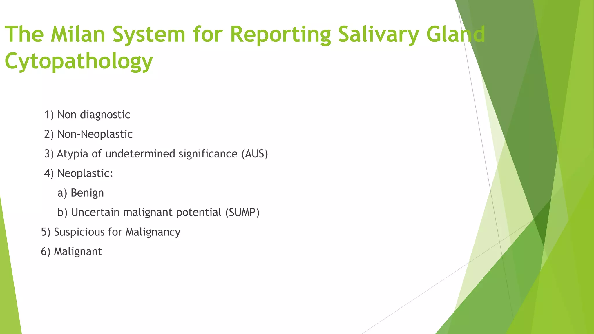
SlideShare a Scribd company logo
Milan’s system of reporting
salivary gland cytology
Presenter: Dr.Argha Baruah
Moderator: Dr.ML Harendra Kumar
Introduction:
 Salivary gland fine-needle aspiration (FNA) cytology has become an accepted method of evaluating
salivary gland tumors preoperatively
 FNA of salivary gland tumors was shown to have a high specificity in differentiating benign and
malignant lesions (97%)
 FNA falls flat in terms of sensitivity (80%)
Why we need a uniform reporting system?
 Because of the wide diversity of tumors arising in the salivary glands along
with significant morphologic overlap.
 Most pathologists report these specimens by employing heterogeneous
diagnostic terminology, specifically for indeterminate diagnoses, and that
clinician’s act variably based upon these reports.
 Inconsistencies in reporting of salivary gland FNA specimens can have an
effect on the correlation between cytologic interpretation and surgical
outcome and finally has an impact on patient care
 The Bethesda system for reporting was created to address the inconsistencies
and the shortcomings of the terminology used for reporting FNA specimens. A
few authors have suggested that a similar classification is needed for
reporting of salivary gland FNA specimens.
 Griffith et al. proposed a four category scheme that effectively stratified
salivary gland FNA specimens by overall risk of malignancy and risk of high
grade malignancy.The scheme was comprised of the following diagnostic
categories: benign, neoplasm of uncertain malignant potential (NUMP),
suspicious for malignancy and positive for malignancy; the overall risk of
malignancy for each category was 2%, 18%, 75.8%, and 100%, respectively.
 Rossi et al. based on the experience of two academic centres with 709
salivary gland FNA specimens, stratified their cytologic diagnosis into the
following diagnostic categories with associated risk of malignancy:
nondiagnostic/inadequate—17%, benign—16%, neoplasm—6%, atypical—
53%,suspicious for malignancy/malignant—94%.
 This led a group of experienced cytopathologists and histopathologists to attempt to create
a uniform cytological reporting scheme. The proposed system is based on key principles
developed for standardized terminology systems adopted for other body sites (ie, uterine
cervix, pancreas,thyroid, and urinary system)
 The standardization effort started in September 2015 at the European Congress of Cytology,
which was held in Milan, Italy, under the umbrella of the American Society of Cytopathology
(ASC) and the International Academy of Cytology (IAC).
 Subsequently, an international panel of 49 cytopathologists and histopathologists (the Milan
group) was organized to establish a unified effort under the title of “The Milan System for
Reporting Salivary Gland Cytopathology.”
 Before starting the task of constructing a taxonomy, the Milan group invited the
international cytology and histology communities to answer a series of questions .Questions
were generated in accord with the current literature and experience of the authors in the
core group. The preliminary version was presented by Dr. William Faquin at USCAP 2016 in
Seattle
THE BENEFITS OF A UNIFORM REPORTING SYSTEM FOR SALIVARY
GLAND CYTOPATHOLOGY:
 1. Improve communication between pathologists and clinicians
 2. Improve patient care
 3.Facilitate cytologic-histologic correlation
 4.Promote research into the epidemiology, molecular biology,
pathology
 5.Foster sharing of data from different laboratories for collaborative
studies
The Milan System for Reporting Salivary Gland
Cytopathology
1) Non diagnostic
2) Non-Neoplastic
3) Atypia of undetermined significance (AUS)
4) Neoplastic:
a) Benign
b) Uncertain malignant potential (SUMP)
5) Suspicious for Malignancy
6) Malignant
Sample adequacy and evaluation:
 Minimum number of lesional and nonlesional cells that cytopathologist require
to consider a aspirate as benign vs nondiagnostic/unsatisfactory was not
established.
Category I:Non-Diagnostic:
 1)Insufficient quantitative and/or qualitative cellular material to make a
cytologic diagnosis
 2)Includes aspirates with benign elements only
 3)Includes non-mucinous cyst contents
 4)Aspirate smears with cyst contents only (ie. granular debris and histiocytes
and few to no epithelial cells)
Benign salivary gland elements only
 For aspirates containing only normal salivary gland elements, a cautionary
note is recommended.
 Note: Clinical and radiologic correlations are recommended to ensure that
the aspirate is representative of the lesion; the findings in this aspirate do not
explain the presence of a salivary gland mass.
Non-mucinous cyst contents
DDX: Ductal cyst, pseudocyst, cystic neoplasm
Absence of an epithelial component
Category II: Non-Neoplastic:
Specimens lacking evidence of a neoplastic process:
1)Inflammatory, metaplastic, and reactive(i.e acute, chronic, and granulomatous
sialadenitis,sialadenosis, etc…)
2)Reactive lymph nodes (flow cytometry is needed)
Note: Clinico-radiological correlation is essential to ensure that the specimen is
representative of the lesion
Reactive Lymph Node
REACTIVE PROCESS VS LYMPHOMA
IMMUNOPHENOTYPING combined with cytomorphology is the key to diagnosing
and subtyping reactive conditions vs lymphoma.
NOTE: For negative lymph nodes, caution is warranted: A note suggesting
repeat FNA or tissue biopsy if lymphadenopathy persists
Chronic Sialadenitis
Hypocellular, cohesive basaloid groups, inflammation
Granulomatous Sialadenitis
Category III: Atypia of
Undetermined Significance (AUS):
1. Cannot entirely exclude a neoplasm.
2. Heterogeneous category
3. A majority will be reactive atypia or poorly sampled neoplasms
4. Specimens are often compromised (eg, air-drying, blood clot)
5. Should be used rarely (<10 % of all salivary gland FNAs)
The diagnosis of AUS can be used in the following scenarios:
 •Squamous, oncocytic, or other metaplastic changes indefinite for a neoplasm
 •Low cellularity specimens that are suggestive of, but not diagnostic of a neoplasm
 •Specimens with preparation artifacts hampering distinction between a non-neoplastic and
neoplastic process
 •Mucinous cystic lesions with an absent or very scant epithelial component
 •Salivary gland lymph nodes or lymphoid lesions which are indefinite for a
lymphoproliferative disorder
 •Reactive and reparative atypia indefinite for a neoplasm
Mucinous Cyst Contents Only- Cannot
exclude MEC
Oncocytic metaplasia vs Neoplasm
Reactive vs basaloid neoplasm
Category IV: NEOPLASM
Diagnostic category reserved for benign neoplasms diagnosed based on established
cytomorphologic criteria
BENIGN NEOPLASM ENTITIES:
 Pleomorphic Adenoma
 Warthin Tumor
 Oncocytoma
 Soft Tissue Tumors
 Lipoma
 Schwannoma
 Lymphangioma
 Hemangioma
Benign
SUMP
Warthin Tumor
Oncocytes, chronic inflammation, and cystic debris
Pleomorphic Adenoma
Matrix-rich types of PA are the easiest.
SUMP(Salivary gland neoplasm of uncertain
malignant potential)
Reserved for: Diagnostic of a neoplasm; however, a diagnosis of a specific entity
cannot be made.Amalignant neoplasm cannot be excluded.
SUMP ENTITIES:
 Cellular Basaloid Neoplasm
 Oncocytoid Neoplasm
 Neoplasm with Granular and/or Vacuolated or clear cells
Basal Cell Neoplasm-
DDX basal cell adenoma, AdCC
Oncocytoid neoplasm
Pleomorphic Adenoma With Atypia or
Metaplasia
 The inability to separate these tumor types with greater accuracy
preoperatively is made more frustrating by the fact that the surgical approach
can be quite different.
 The application of a monomorphic cellular basaloid neoplasm with fibrillary
stroma was meant to capture pleomorphic adenomas .
 The finding of hyaline stroma was predicted to identify those neoplasms of
higher risk, specifically adenoid cystic carcinoma
Category V: Suspicious for Malignancy
 Aspirates which are highly suggestive of malignancy but not definitive.Atypia
concerns malignancy but sample falls short for clear diagnosis.
 Often high grade carcinomas with limited sampling or other limitation
 Markedly atypical cells with poor smear preparation, poor cell preservation,
fixation artifact, or obscuring inflammation and blood
 Presence of limited cytologic features of a specific malignant lesion (e.g.
adenoid cystic carcinoma, mucoepidermoid carcinoma, acinic cell carcinoma)
in an otherwise sparsely cellular aspirate
 Presence of markedly atypical and/or suspicious cytologic features in a subset
of cells but admixed with features of a benign salivary gland lesion.
 Atypical features can include prominent nucleoli or macronucleoli,
anisonucleosis, increased nuclear to cytoplasmic ratio, nuclear molding,
prominent nuclear pleomorphism, atypical mitosis, and clumped, coarse
chromatin
Malignancy:
 Aspirates which are diagnostic of malignancy
 Sub-classify into specific types and grades of carcinoma: e.g. low grade vs
high grade
 "Other" malignancies such as lymphomas, sarcomas and metastases are also
included in this category and should be specifically designated.
Mucoepidermoid Carcinoma
HG B-Cell Lymphoma
Ancillary Studies:
 Immunocytochemistry
LBC
Smears
Cell block
 FISH
 RT-PCR
 Next Generation Sequencing
 Rarely there was enough material on slides or in cell blocks to perform
ancillary studies.
 The use of p63/calponin for myoepithelial cells, mucicarmine/D-PAS for MEC,
and CD117/DOG-1 have been shown to have some usefulness in differentiating
ACC from polymorphous adenocarcinoma and PA.
 Greater than 90% of the ancillary studies are performed on cell block material
and very less are performed directly on cytologic preparations.
Ancillary studies useful in:
Basaloid neoplasm
Oncocytic neoplasm
Metastatic lesions
Rule out lymphoma
Subtype a high grade carcinoma
Subtype a low grade carcinoma
t(6:9) MYB oncogene-NFIB transcription factor
In salivary gland, this finding by FISH is specific for AdCC
Nuclear β catenin in basal cell adenoma:
ETV6-NTRK3 rearrangement for difficult cases
DOG1 and Acinic Cell Carcinoma SOX-10 and Acinic Cell Carcinoma
- Squamous cell carcinoma
Mucoepidermoid Carcinoma:
 IHC is non-specific
 t(11:19) translocation MECT1/MAML2 –FISH
Pitfalls in Milan’s
 It should be noted that the cumulative risk of malignancy for each diagnostic
category may represent an unintended overestimation due to: institutional
referral patterns (large academic and referral centers vs.community
practice), surgical selection bias based on factors such as large size of the
mass, high risk clinical radiologic features.
 The category of atypia of undetermined significance (AUS) is not well
defined. The cases we classified as AUS were those in which the differential
was between a retention cyst (ie, a nonneoplastic lesion) and a low-grade
mucoepidermoid carcinoma (ie, a malignant neoplastic lesion).
 Therefore, these cases were categorized as atypical because we were not
sure even about the neoplastic nature of the lesion.
 Even members of the Milan group are concerned about the overuse of the
term “AUS” as noted in other cytological systems.
 Still the categories of division are very subjective.
 Robust testing is still going on in terms of validity and reproducibility
Summary:
 Salivary gland cytology presents many diagnostic challenges
 The Milan System for Reporting Salivary Gland Cytopathology will help to produce
a more uniform diagnostic structure. It is a practical classification system that is
user friendly and can be internationally accepted with a useful format for
clinicians.
 Improved communication between treating clinician and pathologist
 Improved patient care and help in multicentric collaborative study .
 Availability of IHC and molecular markers can greatly improve the accuracy of
salivary gland FNA

Reference:
 Griffith CC, Pai RK, Schneider F, et al. Salivary gland tumor fineneedle
aspiration cytology: a proposal for a risk stratification classification. Am J Clin
Pathol. 2015;143:839-853
 Rossi ED, Faquin WC ; Baloch Z ,Barkan CA, Foschini MP,Pusztaszeri M, Vielh P,
Daniel FI.The Milan System for Reporting Salivary Gland Cytopathology:
Analysis and Suggestions of Initial Survey. Cancer Cytopathology.2017;1-10
 Wei S,Lester J,Kathleen T,Zubair WB.Reporting of fine needle aspiration (FNA)
specimens of salivary gland lesions: A comprehensive review. Diagnostic
Cytopathology. 2017;1–8.
The Milan System for Reporting Salivary
Gland Cytopathology 1st ed. 2018 Edition

More Related Content

What's hot

THYROID - cytology pptx
THYROID - cytology pptxTHYROID - cytology pptx
THYROID - cytology pptx
KalaivaniGanapathy
 
approach to lymph node cytology part 1
approach to lymph node cytology part 1approach to lymph node cytology part 1
approach to lymph node cytology part 1
Kamalesh Lenka
 
Interpretation of endoscopic gastrointestinal biopsy
Interpretation of endoscopic gastrointestinal biopsyInterpretation of endoscopic gastrointestinal biopsy
Interpretation of endoscopic gastrointestinal biopsy
Appy Akshay Agarwal
 
grossing of Colorectal specimens
grossing of Colorectal specimensgrossing of Colorectal specimens
grossing of Colorectal specimens
Anam Khurshid
 
Patterns in histopathology
Patterns in histopathologyPatterns in histopathology
Patterns in histopathology
Ankita Baghel
 
CYTOLOGY OF BREAST LESIONS??!
CYTOLOGY OF BREAST LESIONS??! CYTOLOGY OF BREAST LESIONS??!
CYTOLOGY OF BREAST LESIONS??!
Ashish Jawarkar
 
Squash smear cytology - By Anamika dev
Squash smear cytology - By Anamika devSquash smear cytology - By Anamika dev
Squash smear cytology - By Anamika dev
Anamika Dev
 
Lymphoma spillover.pptx
Lymphoma spillover.pptxLymphoma spillover.pptx
Lymphoma spillover.pptx
Tamil Mahizhenthi
 
Giant cell lesions of bone
Giant cell lesions of boneGiant cell lesions of bone
Giant cell lesions of bone
Shreya D Prabhu
 
Salivary gland-- cytology .pptx
Salivary gland-- cytology .pptxSalivary gland-- cytology .pptx
Salivary gland-- cytology .pptx
KalaivaniGanapathy
 
Interpretation of testicular biopsy
Interpretation of testicular biopsyInterpretation of testicular biopsy
Interpretation of testicular biopsy
Appy Akshay Agarwal
 
Pitfalls in diagnosis of soft tissue tumors of childhood
Pitfalls in diagnosis of soft tissue tumors of childhoodPitfalls in diagnosis of soft tissue tumors of childhood
Pitfalls in diagnosis of soft tissue tumors of childhood
Sonic V S
 
Interpretation of endoscopic intestinal biopsy
Interpretation of endoscopic intestinal biopsyInterpretation of endoscopic intestinal biopsy
Interpretation of endoscopic intestinal biopsy
Appy Akshay Agarwal
 
IHC in breast pathology
IHC in breast pathologyIHC in breast pathology
IHC in breast pathology
namrathrs87
 
Minimal residual disease
Minimal residual diseaseMinimal residual disease
Minimal residual disease
Appy Akshay Agarwal
 
Bal fluid analysis
Bal fluid analysisBal fluid analysis
Bal fluid analysis
UTTAM KUMAR DAS
 
Effusion cytology - Diagnosis.
Effusion cytology - Diagnosis.Effusion cytology - Diagnosis.
Effusion cytology - Diagnosis.
namrathrs87
 
Non Hodgkin Lymphoma, cytology.
Non Hodgkin Lymphoma, cytology.Non Hodgkin Lymphoma, cytology.
Non Hodgkin Lymphoma, cytology.
DrPradeep Shekhawat
 
Pancreas cytology
Pancreas cytologyPancreas cytology
Pancreas cytology
Sansar Babu Tiwari
 
Cell block in cytology
Cell block in cytologyCell block in cytology
Cell block in cytology
Anam Khurshid
 

What's hot (20)

THYROID - cytology pptx
THYROID - cytology pptxTHYROID - cytology pptx
THYROID - cytology pptx
 
approach to lymph node cytology part 1
approach to lymph node cytology part 1approach to lymph node cytology part 1
approach to lymph node cytology part 1
 
Interpretation of endoscopic gastrointestinal biopsy
Interpretation of endoscopic gastrointestinal biopsyInterpretation of endoscopic gastrointestinal biopsy
Interpretation of endoscopic gastrointestinal biopsy
 
grossing of Colorectal specimens
grossing of Colorectal specimensgrossing of Colorectal specimens
grossing of Colorectal specimens
 
Patterns in histopathology
Patterns in histopathologyPatterns in histopathology
Patterns in histopathology
 
CYTOLOGY OF BREAST LESIONS??!
CYTOLOGY OF BREAST LESIONS??! CYTOLOGY OF BREAST LESIONS??!
CYTOLOGY OF BREAST LESIONS??!
 
Squash smear cytology - By Anamika dev
Squash smear cytology - By Anamika devSquash smear cytology - By Anamika dev
Squash smear cytology - By Anamika dev
 
Lymphoma spillover.pptx
Lymphoma spillover.pptxLymphoma spillover.pptx
Lymphoma spillover.pptx
 
Giant cell lesions of bone
Giant cell lesions of boneGiant cell lesions of bone
Giant cell lesions of bone
 
Salivary gland-- cytology .pptx
Salivary gland-- cytology .pptxSalivary gland-- cytology .pptx
Salivary gland-- cytology .pptx
 
Interpretation of testicular biopsy
Interpretation of testicular biopsyInterpretation of testicular biopsy
Interpretation of testicular biopsy
 
Pitfalls in diagnosis of soft tissue tumors of childhood
Pitfalls in diagnosis of soft tissue tumors of childhoodPitfalls in diagnosis of soft tissue tumors of childhood
Pitfalls in diagnosis of soft tissue tumors of childhood
 
Interpretation of endoscopic intestinal biopsy
Interpretation of endoscopic intestinal biopsyInterpretation of endoscopic intestinal biopsy
Interpretation of endoscopic intestinal biopsy
 
IHC in breast pathology
IHC in breast pathologyIHC in breast pathology
IHC in breast pathology
 
Minimal residual disease
Minimal residual diseaseMinimal residual disease
Minimal residual disease
 
Bal fluid analysis
Bal fluid analysisBal fluid analysis
Bal fluid analysis
 
Effusion cytology - Diagnosis.
Effusion cytology - Diagnosis.Effusion cytology - Diagnosis.
Effusion cytology - Diagnosis.
 
Non Hodgkin Lymphoma, cytology.
Non Hodgkin Lymphoma, cytology.Non Hodgkin Lymphoma, cytology.
Non Hodgkin Lymphoma, cytology.
 
Pancreas cytology
Pancreas cytologyPancreas cytology
Pancreas cytology
 
Cell block in cytology
Cell block in cytologyCell block in cytology
Cell block in cytology
 

Similar to Milan cytology reporting

Milan system TUMS (1).pdf
Milan system TUMS (1).pdfMilan system TUMS (1).pdf
Milan system TUMS (1).pdf
FereshtehAmeli1
 
The bethesda system for reporting thyroid cytopathology
The bethesda system for reporting thyroid cytopathologyThe bethesda system for reporting thyroid cytopathology
The bethesda system for reporting thyroid cytopathology
Indira Shastry
 
Peritoneal malignancies
Peritoneal malignanciesPeritoneal malignancies
Peritoneal malignancies
Mebanshanbor Garod
 
Bethesda system for cervix cytology
Bethesda system for cervix cytologyBethesda system for cervix cytology
Bethesda system for cervix cytology
Ravi Kumar Meena
 
What is new in Genitourinary pathology
What is new in Genitourinary pathology What is new in Genitourinary pathology
What is new in Genitourinary pathology
Argha Baruah
 
NEOPLASIA: Clinical Features of Tumors, Grading and Staging & Laboratory Diag...
NEOPLASIA: Clinical Features of Tumors, Grading and Staging & Laboratory Diag...NEOPLASIA: Clinical Features of Tumors, Grading and Staging & Laboratory Diag...
NEOPLASIA: Clinical Features of Tumors, Grading and Staging & Laboratory Diag...
Dr. Roopam Jain
 
Neoplasia - Patholgy
Neoplasia - Patholgy Neoplasia - Patholgy
Neoplasia - Patholgy
Jonathan Bondes
 
Bethesda updates with Thyroid path 2023.pptx
Bethesda updates with Thyroid path 2023.pptxBethesda updates with Thyroid path 2023.pptx
Bethesda updates with Thyroid path 2023.pptx
DivyaTiwari722899
 
Staging an tumor markers
Staging an tumor markersStaging an tumor markers
Staging an tumor markers
DR DAVIS NADAKKAVUKARAN
 
Tumor Board Meeting on 24/04/2018
Tumor Board Meeting on 24/04/2018Tumor Board Meeting on 24/04/2018
Tumor Board Meeting on 24/04/2018
Dr. Varughese George
 
Bethesda thyroid
Bethesda thyroidBethesda thyroid
Bethesda thyroid
Pankaj Gupta
 
Presentation dr rahul seminar (2)[1387]
Presentation dr rahul  seminar (2)[1387]Presentation dr rahul  seminar (2)[1387]
Presentation dr rahul seminar (2)[1387]
rahulraj956
 
Carcinoma Cervix.pptx
Carcinoma Cervix.pptxCarcinoma Cervix.pptx
Carcinoma Cervix.pptx
AeyshaBegum
 
CARCINOMA URINARY BLADDER
CARCINOMA URINARY BLADDERCARCINOMA URINARY BLADDER
CARCINOMA URINARY BLADDER
Vikas Kumar
 
Colorectal Polyp.pptx
Colorectal Polyp.pptxColorectal Polyp.pptx
Colorectal Polyp.pptx
Dr. Awadhesh
 
Surgical oncology ( malignancies )
Surgical oncology  ( malignancies )Surgical oncology  ( malignancies )
Surgical oncology ( malignancies )
Hristo Rahman
 
Management of Suspected Ovarian Masses in Premenopausal Women RCOG, 2011
Management of Suspected  Ovarian Masses  in Premenopausal Women   RCOG, 2011Management of Suspected  Ovarian Masses  in Premenopausal Women   RCOG, 2011
Management of Suspected Ovarian Masses in Premenopausal Women RCOG, 2011
Aboubakr Elnashar
 
2022 WHO Salivary gland tumors update.pdf
2022 WHO Salivary gland tumors update.pdf2022 WHO Salivary gland tumors update.pdf
2022 WHO Salivary gland tumors update.pdf
KavithaLoganathan6
 
WHO CLASSIFICATION 2016 RENAL CELL CARCINOMA.pptx
WHO CLASSIFICATION 2016 RENAL CELL CARCINOMA.pptxWHO CLASSIFICATION 2016 RENAL CELL CARCINOMA.pptx
WHO CLASSIFICATION 2016 RENAL CELL CARCINOMA.pptx
SURAJ PANCHAL
 

Similar to Milan cytology reporting (20)

Milan system TUMS (1).pdf
Milan system TUMS (1).pdfMilan system TUMS (1).pdf
Milan system TUMS (1).pdf
 
The bethesda system for reporting thyroid cytopathology
The bethesda system for reporting thyroid cytopathologyThe bethesda system for reporting thyroid cytopathology
The bethesda system for reporting thyroid cytopathology
 
Peritoneal malignancies
Peritoneal malignanciesPeritoneal malignancies
Peritoneal malignancies
 
Bethesda system for cervix cytology
Bethesda system for cervix cytologyBethesda system for cervix cytology
Bethesda system for cervix cytology
 
What is new in Genitourinary pathology
What is new in Genitourinary pathology What is new in Genitourinary pathology
What is new in Genitourinary pathology
 
Ovarian tumours
 	Ovarian tumours			 	Ovarian tumours
Ovarian tumours
 
NEOPLASIA: Clinical Features of Tumors, Grading and Staging & Laboratory Diag...
NEOPLASIA: Clinical Features of Tumors, Grading and Staging & Laboratory Diag...NEOPLASIA: Clinical Features of Tumors, Grading and Staging & Laboratory Diag...
NEOPLASIA: Clinical Features of Tumors, Grading and Staging & Laboratory Diag...
 
Neoplasia - Patholgy
Neoplasia - Patholgy Neoplasia - Patholgy
Neoplasia - Patholgy
 
Bethesda updates with Thyroid path 2023.pptx
Bethesda updates with Thyroid path 2023.pptxBethesda updates with Thyroid path 2023.pptx
Bethesda updates with Thyroid path 2023.pptx
 
Staging an tumor markers
Staging an tumor markersStaging an tumor markers
Staging an tumor markers
 
Tumor Board Meeting on 24/04/2018
Tumor Board Meeting on 24/04/2018Tumor Board Meeting on 24/04/2018
Tumor Board Meeting on 24/04/2018
 
Bethesda thyroid
Bethesda thyroidBethesda thyroid
Bethesda thyroid
 
Presentation dr rahul seminar (2)[1387]
Presentation dr rahul  seminar (2)[1387]Presentation dr rahul  seminar (2)[1387]
Presentation dr rahul seminar (2)[1387]
 
Carcinoma Cervix.pptx
Carcinoma Cervix.pptxCarcinoma Cervix.pptx
Carcinoma Cervix.pptx
 
CARCINOMA URINARY BLADDER
CARCINOMA URINARY BLADDERCARCINOMA URINARY BLADDER
CARCINOMA URINARY BLADDER
 
Colorectal Polyp.pptx
Colorectal Polyp.pptxColorectal Polyp.pptx
Colorectal Polyp.pptx
 
Surgical oncology ( malignancies )
Surgical oncology  ( malignancies )Surgical oncology  ( malignancies )
Surgical oncology ( malignancies )
 
Management of Suspected Ovarian Masses in Premenopausal Women RCOG, 2011
Management of Suspected  Ovarian Masses  in Premenopausal Women   RCOG, 2011Management of Suspected  Ovarian Masses  in Premenopausal Women   RCOG, 2011
Management of Suspected Ovarian Masses in Premenopausal Women RCOG, 2011
 
2022 WHO Salivary gland tumors update.pdf
2022 WHO Salivary gland tumors update.pdf2022 WHO Salivary gland tumors update.pdf
2022 WHO Salivary gland tumors update.pdf
 
WHO CLASSIFICATION 2016 RENAL CELL CARCINOMA.pptx
WHO CLASSIFICATION 2016 RENAL CELL CARCINOMA.pptxWHO CLASSIFICATION 2016 RENAL CELL CARCINOMA.pptx
WHO CLASSIFICATION 2016 RENAL CELL CARCINOMA.pptx
 

More from Argha Baruah

Molecular Genetics and Recent updates of Soft tissue tumours Dr.Argha Baruah
Molecular Genetics and Recent updates of Soft tissue tumours Dr.Argha BaruahMolecular Genetics and Recent updates of Soft tissue tumours Dr.Argha Baruah
Molecular Genetics and Recent updates of Soft tissue tumours Dr.Argha Baruah
Argha Baruah
 
Recent diagnostic and Prognostic indices of Endometrial Cancers Dr.Argha Baruah
Recent diagnostic and Prognostic indices of Endometrial Cancers Dr.Argha BaruahRecent diagnostic and Prognostic indices of Endometrial Cancers Dr.Argha Baruah
Recent diagnostic and Prognostic indices of Endometrial Cancers Dr.Argha Baruah
Argha Baruah
 
Radiological and pathological correlation of bone tumours Dr.Argha Baruah
Radiological and pathological correlation of bone tumours  Dr.Argha BaruahRadiological and pathological correlation of bone tumours  Dr.Argha Baruah
Radiological and pathological correlation of bone tumours Dr.Argha Baruah
Argha Baruah
 
Thymic epithelial tumours and recent updates
Thymic epithelial tumours and recent updatesThymic epithelial tumours and recent updates
Thymic epithelial tumours and recent updates
Argha Baruah
 
Unusual type and site of Lymphoma(Extranodal Lymphoma) Dr.Argha Baruah
Unusual type and site of Lymphoma(Extranodal Lymphoma) Dr.Argha BaruahUnusual type and site of Lymphoma(Extranodal Lymphoma) Dr.Argha Baruah
Unusual type and site of Lymphoma(Extranodal Lymphoma) Dr.Argha Baruah
Argha Baruah
 
Recent updates and reporting of testicular tumors Dr.Argha Baruah
Recent updates and reporting  of testicular tumors  Dr.Argha BaruahRecent updates and reporting  of testicular tumors  Dr.Argha Baruah
Recent updates and reporting of testicular tumors Dr.Argha Baruah
Argha Baruah
 
WHO GIT RECENT UPDATES
WHO GIT RECENT UPDATESWHO GIT RECENT UPDATES
WHO GIT RECENT UPDATES
Argha Baruah
 
SPECIAL STAINS USED IN DIAGNOSING LIVER PATHOLOGY
SPECIAL STAINS USED IN DIAGNOSING LIVER PATHOLOGYSPECIAL STAINS USED IN DIAGNOSING LIVER PATHOLOGY
SPECIAL STAINS USED IN DIAGNOSING LIVER PATHOLOGY
Argha Baruah
 
Mediastinal cyst
Mediastinal cyst Mediastinal cyst
Mediastinal cyst
Argha Baruah
 
CNS papillary neoplasm
CNS papillary neoplasm CNS papillary neoplasm
CNS papillary neoplasm
Argha Baruah
 
MALToma
MALTomaMALToma
MALToma
Argha Baruah
 
Cystic diseases of bone
Cystic diseases of boneCystic diseases of bone
Cystic diseases of bone
Argha Baruah
 

More from Argha Baruah (12)

Molecular Genetics and Recent updates of Soft tissue tumours Dr.Argha Baruah
Molecular Genetics and Recent updates of Soft tissue tumours Dr.Argha BaruahMolecular Genetics and Recent updates of Soft tissue tumours Dr.Argha Baruah
Molecular Genetics and Recent updates of Soft tissue tumours Dr.Argha Baruah
 
Recent diagnostic and Prognostic indices of Endometrial Cancers Dr.Argha Baruah
Recent diagnostic and Prognostic indices of Endometrial Cancers Dr.Argha BaruahRecent diagnostic and Prognostic indices of Endometrial Cancers Dr.Argha Baruah
Recent diagnostic and Prognostic indices of Endometrial Cancers Dr.Argha Baruah
 
Radiological and pathological correlation of bone tumours Dr.Argha Baruah
Radiological and pathological correlation of bone tumours  Dr.Argha BaruahRadiological and pathological correlation of bone tumours  Dr.Argha Baruah
Radiological and pathological correlation of bone tumours Dr.Argha Baruah
 
Thymic epithelial tumours and recent updates
Thymic epithelial tumours and recent updatesThymic epithelial tumours and recent updates
Thymic epithelial tumours and recent updates
 
Unusual type and site of Lymphoma(Extranodal Lymphoma) Dr.Argha Baruah
Unusual type and site of Lymphoma(Extranodal Lymphoma) Dr.Argha BaruahUnusual type and site of Lymphoma(Extranodal Lymphoma) Dr.Argha Baruah
Unusual type and site of Lymphoma(Extranodal Lymphoma) Dr.Argha Baruah
 
Recent updates and reporting of testicular tumors Dr.Argha Baruah
Recent updates and reporting  of testicular tumors  Dr.Argha BaruahRecent updates and reporting  of testicular tumors  Dr.Argha Baruah
Recent updates and reporting of testicular tumors Dr.Argha Baruah
 
WHO GIT RECENT UPDATES
WHO GIT RECENT UPDATESWHO GIT RECENT UPDATES
WHO GIT RECENT UPDATES
 
SPECIAL STAINS USED IN DIAGNOSING LIVER PATHOLOGY
SPECIAL STAINS USED IN DIAGNOSING LIVER PATHOLOGYSPECIAL STAINS USED IN DIAGNOSING LIVER PATHOLOGY
SPECIAL STAINS USED IN DIAGNOSING LIVER PATHOLOGY
 
Mediastinal cyst
Mediastinal cyst Mediastinal cyst
Mediastinal cyst
 
CNS papillary neoplasm
CNS papillary neoplasm CNS papillary neoplasm
CNS papillary neoplasm
 
MALToma
MALTomaMALToma
MALToma
 
Cystic diseases of bone
Cystic diseases of boneCystic diseases of bone
Cystic diseases of bone
 

Recently uploaded

Superficial & Deep Fascia of the NECK.pptx
Superficial & Deep Fascia of the NECK.pptxSuperficial & Deep Fascia of the NECK.pptx
Superficial & Deep Fascia of the NECK.pptx
Dr. Rabia Inam Gandapore
 
Alcohol_Dr. Jeenal Mistry MD Pharmacology.pdf
Alcohol_Dr. Jeenal Mistry MD Pharmacology.pdfAlcohol_Dr. Jeenal Mistry MD Pharmacology.pdf
Alcohol_Dr. Jeenal Mistry MD Pharmacology.pdf
Dr Jeenal Mistry
 
Ozempic: Preoperative Management of Patients on GLP-1 Receptor Agonists
Ozempic: Preoperative Management of Patients on GLP-1 Receptor Agonists  Ozempic: Preoperative Management of Patients on GLP-1 Receptor Agonists
Ozempic: Preoperative Management of Patients on GLP-1 Receptor Agonists
Saeid Safari
 
Surat @ℂall @Girls ꧁❤8527049040❤꧂@ℂall @Girls Service Vip Top Model Safe
Surat @ℂall @Girls ꧁❤8527049040❤꧂@ℂall @Girls Service Vip Top Model SafeSurat @ℂall @Girls ꧁❤8527049040❤꧂@ℂall @Girls Service Vip Top Model Safe
Surat @ℂall @Girls ꧁❤8527049040❤꧂@ℂall @Girls Service Vip Top Model Safe
Savita Shen $i11
 
Pharynx and Clinical Correlations BY Dr.Rabia Inam Gandapore.pptx
Pharynx and Clinical Correlations BY Dr.Rabia Inam Gandapore.pptxPharynx and Clinical Correlations BY Dr.Rabia Inam Gandapore.pptx
Pharynx and Clinical Correlations BY Dr.Rabia Inam Gandapore.pptx
Dr. Rabia Inam Gandapore
 
Hemodialysis: Chapter 3, Dialysis Water Unit - Dr.Gawad
Hemodialysis: Chapter 3, Dialysis Water Unit - Dr.GawadHemodialysis: Chapter 3, Dialysis Water Unit - Dr.Gawad
Hemodialysis: Chapter 3, Dialysis Water Unit - Dr.Gawad
NephroTube - Dr.Gawad
 
ACUTE SCROTUM.....pdf. ACUTE SCROTAL CONDITIOND
ACUTE SCROTUM.....pdf. ACUTE SCROTAL CONDITIONDACUTE SCROTUM.....pdf. ACUTE SCROTAL CONDITIOND
ACUTE SCROTUM.....pdf. ACUTE SCROTAL CONDITIOND
DR SETH JOTHAM
 
Prix Galien International 2024 Forum Program
Prix Galien International 2024 Forum ProgramPrix Galien International 2024 Forum Program
Prix Galien International 2024 Forum Program
Levi Shapiro
 
Non-respiratory Functions of the Lungs.pdf
Non-respiratory Functions of the Lungs.pdfNon-respiratory Functions of the Lungs.pdf
Non-respiratory Functions of the Lungs.pdf
MedicoseAcademics
 
ARTIFICIAL INTELLIGENCE IN HEALTHCARE.pdf
ARTIFICIAL INTELLIGENCE IN  HEALTHCARE.pdfARTIFICIAL INTELLIGENCE IN  HEALTHCARE.pdf
ARTIFICIAL INTELLIGENCE IN HEALTHCARE.pdf
Anujkumaranit
 
Triangles of Neck and Clinical Correlation by Dr. RIG.pptx
Triangles of Neck and Clinical Correlation by Dr. RIG.pptxTriangles of Neck and Clinical Correlation by Dr. RIG.pptx
Triangles of Neck and Clinical Correlation by Dr. RIG.pptx
Dr. Rabia Inam Gandapore
 
basicmodesofventilation2022-220313203758.pdf
basicmodesofventilation2022-220313203758.pdfbasicmodesofventilation2022-220313203758.pdf
basicmodesofventilation2022-220313203758.pdf
aljamhori teaching hospital
 
BRACHYTHERAPY OVERVIEW AND APPLICATORS
BRACHYTHERAPY OVERVIEW  AND  APPLICATORSBRACHYTHERAPY OVERVIEW  AND  APPLICATORS
BRACHYTHERAPY OVERVIEW AND APPLICATORS
Krishan Murari
 
KDIGO 2024 guidelines for diabetologists
KDIGO 2024 guidelines for diabetologistsKDIGO 2024 guidelines for diabetologists
KDIGO 2024 guidelines for diabetologists
د.محمود نجيب
 
Report Back from SGO 2024: What’s the Latest in Cervical Cancer?
Report Back from SGO 2024: What’s the Latest in Cervical Cancer?Report Back from SGO 2024: What’s the Latest in Cervical Cancer?
Report Back from SGO 2024: What’s the Latest in Cervical Cancer?
bkling
 
Ophthalmology Clinical Tests for OSCE exam
Ophthalmology Clinical Tests for OSCE examOphthalmology Clinical Tests for OSCE exam
Ophthalmology Clinical Tests for OSCE exam
KafrELShiekh University
 
Tom Selleck Health: A Comprehensive Look at the Iconic Actor’s Wellness Journey
Tom Selleck Health: A Comprehensive Look at the Iconic Actor’s Wellness JourneyTom Selleck Health: A Comprehensive Look at the Iconic Actor’s Wellness Journey
Tom Selleck Health: A Comprehensive Look at the Iconic Actor’s Wellness Journey
greendigital
 
BENIGN PROSTATIC HYPERPLASIA.BPH. BPHpdf
BENIGN PROSTATIC HYPERPLASIA.BPH. BPHpdfBENIGN PROSTATIC HYPERPLASIA.BPH. BPHpdf
BENIGN PROSTATIC HYPERPLASIA.BPH. BPHpdf
DR SETH JOTHAM
 
POST OPERATIVE OLIGURIA and its management
POST OPERATIVE OLIGURIA and its managementPOST OPERATIVE OLIGURIA and its management
POST OPERATIVE OLIGURIA and its management
touseefaziz1
 
Are There Any Natural Remedies To Treat Syphilis.pdf
Are There Any Natural Remedies To Treat Syphilis.pdfAre There Any Natural Remedies To Treat Syphilis.pdf
Are There Any Natural Remedies To Treat Syphilis.pdf
Little Cross Family Clinic
 

Recently uploaded (20)

Superficial & Deep Fascia of the NECK.pptx
Superficial & Deep Fascia of the NECK.pptxSuperficial & Deep Fascia of the NECK.pptx
Superficial & Deep Fascia of the NECK.pptx
 
Alcohol_Dr. Jeenal Mistry MD Pharmacology.pdf
Alcohol_Dr. Jeenal Mistry MD Pharmacology.pdfAlcohol_Dr. Jeenal Mistry MD Pharmacology.pdf
Alcohol_Dr. Jeenal Mistry MD Pharmacology.pdf
 
Ozempic: Preoperative Management of Patients on GLP-1 Receptor Agonists
Ozempic: Preoperative Management of Patients on GLP-1 Receptor Agonists  Ozempic: Preoperative Management of Patients on GLP-1 Receptor Agonists
Ozempic: Preoperative Management of Patients on GLP-1 Receptor Agonists
 
Surat @ℂall @Girls ꧁❤8527049040❤꧂@ℂall @Girls Service Vip Top Model Safe
Surat @ℂall @Girls ꧁❤8527049040❤꧂@ℂall @Girls Service Vip Top Model SafeSurat @ℂall @Girls ꧁❤8527049040❤꧂@ℂall @Girls Service Vip Top Model Safe
Surat @ℂall @Girls ꧁❤8527049040❤꧂@ℂall @Girls Service Vip Top Model Safe
 
Pharynx and Clinical Correlations BY Dr.Rabia Inam Gandapore.pptx
Pharynx and Clinical Correlations BY Dr.Rabia Inam Gandapore.pptxPharynx and Clinical Correlations BY Dr.Rabia Inam Gandapore.pptx
Pharynx and Clinical Correlations BY Dr.Rabia Inam Gandapore.pptx
 
Hemodialysis: Chapter 3, Dialysis Water Unit - Dr.Gawad
Hemodialysis: Chapter 3, Dialysis Water Unit - Dr.GawadHemodialysis: Chapter 3, Dialysis Water Unit - Dr.Gawad
Hemodialysis: Chapter 3, Dialysis Water Unit - Dr.Gawad
 
ACUTE SCROTUM.....pdf. ACUTE SCROTAL CONDITIOND
ACUTE SCROTUM.....pdf. ACUTE SCROTAL CONDITIONDACUTE SCROTUM.....pdf. ACUTE SCROTAL CONDITIOND
ACUTE SCROTUM.....pdf. ACUTE SCROTAL CONDITIOND
 
Prix Galien International 2024 Forum Program
Prix Galien International 2024 Forum ProgramPrix Galien International 2024 Forum Program
Prix Galien International 2024 Forum Program
 
Non-respiratory Functions of the Lungs.pdf
Non-respiratory Functions of the Lungs.pdfNon-respiratory Functions of the Lungs.pdf
Non-respiratory Functions of the Lungs.pdf
 
ARTIFICIAL INTELLIGENCE IN HEALTHCARE.pdf
ARTIFICIAL INTELLIGENCE IN  HEALTHCARE.pdfARTIFICIAL INTELLIGENCE IN  HEALTHCARE.pdf
ARTIFICIAL INTELLIGENCE IN HEALTHCARE.pdf
 
Triangles of Neck and Clinical Correlation by Dr. RIG.pptx
Triangles of Neck and Clinical Correlation by Dr. RIG.pptxTriangles of Neck and Clinical Correlation by Dr. RIG.pptx
Triangles of Neck and Clinical Correlation by Dr. RIG.pptx
 
basicmodesofventilation2022-220313203758.pdf
basicmodesofventilation2022-220313203758.pdfbasicmodesofventilation2022-220313203758.pdf
basicmodesofventilation2022-220313203758.pdf
 
BRACHYTHERAPY OVERVIEW AND APPLICATORS
BRACHYTHERAPY OVERVIEW  AND  APPLICATORSBRACHYTHERAPY OVERVIEW  AND  APPLICATORS
BRACHYTHERAPY OVERVIEW AND APPLICATORS
 
KDIGO 2024 guidelines for diabetologists
KDIGO 2024 guidelines for diabetologistsKDIGO 2024 guidelines for diabetologists
KDIGO 2024 guidelines for diabetologists
 
Report Back from SGO 2024: What’s the Latest in Cervical Cancer?
Report Back from SGO 2024: What’s the Latest in Cervical Cancer?Report Back from SGO 2024: What’s the Latest in Cervical Cancer?
Report Back from SGO 2024: What’s the Latest in Cervical Cancer?
 
Ophthalmology Clinical Tests for OSCE exam
Ophthalmology Clinical Tests for OSCE examOphthalmology Clinical Tests for OSCE exam
Ophthalmology Clinical Tests for OSCE exam
 
Tom Selleck Health: A Comprehensive Look at the Iconic Actor’s Wellness Journey
Tom Selleck Health: A Comprehensive Look at the Iconic Actor’s Wellness JourneyTom Selleck Health: A Comprehensive Look at the Iconic Actor’s Wellness Journey
Tom Selleck Health: A Comprehensive Look at the Iconic Actor’s Wellness Journey
 
BENIGN PROSTATIC HYPERPLASIA.BPH. BPHpdf
BENIGN PROSTATIC HYPERPLASIA.BPH. BPHpdfBENIGN PROSTATIC HYPERPLASIA.BPH. BPHpdf
BENIGN PROSTATIC HYPERPLASIA.BPH. BPHpdf
 
POST OPERATIVE OLIGURIA and its management
POST OPERATIVE OLIGURIA and its managementPOST OPERATIVE OLIGURIA and its management
POST OPERATIVE OLIGURIA and its management
 
Are There Any Natural Remedies To Treat Syphilis.pdf
Are There Any Natural Remedies To Treat Syphilis.pdfAre There Any Natural Remedies To Treat Syphilis.pdf
Are There Any Natural Remedies To Treat Syphilis.pdf
 

Milan cytology reporting

  • 1. Milan’s system of reporting salivary gland cytology Presenter: Dr.Argha Baruah Moderator: Dr.ML Harendra Kumar
  • 2. Introduction:  Salivary gland fine-needle aspiration (FNA) cytology has become an accepted method of evaluating salivary gland tumors preoperatively  FNA of salivary gland tumors was shown to have a high specificity in differentiating benign and malignant lesions (97%)  FNA falls flat in terms of sensitivity (80%)
  • 3. Why we need a uniform reporting system?  Because of the wide diversity of tumors arising in the salivary glands along with significant morphologic overlap.  Most pathologists report these specimens by employing heterogeneous diagnostic terminology, specifically for indeterminate diagnoses, and that clinician’s act variably based upon these reports.  Inconsistencies in reporting of salivary gland FNA specimens can have an effect on the correlation between cytologic interpretation and surgical outcome and finally has an impact on patient care
  • 4.
  • 5.  The Bethesda system for reporting was created to address the inconsistencies and the shortcomings of the terminology used for reporting FNA specimens. A few authors have suggested that a similar classification is needed for reporting of salivary gland FNA specimens.  Griffith et al. proposed a four category scheme that effectively stratified salivary gland FNA specimens by overall risk of malignancy and risk of high grade malignancy.The scheme was comprised of the following diagnostic categories: benign, neoplasm of uncertain malignant potential (NUMP), suspicious for malignancy and positive for malignancy; the overall risk of malignancy for each category was 2%, 18%, 75.8%, and 100%, respectively.
  • 6.  Rossi et al. based on the experience of two academic centres with 709 salivary gland FNA specimens, stratified their cytologic diagnosis into the following diagnostic categories with associated risk of malignancy: nondiagnostic/inadequate—17%, benign—16%, neoplasm—6%, atypical— 53%,suspicious for malignancy/malignant—94%.
  • 7.  This led a group of experienced cytopathologists and histopathologists to attempt to create a uniform cytological reporting scheme. The proposed system is based on key principles developed for standardized terminology systems adopted for other body sites (ie, uterine cervix, pancreas,thyroid, and urinary system)
  • 8.  The standardization effort started in September 2015 at the European Congress of Cytology, which was held in Milan, Italy, under the umbrella of the American Society of Cytopathology (ASC) and the International Academy of Cytology (IAC).  Subsequently, an international panel of 49 cytopathologists and histopathologists (the Milan group) was organized to establish a unified effort under the title of “The Milan System for Reporting Salivary Gland Cytopathology.”  Before starting the task of constructing a taxonomy, the Milan group invited the international cytology and histology communities to answer a series of questions .Questions were generated in accord with the current literature and experience of the authors in the core group. The preliminary version was presented by Dr. William Faquin at USCAP 2016 in Seattle
  • 9. THE BENEFITS OF A UNIFORM REPORTING SYSTEM FOR SALIVARY GLAND CYTOPATHOLOGY:  1. Improve communication between pathologists and clinicians  2. Improve patient care  3.Facilitate cytologic-histologic correlation  4.Promote research into the epidemiology, molecular biology, pathology  5.Foster sharing of data from different laboratories for collaborative studies
  • 10. The Milan System for Reporting Salivary Gland Cytopathology 1) Non diagnostic 2) Non-Neoplastic 3) Atypia of undetermined significance (AUS) 4) Neoplastic: a) Benign b) Uncertain malignant potential (SUMP) 5) Suspicious for Malignancy 6) Malignant
  • 11.
  • 12. Sample adequacy and evaluation:  Minimum number of lesional and nonlesional cells that cytopathologist require to consider a aspirate as benign vs nondiagnostic/unsatisfactory was not established.
  • 13. Category I:Non-Diagnostic:  1)Insufficient quantitative and/or qualitative cellular material to make a cytologic diagnosis  2)Includes aspirates with benign elements only  3)Includes non-mucinous cyst contents  4)Aspirate smears with cyst contents only (ie. granular debris and histiocytes and few to no epithelial cells)
  • 14. Benign salivary gland elements only
  • 15.  For aspirates containing only normal salivary gland elements, a cautionary note is recommended.  Note: Clinical and radiologic correlations are recommended to ensure that the aspirate is representative of the lesion; the findings in this aspirate do not explain the presence of a salivary gland mass.
  • 16. Non-mucinous cyst contents DDX: Ductal cyst, pseudocyst, cystic neoplasm Absence of an epithelial component
  • 17. Category II: Non-Neoplastic: Specimens lacking evidence of a neoplastic process: 1)Inflammatory, metaplastic, and reactive(i.e acute, chronic, and granulomatous sialadenitis,sialadenosis, etc…) 2)Reactive lymph nodes (flow cytometry is needed) Note: Clinico-radiological correlation is essential to ensure that the specimen is representative of the lesion
  • 19. REACTIVE PROCESS VS LYMPHOMA IMMUNOPHENOTYPING combined with cytomorphology is the key to diagnosing and subtyping reactive conditions vs lymphoma. NOTE: For negative lymph nodes, caution is warranted: A note suggesting repeat FNA or tissue biopsy if lymphadenopathy persists
  • 20. Chronic Sialadenitis Hypocellular, cohesive basaloid groups, inflammation
  • 22. Category III: Atypia of Undetermined Significance (AUS): 1. Cannot entirely exclude a neoplasm. 2. Heterogeneous category 3. A majority will be reactive atypia or poorly sampled neoplasms 4. Specimens are often compromised (eg, air-drying, blood clot) 5. Should be used rarely (<10 % of all salivary gland FNAs)
  • 23. The diagnosis of AUS can be used in the following scenarios:  •Squamous, oncocytic, or other metaplastic changes indefinite for a neoplasm  •Low cellularity specimens that are suggestive of, but not diagnostic of a neoplasm  •Specimens with preparation artifacts hampering distinction between a non-neoplastic and neoplastic process  •Mucinous cystic lesions with an absent or very scant epithelial component  •Salivary gland lymph nodes or lymphoid lesions which are indefinite for a lymphoproliferative disorder  •Reactive and reparative atypia indefinite for a neoplasm
  • 24. Mucinous Cyst Contents Only- Cannot exclude MEC
  • 27. Category IV: NEOPLASM Diagnostic category reserved for benign neoplasms diagnosed based on established cytomorphologic criteria BENIGN NEOPLASM ENTITIES:  Pleomorphic Adenoma  Warthin Tumor  Oncocytoma  Soft Tissue Tumors  Lipoma  Schwannoma  Lymphangioma  Hemangioma Benign SUMP
  • 28. Warthin Tumor Oncocytes, chronic inflammation, and cystic debris
  • 29. Pleomorphic Adenoma Matrix-rich types of PA are the easiest.
  • 30. SUMP(Salivary gland neoplasm of uncertain malignant potential) Reserved for: Diagnostic of a neoplasm; however, a diagnosis of a specific entity cannot be made.Amalignant neoplasm cannot be excluded. SUMP ENTITIES:  Cellular Basaloid Neoplasm  Oncocytoid Neoplasm  Neoplasm with Granular and/or Vacuolated or clear cells
  • 31. Basal Cell Neoplasm- DDX basal cell adenoma, AdCC
  • 33. Pleomorphic Adenoma With Atypia or Metaplasia
  • 34.  The inability to separate these tumor types with greater accuracy preoperatively is made more frustrating by the fact that the surgical approach can be quite different.  The application of a monomorphic cellular basaloid neoplasm with fibrillary stroma was meant to capture pleomorphic adenomas .  The finding of hyaline stroma was predicted to identify those neoplasms of higher risk, specifically adenoid cystic carcinoma
  • 35. Category V: Suspicious for Malignancy  Aspirates which are highly suggestive of malignancy but not definitive.Atypia concerns malignancy but sample falls short for clear diagnosis.  Often high grade carcinomas with limited sampling or other limitation  Markedly atypical cells with poor smear preparation, poor cell preservation, fixation artifact, or obscuring inflammation and blood  Presence of limited cytologic features of a specific malignant lesion (e.g. adenoid cystic carcinoma, mucoepidermoid carcinoma, acinic cell carcinoma) in an otherwise sparsely cellular aspirate
  • 36.  Presence of markedly atypical and/or suspicious cytologic features in a subset of cells but admixed with features of a benign salivary gland lesion.  Atypical features can include prominent nucleoli or macronucleoli, anisonucleosis, increased nuclear to cytoplasmic ratio, nuclear molding, prominent nuclear pleomorphism, atypical mitosis, and clumped, coarse chromatin
  • 37. Malignancy:  Aspirates which are diagnostic of malignancy  Sub-classify into specific types and grades of carcinoma: e.g. low grade vs high grade  "Other" malignancies such as lymphomas, sarcomas and metastases are also included in this category and should be specifically designated.
  • 38.
  • 41. Ancillary Studies:  Immunocytochemistry LBC Smears Cell block  FISH  RT-PCR  Next Generation Sequencing
  • 42.  Rarely there was enough material on slides or in cell blocks to perform ancillary studies.  The use of p63/calponin for myoepithelial cells, mucicarmine/D-PAS for MEC, and CD117/DOG-1 have been shown to have some usefulness in differentiating ACC from polymorphous adenocarcinoma and PA.  Greater than 90% of the ancillary studies are performed on cell block material and very less are performed directly on cytologic preparations.
  • 43. Ancillary studies useful in: Basaloid neoplasm Oncocytic neoplasm Metastatic lesions Rule out lymphoma Subtype a high grade carcinoma Subtype a low grade carcinoma
  • 44.
  • 45.
  • 46. t(6:9) MYB oncogene-NFIB transcription factor In salivary gland, this finding by FISH is specific for AdCC
  • 47. Nuclear β catenin in basal cell adenoma:
  • 48. ETV6-NTRK3 rearrangement for difficult cases
  • 49. DOG1 and Acinic Cell Carcinoma SOX-10 and Acinic Cell Carcinoma
  • 50. - Squamous cell carcinoma
  • 51. Mucoepidermoid Carcinoma:  IHC is non-specific  t(11:19) translocation MECT1/MAML2 –FISH
  • 52.
  • 53. Pitfalls in Milan’s  It should be noted that the cumulative risk of malignancy for each diagnostic category may represent an unintended overestimation due to: institutional referral patterns (large academic and referral centers vs.community practice), surgical selection bias based on factors such as large size of the mass, high risk clinical radiologic features.
  • 54.  The category of atypia of undetermined significance (AUS) is not well defined. The cases we classified as AUS were those in which the differential was between a retention cyst (ie, a nonneoplastic lesion) and a low-grade mucoepidermoid carcinoma (ie, a malignant neoplastic lesion).  Therefore, these cases were categorized as atypical because we were not sure even about the neoplastic nature of the lesion.  Even members of the Milan group are concerned about the overuse of the term “AUS” as noted in other cytological systems.  Still the categories of division are very subjective.  Robust testing is still going on in terms of validity and reproducibility
  • 55. Summary:  Salivary gland cytology presents many diagnostic challenges  The Milan System for Reporting Salivary Gland Cytopathology will help to produce a more uniform diagnostic structure. It is a practical classification system that is user friendly and can be internationally accepted with a useful format for clinicians.  Improved communication between treating clinician and pathologist  Improved patient care and help in multicentric collaborative study .  Availability of IHC and molecular markers can greatly improve the accuracy of salivary gland FNA 
  • 56. Reference:  Griffith CC, Pai RK, Schneider F, et al. Salivary gland tumor fineneedle aspiration cytology: a proposal for a risk stratification classification. Am J Clin Pathol. 2015;143:839-853  Rossi ED, Faquin WC ; Baloch Z ,Barkan CA, Foschini MP,Pusztaszeri M, Vielh P, Daniel FI.The Milan System for Reporting Salivary Gland Cytopathology: Analysis and Suggestions of Initial Survey. Cancer Cytopathology.2017;1-10  Wei S,Lester J,Kathleen T,Zubair WB.Reporting of fine needle aspiration (FNA) specimens of salivary gland lesions: A comprehensive review. Diagnostic Cytopathology. 2017;1–8.
  • 57. The Milan System for Reporting Salivary Gland Cytopathology 1st ed. 2018 Edition